356:
of cadherin can bind to each other; since the peaks of the neural folds both express N-cadherin, they are able to merge into a continuous sheet of cells. Likewise, it is this diminished affinity between cells expressing different types of cadherin that allows the neural tube precursor cells to separate from the ectoderm, forming the neural tube on the interior of the embryo and the true epidermis on the exterior. Another set of molecules involved with the merging of the neural folds are the ephrin molecules and their Eph receptors, which adhere in a similar manner to the cadherin molecules discussed above.
308:(BMPs). BMPs are a wide family of proteins that perform many functions throughout the growing embryo, including stimulating the growth of cartilage and bone. In order to allow for the growth of precursor neural tissues, as opposed to precursor bone or cartilage tissues, BMP expression is decreased in the neural plate, specifically along the medial line, where the neural groove will soon form. The proteins produced from the
389:
297:
29:
267:
molecules (N-cadherins) that allows these cells to bind to each other. Thus, when the neural tube precursor cells begin expressing N-cadherin in the place of E-cadherin, this causes the neural tube to form and separate from the ectoderm and settle inside the embryo. When the cells fail to associate
355:
and their CAM receptor molecules, for example, are present in two types in the neural precursor tissue: E-cadherin keeps the cells of the neural plate and surrounding ectoderm adhered to each other, while N-cadherin does the same for the cells of the neural fold. Only cells expressing the same kind
347:
gene also plays a role in attenuating BMP expression, forming the medial hinge point while inhibiting the formation of the dorsolateral hinge points, and in ensuring the proper closure of the neural folds. The prechordal plate, notochord, and non-neural ectoderm are believed to be important inducer
418:
and be treated before birth, though in more severe cases the individual may cope with the condition for the rest of his or her life. Depending on the severity and the affected area, individuals can experience a variety of symptoms, including a varying motor function and mobility, bladder control,
288:. As the neural folds continue to extend, dorsolateral hinge points form, allowing the folds to curve into a tube-like structure. When the peaks of the folds (known as the neural crest regions) touch, they merge and involute, creating the neural tube beneath the newly formed epidermal layer.
333:, which alter the genomic expression of these cells, furthering them along the path of neural cell commitment. This process of BMP inhibition allows for the anchoring of the medial hinge point cells, providing the neural folds with the foundation necessary for folding and closure to occur.
208:
198:
polymerization, increasing their height. The thumbnail below shows this process, as well as the subsequent formation of the neural crest cells and the neural tube, which arise from the joining of the neural folds.
263:(specifically E and N-cadherin), types of intercellular binding protein. When the cells at the peaks of the neural folds come in proximity with each other, it is the affinity for similar
1075:
980:
774:
259:
filaments in the outer epithelial tissue, or ectoderm, in order to induce the dynamic cell movements necessary to create the fold. These cells are held together by
402:
There are many potential diseases that can arise from the improper adhesion or merging of the neural folds. During folding, the openings that are formed at the
1068:
667:
Ferreira MC, Hilfer SR (October 1993). "Calcium regulation of neural fold formation: visualization of the actin cytoskeleton in living chick embryos".
146:, meaning that it forms by the coming together of tissue layers, rather than a clustering, and subsequent hollowing out, of individual cells (known as
380:. The neural fold is an extremely important structure in that this mechanism is needed to produce these diverse kinds of cells in the right places.
33:
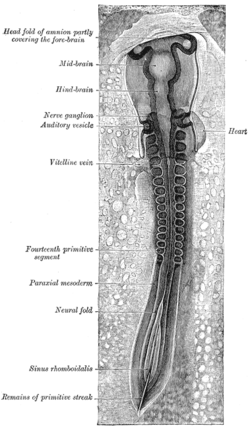
Chick embryo of thirty-three hours’ incubation, viewed from the dorsal aspect. 30x. (Neural fold labeled at center left, third from the bottom.)
1061:
341:
have other roles in the neurulation process, including stimulating the neural crest cells to emigrate from the newly formed neural tube. The
239:, completing the transformation of the embryo from a flattened disk to a three–dimensional body. Cells originating from the fused tips of the
1350:
936:
886:
Kirillova I, Novikova I, Augé J, et al. (May 2000). "Expression of the sonic hedgehog gene in human embryos with neural tube defects".
1010:
899:
758:
486:
721:
280:
The process of folding begins when the cells in the central region of the neural plate, the medial hinge point cells, bind to the
988:
618:
601:
247:) migrate to various locations throughout the embryo, where they will initiate the development of diverse body structures (D).
102:
1178:
407:
403:
351:
The final adhesion of the converging neural folds is due to several different types of intercellular binding proteins.
284:
beneath them. This creates a central anchoring point for the process of folding to occur, and subsequently creates the
305:
1237:
410:
regions are termed the cranial and caudal neuropores. If the caudal neuropore fails to close, a condition called
97:
414:
can occur, in which the bottom of the spinal cord remains exposed. Often this condition can be detected during
364:
The merging of the neural folds gives rise to many structures including the neural tube (the precursor to the
430:, and is subsequently degraded. If the entire neural tube fails to close, the condition is referred to as
790:"Endogenous bone morphogenetic protein antagonists regulate mammalian neural crest generation and survival"
1289:
365:
109:
443:
49:
553:"How to form and close the brain: insight into the mechanism of cranial neural tube closure in mammals"
1185:
1173:
330:
1155:
1108:
768:
533:
397:
244:
1018:
1203:
932:
903:
868:
819:
754:
717:
713:
707:
703:
684:
623:
582:
525:
482:
1294:
1222:
1217:
959:
895:
858:
850:
809:
801:
676:
613:
572:
564:
515:
377:
313:
207:
1266:
1252:
1227:
839:"The BMP antagonist Noggin promotes cranial and spinal neurulation by distinct mechanisms"
325:
220:
44:
255:
from within the cells. The released calcium interacts with proteins that can modify the
1085:
863:
838:
814:
789:
577:
552:
427:
348:
tissues that release these chemical signals, in order to trigger neural plate folding.
343:
219:
directly above it to become the primitive nervous system (i.e., neuroepithelium). The
194:, and consists of epithelial tissue. Here, the epithelial cells elongate by means of
1344:
1279:
1143:
1128:
926:
922:
415:
304:
The molecular mechanism behind this process lies in the expression and repression of
285:
240:
1053:
641:
602:"Cellular mechanisms of neural fold formation and morphogenesis in the chick embryo"
537:
182:
In the embryo, the formation of the neural folds originates from the area where the
1327:
1317:
1299:
1165:
1150:
1133:
1093:
431:
411:
369:
191:
183:
167:
159:
61:
1284:
1274:
1195:
1113:
854:
448:
423:
373:
236:
224:
223:
then folds over (B). As the tips of the folds fuse together, a hollow tube (the
195:
171:
155:
147:
143:
131:
73:
388:
1247:
1232:
1212:
1043:
568:
68:
56:
1242:
1208:
1123:
281:
212:
907:
872:
823:
680:
627:
586:
529:
688:
1322:
900:
10.1002/(SICI)1096-9926(200005)61:5<347::AID-TERA6>3.0.CO;2-#
352:
323:
inhibit these BMPs, and subsequently allow neural commitment genes, like
268:
in a manner that is not part of the normal course of development, severe
264:
260:
232:
228:
216:
187:
163:
151:
150:). In humans, the neural folds are responsible for the formation of the
426:
occurs. In this condition, the brain tissue is directly exposed to the
115:
1118:
805:
619:
10.1002/1097-0185(20010201)262:2<153::AID-AR1021>3.0.CO;2-W
319:
252:
227:) forms (C)—the precursor of the brain and spinal cord. Meanwhile, the
837:
Stottmann RW, Berrong M, Matta K, Choi M, Klingensmith J (July 2006).
520:
503:
1048:
139:
1049:
Anatomy of the Human Body's The Neural Tube and Groove by Henry Gray
788:
Anderson RM, Stottmann RW, Choi M, Klingensmith J (September 2006).
16:
Structure arising during embryonic development of birds and mammals
387:
296:
295:
256:
206:
85:
28:
309:
251:
The formation of the neural fold is initiated by the release of
135:
1057:
504:"Towards a cellular and molecular understanding of neurulation"
753:(4th ed.). Godalming: Springer London. pp. 702–704.
300:
Cross section through the embryonic disc showing the fold.
142:
among other organisms. This structure is associated with
481:(9th ed.). Sunderland, Mass.: Sinauer Associates.
600:
Lawson A, Anderson H, Schoenwolf GC (February 2001).
931:(6th ed.). Sunderland, MA: Sinauer Associates.
422:
If the failure is instead in the cranial neuropore,
190:
converge. This region of the embryo is formed after
1310:
1265:
1194:
1164:
1101:
1092:
749:Khong, Hrsg. Jean W. Keeling; Hrsg. T. Yee (2007).
96:
84:
79:
67:
55:
43:
38:
21:
162:, a preliminary structure consisting of elongated
170:, as well as bringing about the formation of the
235:continue to curve around and fuse to create the
1069:
8:
1044:YouTube Video of Embryonic Chick Neurulation
773:: CS1 maint: multiple names: authors list (
709:Developmental biology protocols, Volume 136
702:Rocky S. Tuan; Cecilia W. Lo, eds. (2000).
103:fold_by_E5.13.1.0.1.0.2 E5.13.1.0.1.0.2
1098:
1076:
1062:
1054:
1011:"7.2: The trilaminar germ disk (3rd week)"
27:
862:
813:
744:
742:
740:
617:
576:
519:
472:
470:
468:
466:
464:
372:(which give rise to a variety of diverse
158:. The neural folds are derived from the
211:A strip of specialized cells called the
551:Yamaguchi Y, Miura M (September 2013).
460:
329:, to be expressed. These genes encode
766:
113:
18:
502:Colas JF, Schoenwolf GC (June 2001).
134:in the embryonic development of both
7:
642:"File:Embryonic Development CNS.gif"
557:Cellular and Molecular Life Sciences
392:A side view of an anencephalic fetus
923:"12: Formation of the Neural Tube"
130:is a structure that arises during
14:
1015:Human Embryology: Embryogenesis
166:cells. The folds give rise to
1:
269:
215:(A) induces the cells of the
1351:Embryology of nervous system
1179:Cardiac neural crest complex
751:Fetal and Neonatal Pathology
855:10.1016/j.ydbio.2006.03.051
306:bone morphogenetic proteins
1367:
477:Gilbert, Scott F. (2010).
395:
569:10.1007/s00018-012-1227-7
108:
26:
985:Learn about Spina Bifida
419:and/or sexual function.
376:cells), and to the true
794:Developmental Dynamics
681:10.1006/dbio.1993.1253
508:Developmental Dynamics
393:
366:central nervous system
301:
248:
110:Anatomical terminology
928:Developmental Biology
843:Developmental Biology
669:Developmental Biology
606:The Anatomical Record
479:Developmental biology
444:Developmental biology
416:prenatal examinations
391:
384:Clinical significance
360:Derivative structures
331:transcription factors
299:
210:
148:secondary neurulation
1186:Truncal neural crest
1174:Cranial neural crest
921:Gilbert, SF (2000).
186:and the surrounding
1084:Development of the
712:. Humama. pp.
144:primary neurulation
1156:Adult neurogenesis
1109:Neural development
1021:on 16 January 2013
981:"SB and the Spine"
806:10.1002/dvdy.20891
432:craniorachischisis
398:Neural tube defect
394:
370:neural crest cells
302:
249:
245:neural crest cells
168:neural crest cells
1338:
1337:
1261:
1260:
1204:Rostral neuropore
938:978-0-87893-243-6
646:Wikimedia Commons
521:10.1002/dvdy.1144
124:
123:
119:
1358:
1295:Surface ectoderm
1223:Cervical flexure
1218:Cephalic flexure
1099:
1078:
1071:
1064:
1055:
1031:
1030:
1028:
1026:
1017:. Archived from
1007:
1001:
1000:
998:
996:
991:on 23 April 2013
987:. Archived from
977:
971:
970:
968:
966:
956:
950:
949:
947:
945:
918:
912:
911:
883:
877:
876:
866:
834:
828:
827:
817:
785:
779:
778:
772:
764:
746:
735:
734:
732:
730:
699:
693:
692:
664:
658:
657:
655:
653:
638:
632:
631:
621:
597:
591:
590:
580:
548:
542:
541:
523:
499:
493:
492:
474:
276:Process overview
116:edit on Wikidata
31:
19:
1366:
1365:
1361:
1360:
1359:
1357:
1356:
1355:
1341:
1340:
1339:
1334:
1306:
1257:
1253:Germinal matrix
1228:Pontine flexure
1190:
1160:
1088:
1082:
1040:
1035:
1034:
1024:
1022:
1009:
1008:
1004:
994:
992:
979:
978:
974:
964:
962:
958:
957:
953:
943:
941:
939:
920:
919:
915:
885:
884:
880:
836:
835:
831:
787:
786:
782:
765:
761:
748:
747:
738:
728:
726:
724:
701:
700:
696:
666:
665:
661:
651:
649:
640:
639:
635:
599:
598:
594:
563:(17): 3171–86.
550:
549:
545:
501:
500:
496:
489:
476:
475:
462:
457:
440:
400:
386:
378:epidermal layer
362:
294:
278:
221:neuroepithelium
205:
180:
120:
34:
17:
12:
11:
5:
1364:
1362:
1354:
1353:
1343:
1342:
1336:
1335:
1333:
1332:
1331:
1330:
1325:
1314:
1312:
1308:
1307:
1305:
1304:
1303:
1302:
1292:
1287:
1282:
1277:
1271:
1269:
1263:
1262:
1259:
1258:
1256:
1255:
1250:
1245:
1240:
1235:
1230:
1225:
1220:
1215:
1206:
1200:
1198:
1192:
1191:
1189:
1188:
1183:
1182:
1181:
1170:
1168:
1162:
1161:
1159:
1158:
1153:
1148:
1147:
1146:
1141:
1131:
1126:
1121:
1116:
1111:
1105:
1103:
1096:
1090:
1089:
1086:nervous system
1083:
1081:
1080:
1073:
1066:
1058:
1052:
1051:
1046:
1039:
1038:External links
1036:
1033:
1032:
1002:
972:
960:"Spina Bifida"
951:
937:
913:
878:
829:
800:(9): 2507–20.
780:
760:978-1846285240
759:
736:
722:
694:
659:
633:
592:
543:
494:
488:978-0878933846
487:
459:
458:
456:
453:
452:
451:
446:
439:
436:
428:amniotic fluid
396:Main article:
385:
382:
361:
358:
344:Sonic hedgehog
293:
290:
277:
274:
204:
201:
179:
176:
122:
121:
112:
106:
105:
100:
94:
93:
91:plica neuralis
88:
82:
81:
77:
76:
71:
65:
64:
59:
53:
52:
47:
45:Carnegie stage
41:
40:
36:
35:
32:
24:
23:
15:
13:
10:
9:
6:
4:
3:
2:
1363:
1352:
1349:
1348:
1346:
1329:
1326:
1324:
1321:
1320:
1319:
1316:
1315:
1313:
1309:
1301:
1298:
1297:
1296:
1293:
1291:
1288:
1286:
1283:
1281:
1280:Optic vesicle
1278:
1276:
1273:
1272:
1270:
1268:
1264:
1254:
1251:
1249:
1246:
1244:
1241:
1239:
1236:
1234:
1231:
1229:
1226:
1224:
1221:
1219:
1216:
1214:
1210:
1207:
1205:
1202:
1201:
1199:
1197:
1193:
1187:
1184:
1180:
1177:
1176:
1175:
1172:
1171:
1169:
1167:
1163:
1157:
1154:
1152:
1149:
1145:
1144:Neural groove
1142:
1140:
1137:
1136:
1135:
1132:
1130:
1129:Neuroectoderm
1127:
1125:
1122:
1120:
1117:
1115:
1112:
1110:
1107:
1106:
1104:
1100:
1097:
1095:
1091:
1087:
1079:
1074:
1072:
1067:
1065:
1060:
1059:
1056:
1050:
1047:
1045:
1042:
1041:
1037:
1020:
1016:
1012:
1006:
1003:
990:
986:
982:
976:
973:
961:
955:
952:
940:
934:
930:
929:
924:
917:
914:
909:
905:
901:
897:
894:(5): 347–54.
893:
889:
882:
879:
874:
870:
865:
860:
856:
852:
849:(2): 647–63.
848:
844:
840:
833:
830:
825:
821:
816:
811:
807:
803:
799:
795:
791:
784:
781:
776:
770:
762:
756:
752:
745:
743:
741:
737:
725:
723:9781592590650
719:
715:
711:
710:
705:
698:
695:
690:
686:
682:
678:
675:(2): 427–40.
674:
670:
663:
660:
647:
643:
637:
634:
629:
625:
620:
615:
612:(2): 153–68.
611:
607:
603:
596:
593:
588:
584:
579:
574:
570:
566:
562:
558:
554:
547:
544:
539:
535:
531:
527:
522:
517:
514:(2): 117–45.
513:
509:
505:
498:
495:
490:
484:
480:
473:
471:
469:
467:
465:
461:
454:
450:
447:
445:
442:
441:
437:
435:
433:
429:
425:
420:
417:
413:
409:
405:
399:
390:
383:
381:
379:
375:
371:
367:
359:
357:
354:
349:
346:
345:
340:
336:
332:
328:
327:
322:
321:
316:
315:
311:
307:
298:
291:
289:
287:
286:neural groove
283:
275:
273:
271:
266:
262:
258:
254:
246:
242:
241:neuroectoderm
238:
234:
230:
226:
222:
218:
214:
209:
202:
200:
197:
193:
189:
185:
177:
175:
173:
169:
165:
161:
157:
153:
149:
145:
141:
137:
133:
129:
117:
111:
107:
104:
101:
99:
95:
92:
89:
87:
83:
78:
75:
72:
70:
69:Gives rise to
66:
63:
60:
58:
54:
51:
48:
46:
42:
37:
30:
25:
20:
1328:Otic vesicle
1318:Otic placode
1300:Lens placode
1166:Neural crest
1151:Neuropoiesis
1138:
1134:Neural plate
1094:Neurogenesis
1023:. Retrieved
1019:the original
1014:
1005:
993:. Retrieved
989:the original
984:
975:
963:. Retrieved
954:
942:. Retrieved
927:
916:
891:
887:
881:
846:
842:
832:
797:
793:
783:
750:
727:. Retrieved
708:
697:
672:
668:
662:
650:. Retrieved
648:. 2012-04-04
645:
636:
609:
605:
595:
560:
556:
546:
511:
507:
497:
478:
421:
412:spina bifida
401:
363:
350:
342:
338:
334:
324:
318:
312:
303:
279:
250:
192:gastrulation
184:neural plate
181:
160:neural plate
127:
125:
90:
62:Neural plate
1285:Optic stalk
1275:Neural tube
1238:Basal plate
1196:Neural tube
1139:Neural fold
1114:Neurulation
944:30 November
449:Neurulation
424:anencephaly
374:mesenchymal
272:can occur.
237:body cavity
225:neural tube
196:microtubule
178:Development
172:neural tube
156:neural tube
154:end of the
132:neurulation
128:neural fold
80:Identifiers
74:Neural tube
22:Neural fold
1248:Neuroblast
1233:Alar plate
1213:Rhombomere
888:Teratology
455:References
1290:Optic cup
1243:Glioblast
1209:Neuromere
1124:Notochord
769:cite book
353:Cadherins
292:Mechanism
282:notochord
261:cadherins
213:notochord
57:Precursor
1345:Category
1323:Otic pit
1025:22 March
908:10777830
873:16712836
824:16894609
628:11169910
587:23242429
538:43666547
530:11376482
438:See also
270:diseases
265:cadherin
233:endoderm
229:ectoderm
217:ectoderm
188:ectoderm
164:ectoderm
152:anterior
1119:Neurula
1102:General
995:1 April
965:1 April
864:3001110
815:6626635
729:1 April
714:125–134
689:8405669
652:1 April
578:3742426
404:cranial
339:Chordin
320:Chordin
253:calcium
203:Folding
140:mammals
39:Details
935:
906:
871:
861:
822:
812:
757:
720:
687:
626:
585:
575:
536:
528:
485:
408:caudal
335:Noggin
314:Noggin
534:S2CID
310:genes
257:actin
136:birds
114:[
86:Latin
1027:2013
997:2013
967:2013
946:2011
933:ISBN
904:PMID
869:PMID
820:PMID
775:link
755:ISBN
731:2013
718:ISBN
704:"15"
685:PMID
654:2013
624:PMID
583:PMID
526:PMID
483:ISBN
406:and
337:and
317:and
231:and
138:and
126:The
1311:Ear
1267:Eye
896:doi
859:PMC
851:doi
847:295
810:PMC
802:doi
798:235
677:doi
673:159
614:doi
610:262
573:PMC
565:doi
516:doi
512:221
368:),
326:SOX
1347::
1211:/
1013:.
983:.
925:.
902:.
892:61
890:.
867:.
857:.
845:.
841:.
818:.
808:.
796:.
792:.
771:}}
767:{{
739:^
716:.
706:.
683:.
671:.
644:.
622:.
608:.
604:.
581:.
571:.
561:70
559:.
555:.
532:.
524:.
510:.
506:.
463:^
434:.
174:.
98:TE
1077:e
1070:t
1063:v
1029:.
999:.
969:.
948:.
910:.
898::
875:.
853::
826:.
804::
777:)
763:.
733:.
691:.
679::
656:.
630:.
616::
589:.
567::
540:.
518::
491:.
243:(
118:]
50:9
Text is available under the Creative Commons Attribution-ShareAlike License. Additional terms may apply.