511:
instigated by a mutation on the ferritin light chain polypeptide (FTL1) and was found to cause iron accumulation in the brain and neurodegeneration. Following the location of the first case of
Neuroferritinopathy, the majority of patients diagnosed with the disease have also been found in Northern and Northeast England. The localization of the majority of cases to Northern and Northeast England suggests that a common ancestor may be responsible for many or possibly all cases. Despite there being fewer than 100 cases reported and the disease's general location of Northern and Northeast England, many more cases of neuroferritinopathy have been diagnosed around the rest of the world in recent years.
72:
503:
224:, found in 7.5% of patients. Full control of upper limbs on the body generally remains until late onset of the disease. Over time, symptoms seen in a patient can change from one side of the body to the opposite side of the body, jumping from left to right or vice versa. Another route that the physically visible symptoms have been observed to take is the appearance, disappearance, and then reappearance once more of specific symptoms.
48:
220:. The symptoms accompanying neuroferritinopathy affecting movement are also progressive, becoming more generalized with time. Usually during the first ten years of onset of the disease only one or two limbs are directly affected. Distinctive symptoms of neuroferritinopathy are chorea, found in 50% of diagnosed patients, dystonia, found in 43% of patients, and
387:
354:(NBIA) disorders which share similar symptoms and imaging findings. Over time single-gene causes have been found for many NBIA disorders, like neuroferritinopathy. Before the availability of genetic testing, all such disorders were considered together and known as Hallervorden-Spatz syndrome, a term which is no longer used due to the
317:
subunits. In neuroferritinopathy, the gene encoding the light chain is mutated. Several different mutation variations have led to diagnosis as neuroferritinopathy; all of these mutations occur in the light chain. A mutated light chain is believed to inhibit ferritin's ability to effectively sequester
510:
Neuroferritinopathy was first discovered in 2001, with its first case being reported in
Cumbria from Northern England. The discovery of neuroferritinopathy was mediated by a study done on a large family suffering from a dominantly inherited basal ganglia disease. It was reported that the disease was
191:
Neuroferritinopathy is mainly seen in those who have reached late adulthood and is generally seen to slowly progress throughout many decades in a lifetime with the mean age of onset being 39 years old. A loss of cognition is generally only seen with late stages of the disease. Diagnosed patients are
142:
Treatment of neuroferritinopathy is focused on managing symptoms associated with chorea and dystonia using standard medications for each. The disorder is progressive and symptoms become worse with age. Fewer than 100 cases of neuroferritinopathy have been reported since its identification in 2001.
235:
Neuroferritinopathy results from abnormal brain iron accumulation. This iron accumulation is due to mutations in the FTL polypeptide, which is responsible for encoding proteins involved in iron metabolism. Neuroferritinopathy is most commonly caused by a single insertion of the nucleotide adenine
183:
and cerebellar cortices. Along with the accumulation of iron in the brain, neuroferritinopathy typically causes severe neuronal loss as well. Secondary symptoms may also arise. It is possible that the initial iron accumulation will cause additional neuronal damage and neuronal death. The damaged
321:
The concentration of iron in a healthy brain varies greatly from region to region. The specific regions of the brain that are associated with motor functions appear to have larger accumulations of iron than non-motor-related regions. This observation of varying iron concentrations is a possible
453:
Due to neuroferritinopathy's genetic etiology, the disorder is not currently curable. Furthermore, progression of the disorder cannot be effectively halted. Therefore current treatment focuses on managing symptoms of the disorder. No medication is available to treat all symptoms.
272:
functions to sequester and release iron, acting as an iron buffering system in cells. Iron is essential to brain function in oxygen transport and cellular metabolism for example. However, careful control of iron is important as increased brain iron levels catalyze the formation of
244:
varies by family. Neuroferritinopathy may also be caused by the insertion of two extra nucleotide bases. The insertion of bases into the L-chain ferritin gene causes the chain to lengthen and alter the sequence of the amino acids found in the gene, also known as a
362:, genetic systemic iron accumulation with neurologic features, and acquired diseases associated with iron excess or iron deficiency. Neuroferritinopathy is classified under the first category. Neuroferritinopathy is classified as a late-onset
1022:
423:
ferritin levels. However this is unreliable as method of diagnosis since some patients show typical serum ferritin levels even at the latest stages of neuroferritinopathy. Cerebral spinal fluid tests also are typically normal. Ferritin
134:
protein. Wild type ferritin functions as a buffer for iron, sequestering it and controlling its release. Thus, mutations in the light chain of ferritin result in the accumulation of iron in the brain which can be imaged using
398:
and other neuroimaging techniques. MRIs help identify the iron deposits in the cerebellum, basal ganglia, and motor cortex common to neuroferritinopathy. MRIs of affected individuals also show mild cerebellar and
444:
Genetic testing can confirm a neuroferritinopathy diagnosis. A diagnosis can be made by analyzing the protein sequences of affected individuals and comparing them to known neuroferritinopathy sequences.
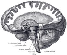
260:, which have damaging effects to the brain. The iron accumulation characteristic of neuroferritinopathy particularly affects the cerebellum, basal ganglia, and motor cortex regions of the brain.
407:. Most importantly, the MRIs show misfolded ferritin proteins and iron deposits in the glial cells of the caudate, putamen, globus pallidus, cerebral cortex, thalamus, and
350:
Neuroferritinopathy was originally described with hallmark features of neurodegeneration and iron accumulation in the brain, leading it to be classified with other
359:
351:
338:, as iron accumulates in the brain over long periods of time. Neuroferritinopathy is diagnosed using either neuroimaging techniques, physiological tests, or
139:. Currently, neuroferritinopathy is the only neurodegenerative disease with an iron accumulation in the brain classified as an autosomal dominant syndrome.
184:
neurons may be replaced by other cells in an effort to reverse the neurodegeneration. These cells often have a higher iron content. The breakdown of the
155:
Neuroferritinopathy has several distinguishing signs and symptoms. These fall into two categories: diagnostic findings and physically visible symptoms.
419:
Blood tests usually come back normal in affected individuals so they do not serve as a reliable means of diagnosis. Blood tests can show low
1037:
917:
778:
685:
Zecca, L; Youdim, MB; Riederer, P; Connor, JR; Crichton, RR (November 2004). "Iron, brain ageing and neurodegenerative disorders".
1110:
256:, or programmed cell death. Accumulation of iron in the brain is extremely dangerous as excess iron catalyzes the formation of
188:
may also occur due to the loss of neurons and will subsequently allow more iron to access the brain and accumulate over time.
147:. Due to its genetic nature, current research is focused on therapeutic management of the symptoms caused by the disorder.
322:
explanation for the correlation between movement disorders and the iron imbalance within the central nervous system.
227:
While these symptoms are the classic indicators of neuroferritinopathy, symptoms will vary from patient to patient.
395:
519:
New potential treatment options being researched are
Venesection (removing red blood cells), Iron chelation with
252:
These mutations result in decreased iron-binding ability. The oxidative damage caused by increased iron leads to
641:
Chinnery, PF; Pagon, RA; Adam, MP; Ardinger, HH; Bird, TD; Dolan, CR; Fong, CT; Smith, RJH; Stephens, K (2010).
143:
Its incidence has been largely localized to
Northwest England, significantly in the Cumbria region suggesting a
1048:
802:
Batista-Nascimento, Liliana; Pimentel, Catarina; Andrade
Menezes, Regina; Rodrigues-Pousada, Claudina (2012).
171:. Patients who are diagnosed with neuroferritinopathy have abnormal iron accumulation in the brain within the
462:
shown to help with involuntary movements. Symptoms affecting movement (dystonia) have also been treated with
281:. These oxidative molecules can cause oxidative brain damage. Iron that is bound to ferritin in nonreactive.
192:
seen to retain most of their cognitive functioning until the most progressive stages of the illness sets in.
163:
Symptoms categorized as medically tested and diagnosed include iron accumulation in the brain, basal ganglia
425:
105:
1088:
335:
331:
108:
in nature, progress slowly and generally do not become apparent until adulthood. These symptoms include
318:
and hold iron. Without control of iron, it is free to cause oxidative brain damage as described above.
429:
428:
found in the skin, liver, kidney and muscle tissues may help in diagnosing neuroferritinopathy. More
375:
314:
213:
127:
123:
71:
278:
274:
246:
185:
882:
710:
217:
17:
502:
330:
Neuroferritinopathy is primarily diagnosed in older adults, specifically in adults affected by
985:
923:
913:
874:
835:
784:
774:
702:
646:
614:
168:
85:
60:
975:
965:
905:
866:
825:
815:
766:
694:
606:
400:
597:
Lehn, A; Boyle, R; Brown, H; Airey, C; Mellick, G (September 2012). "Neuroferritinopathy".
455:
339:
257:
237:
378:(FTL) polypeptide gene while the fourth arises from a missense mutation in the FTL gene.
980:
953:
909:
830:
803:
770:
408:
144:
1104:
714:
610:
459:
420:
363:
93:
886:
592:
590:
588:
586:
584:
582:
580:
578:
576:
574:
572:
570:
568:
566:
564:
562:
560:
524:
467:
358:
ties of the namesakes. Brain iron disorders are now divided into three categories:
221:
101:
1053:
558:
556:
554:
552:
550:
548:
546:
544:
542:
540:
954:"Neurodegeneration with brain iron accumulation: update on pathogenic mechanisms"
752:
750:
748:
746:
744:
240:
end of the entire protein chain. However, exact location of the insertion in the
742:
740:
738:
736:
734:
732:
730:
728:
726:
724:
520:
366:
disease and is a dominantly inherited neurodegenerative disease. Four different
1031:
642:
483:
371:
355:
209:
164:
97:
970:
1059:
475:
471:
253:
120:
65:
989:
927:
878:
839:
788:
706:
650:
618:
820:
1083:
855:"Iron metabolism in the CNS: implications for neurodegenerative diseases"
479:
433:
269:
205:
180:
131:
113:
1014:
386:
404:
172:
1026:
491:
487:
463:
367:
201:
109:
870:
854:
698:
757:
Keogh, MJ; Morris, CM; Chinnery, PF (2013). "Neuroferritinopathy".
47:
1064:
904:. Handbook of Clinical Neurology. Vol. 120. pp. 851–64.
804:"Iron and Neurodegeneration: From Cellular Homeostasis to Disease"
501:
458:
has been shown to help with focal dystonia. The dopamine depleter
385:
1042:
947:
945:
943:
941:
939:
937:
636:
634:
632:
630:
628:
310:
241:
176:
89:
136:
236:
into the gene for L-chain ferritin which in turn, alters the
200:
Symptoms categorized as physically visible symptoms include
370:
are responsible for neuroferritinopathy. Three arise from
403:, or tissue breakdown, and gas cavity formation in the
360:
genetic neurodegeneration with brain iron accumulation
394:
Neuroferritinopathy is most commonly diagnosed using
1004:
432:-negative fibers are also often found in the muscle
216:, all physical symptoms of the body associated with
126:
and is caused by mutations in the gene encoding the
1074:
1008:
59:
37:
32:
116:, and cognitive deficits which worsen with age.
490:. Parkinsonian symptoms were not decreased by
352:neurodegeneration with brain iron accumulation
8:
1005:
952:Levi, Sonia; Finazzi, Dario (7 May 2014).
70:
46:
29:
979:
969:
829:
819:
808:Oxidative Medicine and Cellular Longevity
411:, causing neuronal death in these areas.
680:
277:that create oxidative molecules via the
104:of the human brain. Symptoms, which are
678:
676:
674:
672:
670:
668:
666:
664:
662:
660:
536:
494:. Iron supplements should be avoided.
88:characterized by the accumulation of
7:
759:International Review of Neurobiology
599:Parkinsonism & Related Disorders
309:The ferritin protein is made up of
910:10.1016/B978-0-7020-4087-0.00057-7
853:Rouault, Tracey A. (3 July 2013).
771:10.1016/B978-0-12-410502-7.00006-5
25:
900:Woimant, F; Trocello, JM (2014).
41:Adult-onset basal ganglia disease
18:Adult-onset basal ganglia disease
611:10.1016/j.parkreldis.2012.06.021
1:
55:Cerebellum and basal ganglia
859:Nature Reviews Neuroscience
687:Nature Reviews Neuroscience
1127:
86:neurodegenerative disorder
958:Frontiers in Pharmacology
902:Disorders of heavy metals
525:Coenzyme Q10 (ubiquinone)
436:of affected individuals.
54:
45:
1054:C548080 C548080, C548080
971:10.3389/fphar.2014.00099
1111:Neurological disorders
507:
391:
643:"Neuroferritinopathy"
505:
415:Physiological testing
389:
430:cytochrome c oxidase
376:ferritin light chain
821:10.1155/2012/128647
336:Parkinson's disease
332:Alzheimer's disease
247:frameshift mutation
186:blood brain barrier
159:Diagnostic findings
82:Neuroferritinopathy
33:Neuroferritinopathy
1075:External resources
508:
392:
374:insertions in the
218:movement disorders
151:Signs and symptoms
1098:
1097:
196:Physical symptoms
169:neurodegeneration
119:This disorder is
79:
78:
27:Medical condition
16:(Redirected from
1118:
1006:
994:
993:
983:
973:
949:
932:
931:
897:
891:
890:
850:
844:
843:
833:
823:
799:
793:
792:
754:
719:
718:
682:
655:
654:
638:
623:
622:
594:
401:cerebral atrophy
306:→ Fe + HOO• + H
295:→ Fe + HO• + OH
284:Fenton Reaction
75:
74:
50:
30:
21:
1126:
1125:
1121:
1120:
1119:
1117:
1116:
1115:
1101:
1100:
1099:
1094:
1093:
1070:
1069:
1017:
1003:
998:
997:
951:
950:
935:
920:
899:
898:
894:
871:10.1038/nrn3453
852:
851:
847:
801:
800:
796:
781:
756:
755:
722:
699:10.1038/nrn1537
684:
683:
658:
640:
639:
626:
596:
595:
538:
533:
517:
500:
451:
442:
440:Genetic testing
417:
384:
348:
340:genetic testing
328:
315:light chain (L)
311:heavy chain (H)
305:
301:
294:
290:
279:Fenton Reaction
266:
233:
198:
161:
153:
130:subunit of the
69:
28:
23:
22:
15:
12:
11:
5:
1124:
1122:
1114:
1113:
1103:
1102:
1096:
1095:
1092:
1091:
1079:
1078:
1076:
1072:
1071:
1068:
1067:
1056:
1045:
1034:
1018:
1013:
1012:
1010:
1009:Classification
1002:
1001:External links
999:
996:
995:
933:
918:
892:
865:(8): 551–564.
845:
794:
779:
720:
693:(11): 863–73.
656:
624:
535:
534:
532:
529:
516:
513:
506:Map of England
499:
496:
450:
447:
441:
438:
416:
413:
409:purkinje cells
383:
380:
347:
346:Classification
344:
327:
324:
303:
299:
292:
288:
265:
262:
232:
229:
197:
194:
160:
157:
152:
149:
145:founder effect
106:extrapyramidal
77:
76:
63:
57:
56:
52:
51:
43:
42:
39:
35:
34:
26:
24:
14:
13:
10:
9:
6:
4:
3:
2:
1123:
1112:
1109:
1108:
1106:
1090:
1086:
1085:
1081:
1080:
1077:
1073:
1066:
1062:
1061:
1057:
1055:
1051:
1050:
1046:
1044:
1040:
1039:
1035:
1033:
1029:
1028:
1024:
1020:
1019:
1016:
1011:
1007:
1000:
991:
987:
982:
977:
972:
967:
963:
959:
955:
948:
946:
944:
942:
940:
938:
934:
929:
925:
921:
919:9780702040870
915:
911:
907:
903:
896:
893:
888:
884:
880:
876:
872:
868:
864:
860:
856:
849:
846:
841:
837:
832:
827:
822:
817:
813:
809:
805:
798:
795:
790:
786:
782:
780:9780124105027
776:
772:
768:
764:
760:
753:
751:
749:
747:
745:
743:
741:
739:
737:
735:
733:
731:
729:
727:
725:
721:
716:
712:
708:
704:
700:
696:
692:
688:
681:
679:
677:
675:
673:
671:
669:
667:
665:
663:
661:
657:
652:
648:
644:
637:
635:
633:
631:
629:
625:
620:
616:
612:
608:
605:(8): 909–15.
604:
600:
593:
591:
589:
587:
585:
583:
581:
579:
577:
575:
573:
571:
569:
567:
565:
563:
561:
559:
557:
555:
553:
551:
549:
547:
545:
543:
541:
537:
530:
528:
526:
522:
514:
512:
504:
497:
495:
493:
489:
485:
481:
477:
473:
469:
465:
461:
460:Tetrabenazine
457:
448:
446:
439:
437:
435:
431:
427:
422:
414:
412:
410:
406:
402:
397:
388:
381:
379:
377:
373:
369:
365:
364:basal ganglia
361:
357:
353:
345:
343:
341:
337:
333:
325:
323:
319:
316:
312:
307:
296:
285:
282:
280:
276:
275:free radicals
271:
263:
261:
259:
258:free radicals
255:
250:
248:
243:
239:
230:
228:
225:
223:
219:
215:
211:
207:
203:
195:
193:
189:
187:
182:
178:
174:
170:
166:
158:
156:
150:
148:
146:
140:
138:
133:
129:
125:
122:
117:
115:
111:
107:
103:
99:
95:
94:basal ganglia
91:
87:
84:is a genetic
83:
73:
67:
64:
62:
58:
53:
49:
44:
40:
36:
31:
19:
1082:
1058:
1047:
1036:
1021:
961:
957:
901:
895:
862:
858:
848:
811:
807:
797:
762:
758:
690:
686:
602:
598:
518:
509:
498:Epidemiology
468:orphenadrine
452:
443:
418:
393:
382:Neuroimaging
349:
329:
320:
308:
297:
286:
283:
268:The protein
267:
251:
234:
226:
222:parkinsonism
199:
190:
162:
154:
141:
118:
102:motor cortex
81:
80:
521:deferiprone
128:light chain
38:Other names
814:: 128647.
765:: 91–123.
531:References
484:clonazepam
426:aggregates
372:nucleotide
356:Nazi party
298:(2) Fe + H
287:(1) Fe + H
210:spasticity
165:cavitation
98:cerebellum
1065:699299001
1060:SNOMED CT
715:205500060
476:sulpiride
472:benzhexol
449:Treatment
326:Diagnosis
264:Mechanism
254:apoptosis
121:autosomal
66:Neurology
61:Specialty
1105:Category
1084:Orphanet
990:24847269
928:24365357
887:21302204
879:23820773
840:22701145
789:24209436
707:15496864
651:20301320
619:22818529
515:Research
480:diazepam
434:biopsies
270:ferritin
238:carboxyl
214:rigidity
206:dystonia
181:striatum
132:ferritin
124:dominant
114:dystonia
981:4019866
831:3369498
405:putamen
368:alleles
179:of the
173:neurons
92:in the
1089:157846
1043:606159
988:
978:
964:: 99.
926:
916:
885:
877:
838:
828:
787:
777:
713:
705:
649:
617:
523:, and
492:L-Dopa
488:deanol
486:, and
464:L-Dopa
231:Causes
212:, and
202:chorea
167:, and
110:chorea
100:, and
68:
1032:G23.0
883:S2CID
711:S2CID
456:Botox
421:serum
1049:MeSH
1038:OMIM
986:PMID
924:PMID
914:ISBN
875:PMID
836:PMID
812:2012
785:PMID
775:ISBN
703:PMID
647:PMID
615:PMID
313:and
242:exon
177:glia
175:and
90:iron
1023:ICD
976:PMC
966:doi
906:doi
867:doi
826:PMC
816:doi
767:doi
763:110
695:doi
607:doi
396:MRI
390:MRI
334:or
137:MRI
1107::
1087::
1063::
1052::
1041::
1030::
1027:10
984:.
974:.
960:.
956:.
936:^
922:.
912:.
881:.
873:.
863:14
861:.
857:.
834:.
824:.
810:.
806:.
783:.
773:.
761:.
723:^
709:.
701:.
689:.
659:^
645:.
627:^
613:.
603:18
601:.
539:^
527:.
482:,
478:,
474:,
470:,
466:,
342:.
249:.
208:,
204:,
112:,
96:,
1025:-
1015:D
992:.
968::
962:5
930:.
908::
889:.
869::
842:.
818::
791:.
769::
717:.
697::
691:5
653:.
621:.
609::
304:2
302:O
300:2
293:2
291:O
289:2
20:)
Text is available under the Creative Commons Attribution-ShareAlike License. Additional terms may apply.