427:. The nonmotor domain contains output channels involved in cognition and visuospatial function, and projections to the prefrontal and posterior parietal cortical areas within this region are clustered into distinct regions with little overlap. These areas are activated during tasks involving short-term working memory, rule-based learning, and higher executive function-like planning. Although the ventral aspect of the dentate has been shown to be involved in acquisition of information, whether it is involved in retention and storage remains unclear.
236:
334:
functions, such as language and cognition, as well as versatile and coordinated finger movement. While it is generally accepted that the ventral region is more recent on an evolutionary timescale, current 3-Dimensional imaging raises questions regarding this assumption, as a third axis, the rostrocaudal axis, can now be analyzed. In addition, current images show that the ventral region is not physically larger than the dorsal region in humans, as would be predicted if size increases with cognitive function.
290:: The globose and emboliform nuclei together make up the interposed nucleus. The interposed nucleus is the smallest of the cerebellar nuclei. It is located between the dentate and fastigial nuclei. It receives afferent supply from the anterior (toward the front) lobe of the cerebellum and sends output to the contralateral red nucleus through the superior cerebellar peduncle. This nucleus is the origin of the rubrospinal tract that mainly influences limb flexor muscles.
636:
624:
612:
648:
378:: The intermediate fusiform neurons are scattered throughout the dentate, and they have elongated and elliptical somata which are tapered at both ends. They include three to five primary dendrites which divide into several long dendrites. The upper part of the cell body is parallel to the apical dendrites, which are oriented toward the nuclear core.
366:: The border neurons are concentrated at the boundary of the nuclear lamina and have elliptical somata. They have a stout axon directed into the surrounding white matter, as well as four to six primary dendrites that branch from the opposite end. The dendritic fields of these neurons have a tetrahedron shape with the cell body in one corner.
423:
via efferents to
Pontine nuclei, and cortical areas that do not project onto the cerebellum are not targets of dentate output. The motor domain in the dorsal portion of the dentate contains output channels that control both generation and control of movement, as well as neurons that innervate premotor areas in the
36:
481:
The dentate nucleus is responsible for the planning, initiation and control of voluntary movements. The dorsal region of the dentate contains output channels involved in motor function, while the ventral region contains output channels involved in nonmotor function, such as cognition and visuospatial
476:
The whole cerebellum has only one output, which necessarily leads from the deep cerebellar nuclei. There is output from the cerebellar cortex, so this output must go through the cerebellar nuclei and send output to rest of nervous system. Thus, the cerebellum communicates to the outside world via the
422:
areas of the cerebral cortex. The motor and nonmotor domains make up approximately 50–60 percent and 20 percent, respectively, of the dentate. It is estimated that the human dentate proportions are comparable. All cerebral cortical areas that are targeted by the dentate project back on the cerebellum
209:
The dentate nucleus is highly convoluted, with gyri (ridges on the cerebral cortex) and sulci (furrows or grooves on the cerebral cortex). Its formation is coincident with a critical period of extensive growth in the fetal dentate. The dentate nucleus becomes visible in the cerebellar white matter as
443:
Dentate nucleus axons can be divergent or convergent. Convergent branches include those that originate from entirely different axons and traverse different routes, yet overlap on the same terminal field. Divergent pathways originate from the same axon but travel different routes and target different
401:
The dentate contains anatomically separate and functionally distinct motor and nonmotor domains (dorsal and ventral, respectively), and projections are organized from the dentate nucleus to distinct areas in the ventrolateral thalamus. Through the thalamus, the dorsal parts of the dentate project to
460:
specific mechanism of this remains unclear. For example, the act of lifting an object requires sensory input, such as the size of the object and where it is located in space. While the primary role of the dentate nucleus is control of the movement, it does play a role in sensory processing as well.
459:
input, and all of these stimulate the dentate nucleus. The dentate nucleus is mostly responsible for planning and execution of fine movement. Since any motor function requires sensory information, it can be assumed that the dentate nucleus receives and modulates this sensory information, though the
387:
Small local circuit neurons include signaling pathways that are contained within the dentate. These neurons provide feedback to the dentate and allow for fine control of signaling. Currently, less research has been conducted on the specific shape and role of these neurons, as the dentate nucleus is
435:
There are three distinct routes from the dentate nucleus to the ventrolateral thalamus, including a direct, posterior, and anterior route. The direct route passes in the anterolateral direction under the thalamus and enters from the ventral side. Axons following the posterior pathway turn dorsally
333:
The dentate nucleus is highly convoluted and can be divided into dorsal (motor) and ventral (nonmotor) domains. The ventral half is much more developed in humans than in great apes, and it appears to play an important role in fiber connection. Further, the ventral domain mediates higher cerebellar
250:
The architecture of cerebellum has a form that resembles the structure of a crystal, so a slice anywhere in the cerebellum gives repeated architecture. The eight cerebellar nuclei, located within the deep white matter of each cerebellar hemisphere, are grouped into pairs, with one of each pair in
472:
The dentate nucleus is involved in basic circuitry work, including input to the cerebellum from everywhere else. Any function that needs coordination, including thoughts and motor behavior, must go through the cerebellum to be smoothened. This input travels in two parts, to the surface of the
440:. Within the lamina, fibers turn posteriorly and enter the dorsal side of the thalamus. Therefore, as a result of these various pathways, the neurons of the dentate nucleus are able to traverse all thalamic nuclei, with the exception of those at the midline and anterior nuclear groups.
794:
Ristanovic, D., Milosevic, N. T., Stefanovic, B. D., Maric, D. L., & Rajkovic, K. (2010). Morphology and classification of large neurons in the adult human dentate nucleus: A qualitative and quantitative analysis of 2D images. . Neuroscience
Research, 67(1),
444:
terminal fields. Although no point-to-point connectivity has been observed between the dentate nucleus and the thalamus, it is believed that there is a pre-wired connectivity between a single dentate site and several body part representations in the thalamus.
600:: There is an increase in mean volume of large neurons and a decrease in mean volume of small neurons in the dentate nucleus in AD with myoclonus. Morphological changes in the dentate nucleus may contribute to the pathological substrate of myoclonus in AD.
210:
early as 11–12 weeks of gestation, containing only smooth lateral (towards the side(s) or away from the midline) and medial (towards the midline) surfaces. During this time, the neurons of the dentate nucleus are similar in shape and form, being mainly
856:
Mason, A., Ilinsky, I. A., Maldonado, S., & Kultas-Ilinsky, K. (2000). Thalamic terminal fields of individual axons from the ventral part of the dentate nucleus of the cerebellum in Macaca mulatta. . Journal of
Comparative Neurology, 421(3),
372:: The intermediate asymmetrical neurons are evenly distributed throughout the nuclear mass, and they have large, elliptical somata. They have five to nine dendrites branching in all directions, with one or two much longer than the others.
196:
nucleus. The dentate nucleus is responsible for the planning, initiation and control of voluntary movements. The dorsal region of the dentate nucleus contains output channels involved in motor function, which is the movement of
886:
Fukutani, Y., Cairns, N. J., Everall, I. P., Chadwick, A., Isaki, K., & Lantos, P. L. (1999). Cerebellar dentate nucleus in
Alzheimer's disease with myoclonus. . Dementia and Geriatric Cognitive Disorders, 10(2),
721:
Milosevic, N. T., Ristanovic, D., Maric, D. L., & Rajkovic, K. (2010). Morphology and cell classification of large neurons in the adult human dentate nucleus: A quantitative study. . Neuroscience
Letters, 468(1),
271:
most recent of the cerebellar nuclei. It receives afferent, or incoming, signals from the premotor cortex and supplementary motor cortex via the pontocerebellar system. Efferent, or outgoing, signals travel via the
217:
During 22–28 weeks of gestation, which is the critical period in fetal development of the dentate nucleus, gyral formation occurs extensively over the entire surface. Here, neurons mature into various forms of
843:
Mediavilla, C., Molina, F., & Puerto, A. (2000). Retention of concurrent taste aversion learning after electrolytic lesioning of the interpositus-dentate region of the cerebellum. . Brain
Research, 868(2),
312:
in the form of inhibition. Neurons in the cerebellar nuclei generate spontaneous action potentials despite ongoing inhibition from
Purkinje cells. The cerebellar nuclei receive afferent projections from the
40:
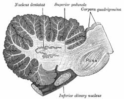
Sagittal section through right cerebellar hemisphere. The right olive, "inferior olivary nucleus", has also been cut sagittally – from front to back. (Dentate nucleus, "nucleus dentatus", labeled at top.)
477:
cerebellar nuclei. Input that reaches the cerebellar cortex is processed in many ways; eventually whatever happens in cerebellar cortex exits the cerebellum through a synapse in the cerebellar nuclei.
436:
from the midline at the posterior side of the thalamus, entering the thalamus from various positions. Axons following the anterior route pass laterally in the subthalamus and enter the external
822:
Matano, S. (2001). Brief communication: Proportions of the ventral half of the cerebellar dentate nucleus in humans and great apes. . American
Journal of Physical Anthropology, 114(2), 163–165.
528:: Clinical and pathological symptoms usually appear in the first year of life and include psychomotor retardation and brain stem dysfunction. Bilaterally symmetric defects are seen in the
738:
Yamaguchi, K., & Goto, N. (1997). Three-dimensional structure of the human cerebellar dentate nucleus: a computerized reconstruction study. . Anatomy and
Embryology, 196(4), 343–348.
877:
McErlean, A., Abdalla, K., Donoghue, V., & Ryan, S. (2010). The dentate nucleus in children: normal development and patterns of disease. . Pediatric
Radiology, 40(3), 326–339.
834:
Dum, R. P., & Strick, P. L. (2003). An unfolded map of the cerebellar dentate nucleus and its projections to the cerebral cortex. . Journal of Neurophysiology, 89(1), 634–639.
588:: LCH is an aggressive disorder due to proliferation of Langerhans cell histiocytes, and the dentate nucleus is believed to be involved in up to 40 percent of patients.
579:. The hallmark of NF1 is the development of numerous tumors. Cerebellar white matter and dentate nucleus lesions usually occur in children less than ten years of age.
144:
911:
172:
is a cluster of neurons, or nerve cells, in the central nervous system that has a dentate – tooth-like or serrated – edge. It is located within the deep
360:, and numerous dendritic trunks emerge from every direction of the soma. These dendrites have complex branching patterns and spherical dendritic fields.
756:
Saab, C. Y., & Willis, W. D. (2003). The cerebellum: organization, functions and its role in nociception. . Brain Research Reviews, 42(1), 85–95.
1410:
345:
The large principal neurons have been classified into four primary types according to position within the dentate, shape of soma (cell body), and
1552:
1500:
1455:
1446:
251:
each of the two hemispheres. As a chunk of tissue, the dentate nucleus with overlying cerebellar cortex makes up a functional unit called the
120:
325:. Together, the deep cerebellar nuclei form a functional unit that provides feedback control of the cerebellar cortex by cerebellar output.
952:
707:
Sultan, F., Hamodeh, S., & Baizer, J. S. (2010). THE HUMAN DENTATE NUCLEUS: A COMPLEX SHAPE UNTANGLED. . Neuroscience, 167(4), 965–968.
337:
The neurons of the adult dentate are divided based on size, morphology, and function into large principal and small local circuit neurons.
805:
201:, while the ventral region contains output channels involved in nonmotor function, such as conscious thought and visuospatial function.
921:
1746:
1562:
925:
2389:
1517:
1331:
996:
151:
2094:
1633:
1537:
356:: The central neurons are located in deeper parts of the nuclear mass, away from the periphery. They have round and prickly
2452:
2426:
1541:
1368:
1020:
583:
437:
139:
912:
https://web.archive.org/web/20150621011739/http://www.mona.uwi.edu/fpas/courses/physiology/neurophysiology/Cerebellum.htm
2457:
2431:
2394:
2360:
2355:
2260:
2227:
2191:
2141:
2006:
1763:
1621:
1364:
1321:
1317:
991:
635:
623:
273:
2398:
1759:
1617:
1403:
986:
2350:
2166:
2064:
1988:
1946:
1353:
1346:
611:
546:
235:
66:
905:
899:
349:
branching. These neurons are responsible for communication between the dentate nucleus and the cerebellar cortex.
2384:
2302:
2136:
1892:
1862:
1645:
1612:
1340:
961:
508:: An inherited disorder of amino acid metabolism in newborns, MSUD results in neurological deterioration. Myelin
503:
318:
252:
184:
to the rest of the brain. It is the largest and most lateral, or farthest from the midline, of the four pairs of
2181:
2068:
1992:
1802:
1654:
1260:
945:
570:
540:
482:
function. The dentate nucleus sends commands and information to the motor and premotor areas in the forebrain.
20:
2282:
2269:
2241:
2174:
2149:
2056:
2024:
1980:
1750:
1521:
1084:
2203:
2170:
2161:
2040:
2036:
2028:
1964:
1769:
1396:
1357:
1142:
1008:
674:
592:
185:
127:
115:
2319:
2256:
2232:
2223:
2195:
2186:
2083:
2044:
1968:
1871:
1779:
1697:
1373:
1326:
1299:
1091:
1055:
177:
2380:
2346:
2297:
1812:
1798:
1742:
1688:
1580:
1529:
1388:
1003:
553:
517:
456:
403:
95:
2491:
2264:
2052:
2032:
1976:
1901:
1896:
1807:
1693:
1683:
1659:
1629:
1624:
1511:
1336:
1309:
1250:
1100:
1077:
1050:
938:
529:
2447:
2421:
2252:
1482:
1478:
1464:
1162:
1156:
419:
189:
2435:
2364:
2307:
2293:
2131:
2048:
1972:
1920:
1866:
1841:
1641:
1604:
1566:
1468:
1174:
1117:
1036:
809:
669:
491:
415:
301:
255:. Thus, there is a part of cerebellum that communicates exclusively with the dentate nucleus.
219:
490:
Several pathological processes involve the dentate nucleus including metabolic, genetic, and
1755:
1731:
1705:
1525:
1473:
1013:
916:
766:
268:
188:, the others being the globose and emboliform nuclei, which together are referred to as the
647:
1915:
1663:
1490:
1486:
1167:
1112:
1072:
679:
411:
198:
1226:
2376:
2342:
2328:
2156:
1816:
1431:
1427:
1270:
1265:
1045:
558:
448:
322:
314:
309:
2485:
2199:
1936:
1608:
1216:
1197:
1067:
533:
524:
211:
2286:
2245:
2072:
2016:
1996:
1956:
1284:
1245:
576:
561:
deficiency. The dentate nucleus is not affected until late in disease progression.
424:
308:
The deep cerebellar nuclei receive the final output from the cerebellar cortex via
173:
243:
296:: The fastigial nucleus is the most medial. It receives afferent input from the
108:
2274:
1836:
1673:
1594:
1423:
1419:
1202:
1133:
452:
277:
2461:
2443:
2417:
2409:
2372:
2338:
2145:
2116:
1240:
1221:
1184:
965:
664:
513:
239:
181:
90:
54:
549:
deficiency. Abnormalities are seen in the basal ganglia and dentate nucleus.
132:
930:
596:
280:
to the contralateral – opposite side – Ventroanterior/Ventrolateral (VA/VL)
193:
222:, and the most frequent neuronal types are medium-sized to large neurons.
2278:
2237:
2098:
2060:
2020:
1984:
1960:
1701:
1637:
1533:
684:
407:
346:
281:
102:
157:
473:
cerebellar cortex as well as collateral input to the cerebellar nuclei.
468:
The role of the dentate nucleus can be described in two basic tenets:
357:
297:
61:
300:, and efferents travel via the inferior cerebellar peduncle to the
35:
509:
234:
78:
19:
This article is about the dentate nucleus. For other dentate, see
1585:
1392:
934:
512:
is seen in the cerebellum, including the dentate nucleus,
917:
http://www.neuroanatomy.wisc.edu/cere/text/P5/dentate.htm
267:: The dentate nucleus is the largest, most lateral, and
447:
Three modalities are received by cerebellum, including
557:: Canavan's disease is a white matter disease due to
321:, upper cervical and lumbar spinal segments, and the
180:, and it is the largest single structure linking the
414:, while the ventral parts of the dentate project to
2408:
2327:
2316:
2212:
2124:
2115:
2080:
2004:
1944:
1935:
1881:
1851:
1825:
1787:
1778:
1730:
1723:
1672:
1593:
1579:
1551:
1499:
1454:
1445:
1438:
1308:
1292:
1283:
1183:
1141:
1132:
1029:
979:
972:
138:
126:
114:
101:
89:
77:
72:
60:
50:
45:
28:
16:
Nucleus in the centre of each cerebellar hemisphere
545:: An autosomal recessive disease, GA1 is due to
388:composed primarily of large principal neurons.
1404:
946:
8:
790:
788:
786:
784:
782:
780:
778:
776:
641:Deep dissection of brain-stem. Lateral view.
629:Deep dissection of brain-stem. Lateral view.
908:at the University of Michigan Health System
902:at the University of Michigan Health System
873:
871:
869:
867:
865:
863:
734:
732:
730:
728:
717:
715:
713:
2324:
2121:
1941:
1784:
1727:
1590:
1451:
1442:
1411:
1397:
1389:
1289:
1222:Bergmann glia cell = Golgi epithelial cell
1138:
976:
953:
939:
931:
34:
2298:Flocculonodular lobe/vestibulocerebellum
852:
850:
703:
701:
699:
2257:Intermediate hemisphere/spinocerebellum
695:
617:Dissection of brain-stem. Lateral view.
607:
494:disorders, as well as some infections.
2427:Ventral/anterior spinocerebellar tract
2356:Dorsal/posterior spinocerebellar tract
752:
750:
748:
746:
744:
155:
25:
830:
828:
7:
767:"Cerebellum - Deep Nuclei - Dentate"
242:of the dentate nucleus (pale pink).
2224:Lateral hemisphere/pontocerebellum
1747:Posterior limb of internal capsule
1563:Posterior limb of internal capsule
926:Neuroscience Information Framework
14:
1518:Posterior external arcuate fibers
547:glutaryl-coenzyme A dehydrogenase
806:"Chapter 8B: Cerebellar Systems"
646:
634:
622:
610:
152:Anatomical terms of neuroanatomy
575:: NF1 is an autosomal dominant
370:Intermediate asymmetric neurons
1:
2453:Rostral spinocerebellar tract
1369:Ventral spinocerebellar tract
584:Langerhans cell histiocytosis
376:Intermediate fusiform neurons
1322:Dorsal spinocerebellar tract
922:NIF Search – Dentate Nucleus
274:superior cerebellar peduncle
2399:Anterior lobe of cerebellum
383:Small local circuit neurons
2508:
2351:Posterior thoracic nucleus
2065:Thalamocortical radiations
1989:Thalamocortical radiations
1347:Trigeminocerebellar fibers
67:superior cerebellar artery
18:
2385:Accessory cuneate nucleus
2303:Vestibulocerebellar tract
2137:Vestibulocerebellar tract
1911:
1646:Posterior parietal cortex
1613:Anterior white commissure
1341:Vestibulocerebellar tract
504:Maple syrup urine disease
319:lateral reticular nucleus
150:
33:
2182:Inferior olivary nucleus
2069:Supplementary motor area
1993:Supplementary motor area
1803:Genu of internal capsule
1655:Spinomesencephalic tract
571:Neurofibromatosis type 1
541:Glutaric aciduria type 1
21:Dentate (disambiguation)
2270:Cerebellothalamic tract
1751:Decussation of pyramids
1522:Internal arcuate fibers
1085:Vallecula of cerebellum
577:neurocutaneous disorder
565:Miscellaneous disorders
536:, and dentate nucleus.
341:Large principal neurons
2204:Deep cerebellar nuclei
2171:Deep cerebellar nuclei
2162:Pontocerebellar fibers
2037:Subthalamic fasciculus
2029:Subthalamic fasciculus
1770:Neuromuscular junction
1358:Pontocerebellar fibers
1143:Deep cerebellar nuclei
675:Deep cerebellar nuclei
259:Deep cerebellar nuclei
247:
186:deep cerebellar nuclei
2390:Cuneocerebellar tract
2233:Dentatothalamic tract
2187:Olivocerebellar tract
2084:nigrostriatal pathway
2045:Lenticular fasciculus
1969:Lenticular fasciculus
1872:Vestibulospinal tract
1374:Dentatothalamic tract
1332:Cuneocerebellar tract
1327:Olivocerebellar tract
486:Clinical significance
238:
178:cerebellar hemisphere
1902:Reticulospinal tract
1813:Facial motor nucleus
1799:Primary motor cortex
1743:Primary motor cortex
1689:Spinoreticular tract
1530:Trigeminal lemniscus
1469:Meissner's corpuscle
1004:Flocculonodular lobe
532:matter, brain stem,
518:corticospinal tracts
2265:Reticular formation
2053:Thalamic fasciculus
2033:Subthalamic nucleus
1977:Thalamic fasciculus
1897:Reticular formation
1893:Vestibulocerebellum
1863:Vestibulocerebellum
1808:Corticobulbar tract
1756:Corticospinal tract
1694:Reticular formation
1684:Group C nerve fiber
1660:Superior colliculus
1625:Spinothalamic tract
1512:sensory decussation
1337:Juxtarestiform body
1251:Unipolar brush cell
1212:Purkinje cell layer
1101:Alar central lobule
906:Atlas image: n2a7p9
900:Atlas image: n2a7p6
593:Alzheimer's disease
530:periaqueductal grey
498:Metabolic disorders
2448:Golgi tendon organ
2422:Golgi tendon organ
2253:Interposed nucleus
1483:Cuneate fasciculus
1479:Gracile fasciculus
1465:Pacinian corpuscle
1236:Granule cell layer
997:Horizontal fissure
420:posterior parietal
248:
190:interposed nucleus
2479:
2478:
2475:
2474:
2471:
2470:
2436:Cerebellar vermis
2365:Cerebellar vermis
2308:Vestibular nuclei
2294:Fastigial nucleus
2132:Vestibular nuclei
2111:
2110:
2107:
2106:
2049:Ansa lenticularis
1973:Ansa lenticularis
1931:
1930:
1924:→ muscles of neck
1921:Tectospinal tract
1867:Vestibular nuclei
1842:Rubrospinal tract
1719:
1718:
1715:
1714:
1642:Postcentral gyrus
1605:Free nerve ending
1575:
1574:
1567:Postcentral gyrus
1386:
1385:
1382:
1381:
1279:
1278:
1185:Cerebellar cortex
1128:
1127:
1118:Cerebellar tonsil
670:Cerebellar cortex
604:Additional images
554:Canavan's disease
492:neurodegenerative
302:vestibular nuclei
253:cerebrocerebellum
166:
165:
161:
2499:
2325:
2122:
2088:
2011:
1951:
1942:
1886:
1856:
1830:
1792:
1785:
1728:
1706:Cingulate cortex
1630:Spinal lemniscus
1591:
1526:Medial lemniscus
1474:Posterior column
1452:
1443:
1413:
1406:
1399:
1390:
1290:
1139:
977:
955:
948:
941:
932:
888:
884:
878:
875:
858:
854:
845:
841:
835:
832:
823:
820:
814:
813:
808:. Archived from
802:
796:
792:
771:
770:
763:
757:
754:
739:
736:
723:
719:
708:
705:
650:
638:
626:
614:
438:medullary lamina
269:phylogenetically
220:multipolar cells
158:edit on Wikidata
84:nucleus dentatus
38:
26:
2507:
2506:
2502:
2501:
2500:
2498:
2497:
2496:
2482:
2481:
2480:
2467:
2404:
2377:muscle spindles
2343:muscle spindles
2330:
2320:Spinocerebellar
2318:
2312:
2220:Dentate nucleus
2208:
2103:
2081:
2076:
2005:
2000:
1945:
1927:
1916:Midbrain tectum
1907:
1882:
1877:
1852:
1847:
1826:
1821:
1788:
1774:
1711:
1668:
1664:Midbrain tectum
1584:
1571:
1547:
1514:/arcuate fibers
1495:
1491:Cuneate nucleus
1487:Gracile nucleus
1434:
1417:
1387:
1378:
1304:
1275:
1193:Molecular layer
1179:
1124:
1113:Biventer lobule
1025:
1021:Primary fissure
968:
959:
896:
891:
885:
881:
876:
861:
855:
848:
842:
838:
833:
826:
821:
817:
804:
803:
799:
793:
774:
765:
764:
760:
755:
742:
737:
726:
720:
711:
706:
697:
693:
680:Cerebral cortex
661:
654:
653:Rhomboid fossa.
651:
642:
639:
630:
627:
618:
615:
606:
567:
500:
488:
466:
433:
412:cerebral cortex
399:
394:
385:
354:Central neurons
343:
331:
261:
233:
228:
207:
199:skeletal muscle
170:dentate nucleus
162:
41:
29:Dentate nucleus
24:
17:
12:
11:
5:
2505:
2503:
2495:
2494:
2484:
2483:
2477:
2476:
2473:
2472:
2469:
2468:
2466:
2465:
2440:
2439:
2414:
2412:
2406:
2405:
2403:
2402:
2369:
2368:
2335:
2333:
2331:proprioception
2322:
2317:Bidirectional:
2314:
2313:
2311:
2310:
2290:
2289:
2249:
2248:
2216:
2214:
2210:
2209:
2207:
2206:
2178:
2177:
2157:Pontine nuclei
2153:
2152:
2128:
2126:
2119:
2113:
2112:
2109:
2108:
2105:
2104:
2102:
2101:
2091:
2089:
2078:
2077:
2014:
2012:
2002:
2001:
1954:
1952:
1939:
1933:
1932:
1929:
1928:
1926:
1925:
1912:
1909:
1908:
1906:
1905:
1889:
1887:
1879:
1878:
1876:
1875:
1859:
1857:
1849:
1848:
1846:
1845:
1833:
1831:
1823:
1822:
1820:
1819:
1817:Facial muscles
1795:
1793:
1782:
1780:Extrapyramidal
1776:
1775:
1773:
1772:
1736:
1734:
1725:
1721:
1720:
1717:
1716:
1713:
1712:
1710:
1709:
1679:
1677:
1670:
1669:
1650:
1649:
1600:
1598:
1588:
1577:
1576:
1573:
1572:
1570:
1569:
1558:
1556:
1549:
1548:
1546:
1545:
1506:
1504:
1497:
1496:
1494:
1493:
1461:
1459:
1449:
1440:
1436:
1435:
1418:
1416:
1415:
1408:
1401:
1393:
1384:
1383:
1380:
1379:
1377:
1376:
1371:
1361:
1360:
1350:
1349:
1344:
1334:
1329:
1324:
1314:
1312:
1306:
1305:
1303:
1302:
1296:
1294:
1287:
1281:
1280:
1277:
1276:
1274:
1273:
1271:Parallel fiber
1268:
1266:Climbing fiber
1263:
1256:
1255:
1254:
1253:
1248:
1243:
1232:
1231:
1230:
1229:
1224:
1219:
1208:
1207:
1206:
1205:
1200:
1189:
1187:
1181:
1180:
1178:
1177:
1172:
1171:
1170:
1165:
1153:
1147:
1145:
1136:
1130:
1129:
1126:
1125:
1123:
1122:
1121:
1120:
1115:
1105:
1104:
1103:
1088:
1087:
1082:
1081:
1080:
1075:
1070:
1060:
1059:
1058:
1053:
1048:
1046:Central lobule
1033:
1031:
1030:Medial/lateral
1027:
1026:
1024:
1023:
1018:
1017:
1016:
1011:
1001:
1000:
999:
992:Posterior lobe
989:
983:
981:
974:
970:
969:
960:
958:
957:
950:
943:
935:
929:
928:
919:
914:
909:
903:
895:
894:External links
892:
890:
889:
879:
859:
846:
836:
824:
815:
812:on 2007-12-08.
797:
772:
758:
740:
724:
709:
694:
692:
689:
688:
687:
682:
677:
672:
667:
660:
657:
656:
655:
652:
645:
643:
640:
633:
631:
628:
621:
619:
616:
609:
605:
602:
566:
563:
559:aspartoacylase
499:
496:
487:
484:
479:
478:
474:
465:
462:
449:proprioception
432:
429:
398:
395:
393:
390:
384:
381:
380:
379:
373:
367:
364:Border neurons
361:
342:
339:
330:
327:
323:Pontine nuclei
315:inferior olive
310:Purkinje cells
306:
305:
291:
285:
260:
257:
232:
229:
227:
224:
206:
203:
164:
163:
154:
148:
147:
142:
136:
135:
130:
124:
123:
118:
112:
111:
106:
99:
98:
93:
87:
86:
81:
75:
74:
70:
69:
64:
58:
57:
52:
48:
47:
43:
42:
39:
31:
30:
15:
13:
10:
9:
6:
4:
3:
2:
2504:
2493:
2490:
2489:
2487:
2463:
2459:
2455:
2454:
2449:
2445:
2442:
2441:
2437:
2433:
2429:
2428:
2423:
2419:
2416:
2415:
2413:
2411:
2407:
2400:
2396:
2392:
2391:
2386:
2382:
2378:
2374:
2371:
2370:
2366:
2362:
2358:
2357:
2352:
2348:
2344:
2340:
2337:
2336:
2334:
2332:
2326:
2323:
2321:
2315:
2309:
2305:
2304:
2299:
2295:
2292:
2291:
2288:
2284:
2280:
2276:
2272:
2271:
2266:
2262:
2258:
2254:
2251:
2250:
2247:
2243:
2239:
2235:
2234:
2229:
2225:
2221:
2218:
2217:
2215:
2211:
2205:
2201:
2200:Purkinje cell
2197:
2193:
2189:
2188:
2183:
2180:
2179:
2176:
2172:
2168:
2164:
2163:
2158:
2155:
2154:
2151:
2147:
2143:
2139:
2138:
2133:
2130:
2129:
2127:
2123:
2120:
2118:
2114:
2100:
2096:
2095:Pars compacta
2093:
2092:
2090:
2087:
2085:
2079:
2074:
2070:
2066:
2062:
2058:
2054:
2050:
2046:
2042:
2038:
2034:
2030:
2026:
2022:
2018:
2013:
2010:
2009:
2003:
1998:
1994:
1990:
1986:
1982:
1978:
1974:
1970:
1966:
1962:
1958:
1953:
1950:
1949:
1943:
1940:
1938:
1937:Basal ganglia
1934:
1923:
1922:
1917:
1914:
1913:
1910:
1904:
1903:
1898:
1894:
1891:
1890:
1888:
1885:
1880:
1874:
1873:
1868:
1864:
1861:
1860:
1858:
1855:
1850:
1844:
1843:
1838:
1835:
1834:
1832:
1829:
1824:
1818:
1814:
1810:
1809:
1804:
1800:
1797:
1796:
1794:
1791:
1786:
1783:
1781:
1777:
1771:
1767:
1765:
1761:
1757:
1752:
1748:
1744:
1741:
1738:
1737:
1735:
1733:
1729:
1726:
1722:
1707:
1703:
1699:
1695:
1691:
1690:
1685:
1681:
1680:
1678:
1675:
1671:
1667:
1665:
1661:
1657:
1656:
1647:
1643:
1639:
1635:
1631:
1627:
1626:
1623:
1619:
1614:
1610:
1609:A delta fiber
1606:
1602:
1601:
1599:
1596:
1592:
1589:
1587:
1582:
1581:Anterolateral
1578:
1568:
1564:
1560:
1559:
1557:
1554:
1550:
1543:
1539:
1535:
1531:
1527:
1523:
1519:
1515:
1513:
1508:
1507:
1505:
1502:
1498:
1492:
1488:
1484:
1480:
1476:
1475:
1470:
1466:
1463:
1462:
1460:
1457:
1453:
1450:
1448:
1444:
1441:
1437:
1433:
1429:
1428:neural tracts
1425:
1421:
1414:
1409:
1407:
1402:
1400:
1395:
1394:
1391:
1375:
1372:
1370:
1366:
1363:
1362:
1359:
1355:
1352:
1351:
1348:
1345:
1342:
1338:
1335:
1333:
1330:
1328:
1325:
1323:
1319:
1316:
1315:
1313:
1311:
1307:
1301:
1298:
1297:
1295:
1291:
1288:
1286:
1282:
1272:
1269:
1267:
1264:
1262:
1258:
1257:
1252:
1249:
1247:
1244:
1242:
1239:
1238:
1237:
1234:
1233:
1228:
1225:
1223:
1220:
1218:
1217:Purkinje cell
1215:
1214:
1213:
1210:
1209:
1204:
1201:
1199:
1198:Stellate cell
1196:
1195:
1194:
1191:
1190:
1188:
1186:
1182:
1176:
1173:
1169:
1166:
1164:
1161:
1160:
1159:
1158:
1154:
1152:
1149:
1148:
1146:
1144:
1140:
1137:
1135:
1131:
1119:
1116:
1114:
1111:
1110:
1109:
1106:
1102:
1099:
1098:
1097:
1093:
1090:
1089:
1086:
1083:
1079:
1076:
1074:
1071:
1069:
1066:
1065:
1064:
1061:
1057:
1054:
1052:
1049:
1047:
1044:
1043:
1042:
1038:
1035:
1034:
1032:
1028:
1022:
1019:
1015:
1012:
1010:
1007:
1006:
1005:
1002:
998:
995:
994:
993:
990:
988:
987:Anterior lobe
985:
984:
982:
978:
975:
971:
967:
963:
956:
951:
949:
944:
942:
937:
936:
933:
927:
923:
920:
918:
915:
913:
910:
907:
904:
901:
898:
897:
893:
883:
880:
874:
872:
870:
868:
866:
864:
860:
853:
851:
847:
840:
837:
831:
829:
825:
819:
816:
811:
807:
801:
798:
791:
789:
787:
785:
783:
781:
779:
777:
773:
768:
762:
759:
753:
751:
749:
747:
745:
741:
735:
733:
731:
729:
725:
718:
716:
714:
710:
704:
702:
700:
696:
690:
686:
683:
681:
678:
676:
673:
671:
668:
666:
663:
662:
658:
649:
644:
637:
632:
625:
620:
613:
608:
603:
601:
599:
598:
594:
589:
587:
585:
580:
578:
574:
572:
564:
562:
560:
556:
555:
550:
548:
544:
542:
537:
535:
534:basal ganglia
531:
527:
526:
525:Leigh disease
521:
519:
515:
511:
507:
505:
497:
495:
493:
485:
483:
475:
471:
470:
469:
463:
461:
458:
454:
450:
445:
441:
439:
430:
428:
426:
421:
417:
413:
410:areas of the
409:
405:
404:primary motor
396:
391:
389:
382:
377:
374:
371:
368:
365:
362:
359:
355:
352:
351:
350:
348:
340:
338:
335:
328:
326:
324:
320:
316:
311:
303:
299:
295:
292:
289:
286:
283:
279:
275:
270:
266:
263:
262:
258:
256:
254:
245:
244:H&E stain
241:
237:
230:
225:
223:
221:
215:
213:
212:bipolar cells
204:
202:
200:
195:
191:
187:
183:
179:
175:
171:
159:
153:
149:
146:
143:
141:
137:
134:
131:
129:
125:
122:
119:
117:
113:
110:
107:
104:
100:
97:
94:
92:
88:
85:
82:
80:
76:
71:
68:
65:
63:
59:
56:
53:
49:
44:
37:
32:
27:
22:
2451:
2425:
2388:
2354:
2301:
2287:Motor cortex
2268:
2246:Motor cortex
2231:
2219:
2185:
2175:Granule cell
2160:
2150:Granule cell
2135:
2082:
2073:Motor cortex
2017:Motor cortex
2007:
1997:Motor cortex
1957:Motor cortex
1947:
1919:
1900:
1883:
1870:
1853:
1840:
1827:
1806:
1789:
1754:
1739:
1687:
1653:
1651:
1616:
1510:
1472:
1367:(midbrain):
1285:White matter
1261:Mossy fibers
1246:Granule cell
1235:
1227:Fañanas cell
1211:
1192:
1155:
1150:
1107:
1095:
1062:
1040:
882:
839:
818:
810:the original
800:
761:
591:
590:
582:
581:
569:
568:
552:
551:
539:
538:
523:
522:
502:
501:
489:
480:
467:
446:
442:
434:
425:frontal lobe
400:
386:
375:
369:
363:
353:
344:
336:
332:
307:
293:
287:
276:through the
264:
249:
216:
208:
174:white matter
169:
167:
121:A14.1.07.407
109:birnlex_1171
83:
2329:Unconscious
2275:Red nucleus
1837:Red nucleus
1424:spinal cord
1320:(medulla):
1300:Arbor vitae
1203:Basket cell
1134:Grey matter
453:nociception
397:Projections
278:red nucleus
205:Development
73:Identifiers
2492:Cerebellum
2462:Cerebellum
2444:upper limb
2418:lower limb
2410:Reflex arc
2373:upper limb
2339:lower limb
2196:Hemisphere
2146:Cerebellum
2117:Cerebellar
1995:) → 5° (
1884:extension:
1854:extension:
1241:Golgi cell
1163:Emboliform
1157:interposed
1092:Hemisphere
966:cerebellum
691:References
665:Cerebellum
595:(AD) with
514:brain stem
416:prefrontal
329:Morphology
288:Interposed
240:Micrograph
192:, and the
182:cerebellum
91:NeuroNames
55:cerebellum
2071:) → 7° (
2008:indirect:
1732:Pyramidal
1432:fasciculi
1310:Peduncles
1175:Fastigial
1108:posterior
1063:posterior
1009:Flocculus
597:myoclonus
347:dendritic
294:Fastigial
226:Structure
194:fastigial
2486:Category
2450:) → 2° (
2424:) → 2° (
2383:) → 2° (
2349:) → 2° (
2279:Thalamus
2238:Thalamus
2213:Efferent
2125:Afferent
2099:Striatum
2063:) → 6° (
2061:Thalamus
2043:) → 5° (
2035:) → 4° (
2027:) → 3° (
2023:) → 2° (
2021:Striatum
1987:) → 4° (
1985:Thalamus
1967:) → 3° (
1963:) → 2° (
1961:Striatum
1828:flexion:
1790:flexion:
1764:Anterior
1740:flexion:
1704:) → 3° (
1702:Thalamus
1696:) → 2° (
1644:) → 4° (
1640:) → 3° (
1638:Thalamus
1622:Anterior
1611:) → 2° (
1597:/lateral
1534:Thalamus
1365:Superior
1356:(pons):
1318:Inferior
1293:Internal
1259:Fibers:
1096:anterior
1041:anterior
924:via the
857:412–428.
844:329–337.
685:Thalamus
659:See also
431:Pathways
408:premotor
392:Function
282:thalamus
231:Location
176:of each
103:NeuroLex
2267:, or →
1948:direct:
1760:Lateral
1676:/medial
1618:Lateral
1439:Sensory
1168:Globose
1151:Dentate
1056:Lingula
973:Surface
964:of the
962:Anatomy
457:somatic
265:Dentate
51:Part of
46:Details
2446:→ 1° (
2420:→ 1° (
2375:→ 1° (
2341:→ 1° (
1354:Middle
1068:Folium
1051:Culmen
1037:Vermis
1014:Nodule
887:81–88.
722:59–63.
516:, and
506:(MSUD)
455:, and
358:somata
298:vermis
62:Artery
1724:Motor
1420:Brain
1078:Uvula
1073:Tuber
980:Lobes
586:(LCH)
573:(NF1)
543:(GA1)
510:edema
156:[
145:72260
79:Latin
2285:) →
2244:) →
2015:1° (
1955:1° (
1682:1° (
1674:Slow
1652:2° (
1620:and
1603:1° (
1595:Fast
1586:pain
1524:) →
1485:) →
1447:DCML
1430:and
1422:and
795:1–7.
464:Role
418:and
406:and
402:the
168:The
133:5836
116:TA98
2458:ICP
2432:SCP
2395:ICP
2381:DRG
2361:ICP
2347:DRG
2296:in
2261:SCP
2255:in
2228:SCP
2222:in
2192:ICP
2184:→
2167:MCP
2142:ICP
2059:of
2041:GPi
2025:GPe
1983:of
1965:GPi
1865:→
1700:of
1662:of
1636:of
1634:VPL
1542:VPM
1538:VPL
140:FMA
128:TA2
96:683
2488::
2460:→
2456:→
2434:→
2430:→
2397:→
2393:→
2387:→
2379:→
2363:→
2359:→
2353:→
2345:→
2306:→
2300:→
2283:VL
2277:→
2273:→
2263:→
2259:→
2242:VL
2236:→
2230:→
2226:→
2202:→
2198:→
2194:→
2190:→
2173:→
2169:→
2165:→
2159:→
2148:→
2144:→
2140:→
2134:→
2097:→
2067:→
2057:VL
2055:→
2051:→
2039:→
2031:→
2019:→
1991:→
1981:VL
1979:→
1975:→
1959:→
1918:→
1899:→
1895:→
1869:→
1839:→
1815:→
1811:→
1805:→
1801:→
1768:→
1762:,
1753:→
1749:→
1745:→
1698:MD
1692:→
1686:→
1666:)
1658:→
1632:→
1628:→
1615:→
1607:→
1565:→
1561:→
1553:3°
1540:,
1532:→
1520:,
1509:→
1501:2°
1471:→
1456:1°
1426::
1094::
1039::
862:^
849:^
827:^
775:^
743:^
727:^
712:^
698:^
520:.
451:,
317:,
214:.
105:ID
2464:)
2438:)
2401:)
2367:)
2281:(
2240:(
2086::
2075:)
2047:/
1999:)
1971:/
1766:)
1758:(
1708:)
1648:)
1583:/
1555::
1544:)
1536:(
1528:/
1516:(
1503::
1489:/
1481:/
1477:(
1467:/
1458::
1412:e
1405:t
1398:v
1343:)
1339:(
954:e
947:t
940:v
769:.
304:.
284:.
246:.
160:]
23:.
Text is available under the Creative Commons Attribution-ShareAlike License. Additional terms may apply.