43:
211:
256:
284:
290:
95:
620:
590:
560:
749:
311:
by waiving all of their rights to the work worldwide under copyright law, including all related and neighboring rights, to the extent allowed by law. You can copy, modify, distribute and perform the work, even for commercial purposes, all without asking permission.
69:
65:
61:
57:
53:
47:
106:
719:
78:
769:
729:
619:
614:
589:
584:
689:
559:
554:
42:
505:
399:
435:
792:
This file contains additional information, probably added from the digital camera or scanner used to create or digitize it.
489:
383:
226:
455:
419:
366:
630:
600:
570:
177:
149:
165:
661:
656:
795:
If the file has been modified from its original state, some details may not fully reflect the modified file.
210:
647:
The following pages on the
English Knowledge (XXG) use this file (pages on other projects are not listed):
235:
214:
296:
169:
160:
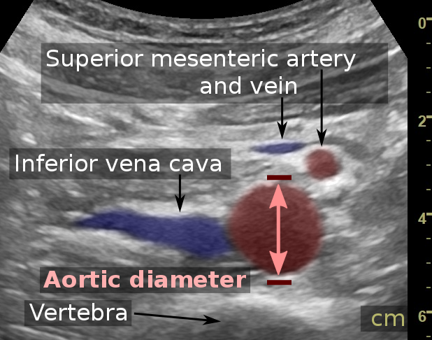
of a 31 year old man, showing normal anatomy. It uses compression to decrease the distance to the
666:
114:
221:
684:
308:
299:
779:
651:
246:: Written informed consent was obtained from the individual, including online publication.
239:
218:
255:
101:
275:
I, the copyright holder of this work, hereby publish it under the following license:
153:
739:
189:
307:
The person who associated a work with this deed has dedicated the work to the
709:
173:
161:
157:
759:
527:
Click on a date/time to view the file as it appeared at that time.
254:
139:
Ultrasonographic measurement of aortic diameter at the navel.svg
699:
400:
copyrighted, dedicated to the public domain by copyright holder
278:
89:
164:
for better visualization, resulting in flat shapes of the
83:(SVG file, nominally 618 × 486 pixels, file size: 124 KB)
315:
http://creativecommons.org/publicdomain/zero/1.0/deed.en
350:
Add a one-line explanation of what this file represents
133:
113:
Commons is a freely licensed media file repository.
321:Creative Commons Zero, Public Domain Dedication
94:
8:
46:Size of this PNG preview of this SVG file:
797:
529:
346:
300:CC0 1.0 Universal Public Domain Dedication
677:The following other wikis use this file:
283:
209:
807:
799:
649:
503:
487:
469:
453:
433:
417:
397:
381:
364:
361:
342:
335:
295:This file is made available under the
7:
637:User created page with UploadWizard
518:
790:
355:
349:
274:
143:
126:
76:
354:
340:
288:
282:
93:
31:
21:
337:
289:
144:
14:
336:
172:. It shows measurement of the
26:
1:
506:original creation by uploader
36:
720:Որովայնային աորտայի անևրիզմա
436:Creative Commons CC0 License
362:Items portrayed in this file
180:(not present in this case).
829:
776:Usage on zh.wikipedia.org
766:Usage on th.wikipedia.org
756:Usage on ms.wikipedia.org
750:അബ്ഡൊമിനൽ അൾട്രാസോണോഗ്രാഫി
746:Usage on ml.wikipedia.org
736:Usage on ko.wikipedia.org
726:Usage on ja.wikipedia.org
716:Usage on hy.wikipedia.org
706:Usage on fr.wikipedia.org
696:Usage on bs.wikipedia.org
681:Usage on ar.wikipedia.org
662:Abdominal ultrasonography
657:Abdominal aortic aneurysm
519:
281:
199:
178:abdominal aortic aneurysm
16:
339:
230:- Conflicts of interest:
176:as applied in detecting
166:inferior mesenteric vein
100:This is a file from the
636:
606:
576:
250:
205:
195:
188:
185:
136:
104:. Information from its
760:Ultrasonografi abdomen
710:Échographie abdominale
615:19:34, 24 January 2018
585:20:02, 24 January 2018
555:20:08, 24 January 2018
260:
233:
107:description page there
258:
213:
259:Without annotations.
156:at the level of the
150:Abdominal ultrasound
70:2,560 × 2,013 pixels
66:1,280 × 1,007 pixels
607:Further annotations
52:Other resolutions:
690:تخطيط الصدى البطني
667:Medical ultrasound
261:
234:
170:inferior vena cava
816:
815:
673:Global file usage
640:
420:copyright license
348:
333:
332:
265:
264:
122:
121:
102:Wikimedia Commons
32:Global file usage
820:
798:
770:เอออร์ตาส่วนท้อง
632:Mikael Häggström
627:
602:Mikael Häggström
597:
572:Mikael Häggström
567:
384:copyright status
328:
325:
322:
319:
316:
297:Creative Commons
292:
291:
286:
285:
279:
236:Mikael Häggström
215:Mikael Häggström
201:
191:
148:
140:
134:
118:
97:
96:
90:
84:
73:
62:977 × 768 pixels
58:611 × 480 pixels
54:305 × 240 pixels
48:618 × 486 pixels
828:
827:
823:
822:
821:
819:
818:
817:
786:
730:利用者:加藤勝憲/腹部大動脈瘤
671:
652:Abdominal aorta
648:
641:
633:
625:
603:
595:
573:
565:
521:
520:
517:
516:
515:
514:
513:
512:
511:
510:
508:
496:
495:
494:
492:
481:
480:
479:
478:
477:
476:
475:
474:
472:
471:24 January 2018
462:
461:
460:
458:
447:
446:
445:
444:
443:
442:
441:
440:
438:
426:
425:
424:
422:
411:
410:
409:
408:
407:
406:
405:
404:
402:
390:
389:
388:
386:
375:
374:
373:
372:
371:
369:
353:
352:
351:
334:
326:
323:
320:
317:
314:
277:
276:
271:
266:
190:24 January 2018
181:
138:
131:
124:
123:
112:
111:
110:is shown below.
86:
82:
75:
74:
51:
12:
11:
5:
826:
824:
814:
813:
810:
806:
805:
802:
789:
785:
784:
783:
782:
774:
773:
772:
764:
763:
762:
754:
753:
752:
744:
743:
742:
734:
733:
732:
724:
723:
722:
714:
713:
712:
704:
703:
702:
694:
693:
692:
687:
675:
674:
670:
669:
664:
659:
654:
646:
645:
644:
639:
638:
635:
631:
628:
622:
617:
612:
609:
608:
605:
601:
598:
592:
587:
582:
579:
578:
575:
571:
568:
562:
557:
552:
548:
547:
544:
541:
538:
535:
532:
525:
524:
509:
504:
502:
501:
500:
499:
498:
497:
493:
490:source of file
488:
486:
485:
484:
483:
482:
473:
470:
468:
467:
466:
465:
464:
463:
459:
454:
452:
451:
450:
449:
448:
439:
434:
432:
431:
430:
429:
428:
427:
423:
418:
416:
415:
414:
413:
412:
403:
398:
396:
395:
394:
393:
392:
391:
387:
382:
380:
379:
378:
377:
376:
370:
365:
363:
360:
359:
358:
357:
356:
345:
344:
341:
338:
331:
330:
304:
303:
293:
273:
272:
270:
267:
263:
262:
252:
251:Other versions
248:
247:
227:Reusing images
207:
203:
202:
197:
193:
192:
187:
183:
182:
141:
132:
130:
127:
125:
120:
119:
98:
88:
87:
45:
41:
40:
39:
34:
29:
24:
19:
13:
10:
9:
6:
4:
3:
2:
825:
811:
808:
803:
800:
796:
793:
787:
781:
778:
777:
775:
771:
768:
767:
765:
761:
758:
757:
755:
751:
748:
747:
745:
741:
738:
737:
735:
731:
728:
727:
725:
721:
718:
717:
715:
711:
708:
707:
705:
701:
700:Trbušna aorta
698:
697:
695:
691:
688:
686:
683:
682:
680:
679:
678:
672:
668:
665:
663:
660:
658:
655:
653:
650:
642:
634:
629:
623:
621:
618:
616:
613:
611:
610:
604:
599:
593:
591:
588:
586:
583:
581:
580:
574:
569:
563:
561:
558:
556:
553:
550:
549:
545:
542:
539:
536:
533:
531:
530:
528:
522:
507:
491:
457:
437:
421:
401:
385:
368:
329:
310:
309:public domain
306:
305:
301:
298:
294:
280:
268:
257:
253:
249:
245:
242:
241:
237:
231:
229:
228:
224:
223:
220:
216:
212:
208:
204:
198:
194:
184:
179:
175:
171:
167:
163:
159:
155:
151:
147:
142:
135:
128:
116:
109:
108:
103:
99:
92:
91:
85:
80:
79:Original file
71:
67:
63:
59:
55:
49:
44:
38:
35:
33:
30:
28:
25:
23:
20:
18:
15:
794:
791:
676:
526:
523:File history
313:
244:Consent note
243:
145:
115:You can help
105:
77:
22:File history
232: None
222:Author info
154:axial plane
137:Description
643:File usage
624:618 × 486
594:618 × 486
564:618 × 486
540:Dimensions
27:File usage
685:أبهر بطني
537:Thumbnail
534:Date/Time
456:inception
269:Licensing
146:English:
788:Metadata
626:(118 KB)
596:(124 KB)
566:(124 KB)
343:Captions
200:Own work
168:and the
37:Metadata
551:current
546:Comment
367:depicts
347:English
152:in the
129:Summary
81:
809:Height
577:Aortic
287:
206:Author
196:Source
801:Width
780:腹部超聲波
327:false
324:false
174:aorta
162:aorta
158:navel
740:배대동맥
543:User
240:M.D.
219:M.D.
186:Date
17:File
812:486
804:618
318:CC0
302:.
238:,
225:-
217:,
68:|
64:|
60:|
56:|
50:.
117:.
72:.
Text is available under the Creative Commons Attribution-ShareAlike License. Additional terms may apply.

