1235:
281:
478:; Latin, "tail") papillary process of the liver, which arise from its left side. It also has a caudate process (that is not tail-like shaped) arising from its right side, which provides surface continuity between the caudate lobe and the visceral surface of the anatomical right lobe of the liver. The caudate process is a small elevation of the hepatic substance extending obliquely and laterally, from the lower extremity of the caudate lobe to the undersurface of the right lobe.
40:
263:
492:
481:
The caudate lobe has a complex blood supply system. It derives its arterial supply from the caudate arteries, which arise from the right, left, and middle hepatic arteries that are connected to each other. Besides, the caudate lobe also derives its supply from the right and left branches of the
367:
The right lobe is functionally separated from the left lobe by the middle hepatic vein. From a functional perspective (one that takes the arterial, portal venous, and systemic venous anatomy into account) the falciform ligament separates the medial and lateral segments of the left hepatic lobe.
1255:
60:
212:
surface – the other two smaller lobes, the caudate lobe and the quadrate lobe, are also visible. The two smaller lobes, the caudate lobe and the quadrate lobe, are known as superficial or accessory lobes, and both are located on the underside of the right lobe.
220:, visible on the front of the liver, makes a superficial division of the right and left lobes of the liver. From the underside, the two additional lobes are located on the right lobe. A line can be imagined running from the left of the
453:. It looks backward, being nearly vertical in position; it is longer from above downward than from side to side, and is somewhat concave in the transverse direction. It is situated behind the
471:, caused by occlusion of hepatic venous outflow, can lead to hypertrophy of the caudate lobe due to its own caval anastomosis that allows for continued function of this lobe of the liver.
901:
399:
situated on the undersurface of the medial segment left lobe (Couinaud segment IVb), bounded in front by the anterior margin of the liver, behind by the
482:
portal vein. Its venous drainage is through short hepatic veins that drain directly into the inferior vena cava (IVC) due to its proximity to the IVC.
894:
637:
586:
557:
887:
1024:
371:
The right lobe is of a somewhat quadrilateral form. Its under and posterior surfaces being marked by three fossæ: the fossa for the
846:
806:
552:(Eighth ed.). Philadelphia Baltimore New York London Buenos Aires Hong Kong Sydney Tokyo: Wolters Kluwer. pp. 493–498.
328:. Its upper surface is slightly convex and is moulded on to the diaphragm; its under surface presents the gastric impression and
325:
430:) is situated upon the posterosuperior surface of the liver on the right lobe of the liver, opposite the tenth and eleventh
383:. These separate the right lobe into two smaller lobes on its left posterior part: the quadrate lobe and the caudate lobe.
243:(ligamentum teres), which further divide the left side of the liver in two sections. An important anatomical landmark, the
1225:
1174:
256:
969:
1190:
208:
surface – the liver is divided into two lobes: the right lobe and the left lobe. Viewed from the underside – the
468:
1210:
357:
1068:
1053:
947:
833:
827:
353:
240:
80:
1234:
48:
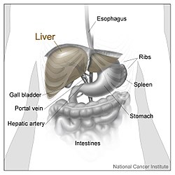
is divided into four lobes. This image shows the large right lobe and a smaller left lobe separated by the
962:
927:
911:
155:
1014:
957:
864:
at the SUNY Downstate
Medical Center - "Abdominal Cavity: Inspection of the Abdominal Viscera in situ"
974:
873:
839:
280:
1085:
867:
861:
821:
345:
120:
879:
1080:
937:
462:
450:
442:
431:
380:
361:
349:
329:
236:
217:
205:
95:
85:
49:
259:, seven segments can be seen, because the eighth segment is only visible in the visceral view.
1205:
1200:
1118:
1075:
784:
733:
684:
633:
625:
582:
553:
502:
229:
574:
1170:
1165:
1160:
1155:
1113:
1108:
1063:
1058:
996:
984:
774:
764:
723:
715:
674:
666:
100:
1256:
Knowledge (XXG) articles incorporating text from the 20th edition of Gray's
Anatomy (1918)
1185:
296:
285:
414:
It is oblong in shape, its antero-posterior diameter being greater than its transverse.
39:
1239:
1128:
979:
854:
814:
779:
752:
728:
703:
679:
654:
446:
408:
252:
185:
105:
59:
17:
1249:
1036:
1001:
942:
520:
497:
454:
438:
400:
322:
292:
244:
181:
173:
1019:
991:
932:
458:
404:
376:
262:
1103:
1095:
704:"Morphology and morphometry of the caudate lobe of the liver in two populations"
603:
372:
225:
125:
115:
110:
1009:
753:"A practical study of the hepatic vascular system anatomy of the caudate lobe"
719:
670:
318:
1045:
221:
788:
737:
688:
870:
at the SUNY Downstate
Medical Center - "The Visceral Surface of the Liver"
824:
at the SUNY Downstate
Medical Center - "The Visceral Surface of the Liver"
161:
1144:
769:
360:
turns around the inferior margin of the liver to come out ventral in the
251:, divides this left portion into four segments, which can be numbered in
209:
255:
starting at the caudate lobe as I in an anticlockwise manner. From this
751:
Mao W, Jiang X, Cao Y, Xiong S, Huang Y, Jiao L, Wang HJ (April 2021).
300:
317:
is smaller and more flattened than the right. It is situated in the
919:
396:
352:
for the cranial (upper) half and by the ligamentum teres hepatis (
279:
261:
177:
143:
45:
474:
The caudate lobe is named after the tail-shaped hepatic tissue (
883:
876:—Plastination Laboratory at the Medical University of Vienna
842:—Plastination Laboratory at the Medical University of Vienna
284:
Diagram showing the segments of the lobes as classified by
232:
and is used to mark the division between the two lobes.
465:. See Adriaan van den Spiegel 1578-1625 Spiegel's lobe.
449:
and the physiological division of the liver, called the
344:
is six times the size of the left lobe. It occupies the
437:
The caudate lobe of the liver is bounded below by the
291:
The lobes of the liver are further divided into eight
1223:
299:. These are also known as hepatic segments that are
1143:
1094:
1044:
1035:
918:
604:"Cantlie's line | Radiology Reference Article"
142:
137:
132:
32:
224:and all the way forward to divide the liver and
235:Other anatomical landmarks exist, such as the
895:
702:Sagoo MG, Aland RC, Gosden E (January 2018).
602:Mudgal P, Hacking C, Di Muzio B, et al.
8:
757:Quantitative Imaging in Medicine and Surgery
632:(3rd ed.). Springer. pp. 900–903.
543:
541:
539:
537:
653:Abdel-Misih SR, Bloomston M (August 2010).
461:from the commencement of the fossa for the
1041:
902:
888:
880:
58:
38:
778:
768:
727:
678:
1230:
577:. In Busuttil RW, Klintmalm GB (eds.).
511:
445:, and on the left by the fossa for the
407:, and on the left by the fossa for the
27:Four gross divisions of the human liver
496:This article incorporates text in the
159:
29:
851:Roche Lexicon - illustrated navigator
811:Roche Lexicon - illustrated navigator
659:The Surgical Clinics of North America
548:Moore KL, Dalley AF, Agur AM (2018).
228:into two halves. This line is called
7:
836:at the SUNY Downstate Medical Center
830:at the SUNY Downstate Medical Center
441:, on the right by the fossa for the
403:, on the right by the fossa for the
874:Cross section image: pembody/body8a
840:Cross section image: pembody/body8a
356:) for the caudal (under) half. The
457:, and separates the fossa for the
348:, on its posterior surface by the
25:
1025:Liver sinusoidal endothelial cell
1233:
912:liver, pancreas and biliary tree
708:Anatomical Science International
490:
575:"Surgical Anatomy of the Liver"
573:Renz JF, Kinkhabwala M (2014).
249:transverse fissure of the liver
847:"Anatomy diagram: 12581.000-1"
807:"Anatomy diagram: 12581.000-1"
630:Hepatology: Textbook and Atlas
1:
581:. Elsevier. pp. 23–39.
579:Transplantation of the Liver
204:. Seen from the front – the
550:Clinically Oriented Anatomy
428:posterior hepatic segment I
241:round ligament of the liver
1272:
970:Fibrous capsule of Glisson
853:. Elsevier. Archived from
813:. Elsevier. Archived from
624:Kuntz E, Kuntz HD (2009).
720:10.1007/s12565-016-0365-7
671:10.1016/j.suc.2010.04.017
154:
57:
37:
868:Anatomy photo:38:12-0204
862:Anatomy photo:37:02-0201
822:Anatomy photo:38:12-0201
665:(4). Elsevier: 643–653.
500:from the 20th edition of
358:ligamentum teres hepatis
1054:Intrahepatic bile ducts
379:and the fossae for the
354:round ligament of liver
81:Round ligament of liver
521:"Anatomy of the Liver"
326:regions of the abdomen
288:
267:
156:Anatomical terminology
76:Quadrate lobe of liver
18:Quadrate lobe of liver
1015:Hepatic stellate cell
301:surgically resectable
283:
265:
91:Caudate lobe of liver
975:Perisinusoidal space
770:10.21037/qims-20-780
469:Budd–Chiari syndrome
375:, the fossa for the
247:, also known as the
1086:Common hepatic duct
346:right hypochondrium
266:Labeled human liver
184:into four parts or
121:Common hepatic duct
66:Right lobe of liver
1081:Right hepatic duct
938:Ligamentum venosum
834:Anatomy image:8189
828:Anatomy image:7768
463:inferior vena cava
451:ligamentum venosum
443:inferior vena cava
432:thoracic vertebrae
395:is an area of the
381:inferior vena cava
362:falciform ligament
350:ligamentum venosum
330:omental tuberosity
289:
268:
237:ligamentum venosum
218:falciform ligament
96:Inferior vena cava
86:Falciform ligament
71:Left lobe of liver
50:falciform ligament
1221:
1220:
1206:Centroacinar cell
1201:Pancreatic islets
1139:
1138:
1119:Sphincter of Oddi
1076:Left hepatic duct
639:978-3-540-76839-5
626:"Liver resection"
606:. Radiopaedia.org
588:978-1-4557-5383-3
559:978-1-4963-5404-4
170:
169:
165:
16:(Redirected from
1263:
1238:
1237:
1229:
1175:Uncinate process
1114:Ampulla of Vater
1109:Common bile duct
1064:Canals of Hering
1059:Bile canaliculus
1042:
997:Lobules of liver
985:Periportal space
904:
897:
890:
881:
858:
818:
793:
792:
782:
772:
763:(4): 1313–1321.
748:
742:
741:
731:
699:
693:
692:
682:
650:
644:
643:
621:
615:
614:
612:
611:
599:
593:
592:
570:
564:
563:
545:
532:
531:
529:
528:
516:
494:
493:
162:edit on Wikidata
101:Common bile duct
62:
42:
30:
21:
1271:
1270:
1266:
1265:
1264:
1262:
1261:
1260:
1246:
1245:
1244:
1232:
1224:
1222:
1217:
1135:
1090:
1031:
914:
910:Anatomy of the
908:
845:
805:
802:
797:
796:
750:
749:
745:
701:
700:
696:
655:"Liver anatomy"
652:
651:
647:
640:
623:
622:
618:
609:
607:
601:
600:
596:
589:
572:
571:
567:
560:
547:
546:
535:
526:
524:
518:
517:
513:
491:
488:
420:
389:
338:
309:
297:Couinaud system
278:
273:
166:
128:
123:
118:
113:
108:
103:
98:
93:
88:
83:
78:
73:
68:
53:
28:
23:
22:
15:
12:
11:
5:
1269:
1267:
1259:
1258:
1248:
1247:
1243:
1242:
1219:
1218:
1216:
1215:
1214:
1213:
1208:
1203:
1195:
1194:
1193:
1188:
1180:
1179:
1178:
1168:
1163:
1158:
1149:
1147:
1141:
1140:
1137:
1136:
1134:
1133:
1132:
1131:
1129:Cholecystocyte
1123:
1122:
1121:
1111:
1106:
1100:
1098:
1092:
1091:
1089:
1088:
1083:
1078:
1073:
1072:
1071:
1066:
1061:
1050:
1048:
1039:
1033:
1032:
1030:
1029:
1028:
1027:
1022:
1017:
1012:
1004:
999:
994:
989:
988:
987:
980:Liver sinusoid
977:
972:
967:
966:
965:
960:
953:Lobes of liver
950:
948:Round ligament
945:
940:
935:
930:
924:
922:
916:
915:
909:
907:
906:
899:
892:
884:
878:
877:
871:
865:
859:
857:on 2012-07-22.
843:
837:
831:
825:
819:
817:on 2012-07-22.
801:
800:External links
798:
795:
794:
743:
694:
645:
638:
616:
594:
587:
565:
558:
533:
510:
509:
503:Gray's Anatomy
487:
484:
447:ductus venosus
419:
416:
409:umbilical vein
388:
385:
337:
334:
308:
305:
293:liver segments
277:
274:
272:
269:
253:Roman numerals
230:Cantlie's line
168:
167:
158:
152:
151:
146:
140:
139:
135:
134:
130:
129:
106:Hepatic artery
63:
55:
54:
43:
35:
34:
33:Lobes of liver
26:
24:
14:
13:
10:
9:
6:
4:
3:
2:
1268:
1257:
1254:
1253:
1251:
1241:
1236:
1231:
1227:
1212:
1211:Stellate cell
1209:
1207:
1204:
1202:
1199:
1198:
1197:Microanatomy
1196:
1192:
1189:
1187:
1184:
1183:
1181:
1176:
1172:
1169:
1167:
1164:
1162:
1159:
1157:
1154:
1153:
1151:
1150:
1148:
1146:
1142:
1130:
1127:
1126:
1124:
1120:
1117:
1116:
1115:
1112:
1110:
1107:
1105:
1102:
1101:
1099:
1097:
1093:
1087:
1084:
1082:
1079:
1077:
1074:
1070:
1067:
1065:
1062:
1060:
1057:
1056:
1055:
1052:
1051:
1049:
1047:
1043:
1040:
1038:
1037:Biliary tract
1034:
1026:
1023:
1021:
1018:
1016:
1013:
1011:
1008:
1007:
1006:Microanatomy
1005:
1003:
1002:Liver segment
1000:
998:
995:
993:
990:
986:
983:
982:
981:
978:
976:
973:
971:
968:
964:
961:
959:
956:
955:
954:
951:
949:
946:
944:
943:Porta hepatis
941:
939:
936:
934:
931:
929:
926:
925:
923:
921:
917:
913:
905:
900:
898:
893:
891:
886:
885:
882:
875:
872:
869:
866:
863:
860:
856:
852:
848:
844:
841:
838:
835:
832:
829:
826:
823:
820:
816:
812:
808:
804:
803:
799:
790:
786:
781:
776:
771:
766:
762:
758:
754:
747:
744:
739:
735:
730:
725:
721:
717:
713:
709:
705:
698:
695:
690:
686:
681:
676:
672:
668:
664:
660:
656:
649:
646:
641:
635:
631:
627:
620:
617:
605:
598:
595:
590:
584:
580:
576:
569:
566:
561:
555:
551:
544:
542:
540:
538:
534:
523:. Liver.co.uk
522:
515:
512:
508:
507:
504:
501:
499:
498:public domain
485:
483:
479:
477:
472:
470:
466:
464:
460:
456:
452:
448:
444:
440:
439:porta hepatis
435:
433:
429:
425:
417:
415:
412:
410:
406:
402:
401:porta hepatis
398:
394:
393:quadrate lobe
387:Quadrate lobe
386:
384:
382:
378:
374:
369:
365:
363:
359:
355:
351:
347:
343:
335:
333:
331:
327:
324:
323:hypochondriac
320:
316:
315:
306:
304:
302:
298:
294:
287:
282:
275:
270:
264:
260:
258:
257:parietal view
254:
250:
246:
245:porta hepatis
242:
238:
233:
231:
227:
223:
219:
214:
211:
207:
206:diaphragmatic
203:
202:quadrate lobe
199:
195:
191:
187:
183:
179:
175:
174:human anatomy
163:
157:
153:
150:
149:lobus hepatis
147:
145:
141:
136:
131:
127:
122:
117:
112:
107:
102:
97:
92:
87:
82:
77:
72:
67:
61:
56:
51:
47:
41:
36:
31:
19:
1069:Interlobular
1020:Kupffer cell
992:Portal triad
952:
933:Cantlie line
855:the original
850:
815:the original
810:
760:
756:
746:
714:(1): 48–57.
711:
707:
697:
662:
658:
648:
629:
619:
608:. Retrieved
597:
578:
568:
549:
525:. Retrieved
519:Karanjia N.
514:
505:
495:
489:
480:
475:
473:
467:
459:gall-bladder
436:
427:
424:caudate lobe
423:
421:
418:Caudate lobe
413:
405:gall-bladder
392:
390:
377:gall-bladder
370:
366:
341:
339:
313:
312:
310:
290:
248:
234:
215:
201:
198:caudate lobe
197:
193:
189:
171:
148:
90:
75:
70:
65:
1104:Cystic duct
1096:Gallbladder
373:portal vein
321:, and left
314:left lobe''
226:gallbladder
180:is divided
138:Identifiers
126:Gallbladder
116:Cystic duct
111:Portal vein
1046:Bile ducts
1010:Hepatocyte
610:2015-06-26
527:2015-06-26
486:References
342:right lobe
336:Right lobe
319:epigastric
200:, and the
190:right lobe
1191:accessory
928:Bare area
307:Left lobe
286:Couinaud.
271:Structure
222:vena cava
194:left lobe
1250:Category
1145:Pancreas
963:Quadrate
789:33816170
738:27586453
689:20637938
276:Segments
239:and the
210:visceral
1240:Anatomy
958:Caudate
780:7930664
729:5741786
680:4038911
295:in the
182:grossly
133:Details
1226:Portal
1182:Ducts
1152:Gross
1125:Cells
787:
777:
736:
726:
687:
677:
636:
585:
556:
506:(1918)
311:The ''
196:, the
192:, the
188:: the
176:, the
920:Liver
476:cauda
455:porta
397:liver
186:lobes
178:liver
160:[
144:Latin
46:liver
1186:main
1171:Head
1166:Neck
1161:Body
1156:Tail
785:PMID
734:PMID
685:PMID
634:ISBN
583:ISBN
554:ISBN
422:The
391:The
340:The
216:The
124:13:
119:12:
114:11:
109:10:
44:The
775:PMC
765:doi
724:PMC
716:doi
675:PMC
667:doi
172:In
104:9:
99:8:
94:7:
89:6:
84:5:
79:4:
74:3:
69:2:
64:1:
1252::
849:.
809:.
783:.
773:.
761:11
759:.
755:.
732:.
722:.
712:93
710:.
706:.
683:.
673:.
663:90
661:.
657:.
628:.
536:^
434:.
411:.
364:.
332:.
303:.
1228::
1177:)
1173:(
903:e
896:t
889:v
791:.
767::
740:.
718::
691:.
669::
642:.
613:.
591:.
562:.
530:.
426:(
164:]
52:.
20:)
Text is available under the Creative Commons Attribution-ShareAlike License. Additional terms may apply.