82:
282:
25:
270:
EEGs and EOGs, due to artifact from the carotid arteries. EKG artifact can be reduced with post-filtering of signals, or by "jumping" (co-referencing) of A/M reference electrodes, if replacement of reference electrodes is not possible, ameliorative, or if other clinical considerations prevent otherwise good placement (such as congenital malformation, or post-surgical considerations such as
Cochlear Implants).
184:(O), and central (C). Note that there is no "central lobe"; due to their placement, and depending on the individual, the "C" electrodes can exhibit/represent EEG activity more typical of frontal, temporal, and some parietal-occipital activity, and are always utilized in polysomnography sleep studies for the purpose of determining stages of sleep.
297:(MCN). This MCN system uses 1, 3, 5, 7, 9 for the left hemisphere which represents 10%, 20%, 30%, 40%, 50% of the inion-to-nasion distance respectively. The introduction of extra letter codes allows the naming of intermediate electrode sites. Note that these new letter codes do not necessarily refer to an area on the underlying cerebral cortex.
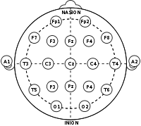
257:). The preauricular point is in front of each ear, and can be more easily located with mild palpation, and if necessary, requesting patient to open mouth slightly. The T3, C3, Cz, C4, and T4 electrodes are placed at marks made at intervals of 10%, 20%, 20%, 20%, 20% and 10%, respectively, measured across the top of the head.
225:
The "A" (sometimes referred to as "M" for mastoid process) refers to the prominent bone process usually found just behind the outer ear (less prominent in children and some adults). In basic polysomnography, F3, F4, Fz, Cz, C3, C4, O1, O2, A1, A2 (M1, M2), are used. Cz and Fz are 'ground' or 'common'
269:
When placing the A (or M) electrodes, palpation is often necessary to determine the most pronounced point of the mastoid process behind either ear; failure to do so, and to place the reference electrodes too low (posterior to the ear pinna, proximal to the throat) may result in "EKG artifact" in the
264:
Measurement methods for placement of the F3, F4, P3, and P4 points differ. If measured front-to-back (Fp1-F3-C3-P3-O1 and Fp2-F4-C4-P4-O2 montages), they can be 25% "up" from the front and back points (Fp1, Fp2, O1, and O2). If measured side-to-side (F7-F3-Fz-F4-F8 and T5-P3-Pz-P4-T6 montages), they
260:
Skull circumference is measured just above the ears (T3 and T4), just above the bridge of the nose (at Fpz), and just above the occipital point (at Oz). The Fp2, F8, T4, T6, and O2 electrodes are placed at intervals of 5%, 10%, 10%, 10%, 10%, and 5%, respectively, measured above the right ear, from
123:
Across all phases of consciousness, brains produce different, objectively recognizable and distinguishable electrical patterns, which can be detected by electrodes on the skin. These patterns vary, and are affected by multiple extrinsic factors, including age, prescription drugs, somatic diagnoses,
265:
can be 25% "up" from the side points (F7, F8, T5, and T6). If measured diagonally, from Nasion to Inion through the C3 and C4 points, they will be 20% in front of and behind the C3 and C4 points. Each of these measurement methods results in different nominal electrode placements.
246:, is the crest point of back of the skull, often indicated by a bump (the prominent occipital ridge, can usually be located with mild palpation). Marks for the Z electrodes are made between these points along the midline, at intervals of 10%, 20%, 20%, 20%, 20% and 10%.
515:
Nuwer, Marc R.; Comi, Giancarlo; Emerson, Ronald; Fuglsang-Frederiksen, Anders; Guérit, Jean-Michel; Hinrichs, Hermann; Ikeda, Akio; Jose C. Luccas, Fransisco; Rappelsburger, Peter (March 1998). "IFCN standards for digital recording of clinical EEG".
226:
reference points for all EEG and EOG electrodes, and A1-A2 are used for contralateral referencing of all EEG electrodes. This EEG montage may be extended to utilize T3-T4, P3-P4, as well as others, if an extended or "seizure montage" is called for.
131:
The "10" and "20" refer to the fact that the actual distances between adjacent electrodes are either 10% or 20% of the total front–back or right–left distance of the skull. For example, a measurement is taken across the top of the head, from the
112:, or voluntary lab research. This method was developed to maintain standardized testing methods ensuring that a subject's study outcomes (clinical or research) could be compiled, reproduced, and effectively analyzed and compared using the
222:) electrodes are more commonly just referred to with "right," "left," and "reference," or "common," as there are usually only three placed, and they can be differentially referenced from the EEG and EOG reference sites.
194:
of the skull, (FpZ, Fz, Cz, Oz) and is present mostly for reference/measurement points. These electrodes will not necessarily reflect or amplify lateral hemispheric cortical activity as they are placed over the
234:
Specific anatomical landmarks are used for the essential measuring and positioning of the EEG electrodes. These are found with a tape measure, and often marked with a grease pencil, or "China marker."
140:. Most other common measurements ('landmarking methods') start at one ear and end at the other, normally over the top of the head. Specific anatomical locations of the ear used include the
210:
Even-numbered electrodes (2,4,6,8) refer to electrode placement on the right side of the head, whereas odd numbers (1,3,5,7) refer to those on the left; this applies to both EEG and EOG (
207:
activity, or possible clinical brain death. Note that the required number of EEG electrodes, and their careful, measured placement, increases with each clinical requirement and modality.
199:, and do not represent either hemisphere adequately. "Z" electrodes are often utilized as 'grounds' or 'references,' especially in polysomnography sleep studies, and diagnostic/clinical
545:
Klem, GH; Lüders, HO; Jasper, HH; Elger, C (1999). "The ten-twenty electrode system of the
International Federation. The International Federation of Clinical Neurophysiology".
425:
445:
G.E. Chatrian, E. Lettich, and P.L. Nelson. Ten percent electrode system for topographic studies of spontaneous and evoked EEG activity. Am J EEG Technol, 25:83-92, 1985.
632:
293:, which fills in intermediate sites halfway between those of the existing 10–20 system. This new electrode-naming-system is more complicated giving rise to the
655:
285:
EEG electrode positions in the 10-10 system using modified combinatorial nomenclature, along with the fiducials and associated lobes of the brain.
582:
886:
625:
46:
680:
363:
116:. The system is based on the relationship between the location of an electrode and the underlying area of the brain, specifically the
574:
149:
68:
565:
Ernst
Niedermeyer, Fernando Lopes da Silva, Electroencephalography: Basic Principles, Clinical Applications, and Related Fields -
261:
front (Fpz) to back (Oz). The same is done for the odd-numbered electrodes on the left side, to complete the full circumference.
907:
618:
81:
706:
675:
741:
294:
39:
33:
281:
912:
566:
358:
50:
917:
871:
756:
792:
660:
701:
670:
641:
372:
665:
340:
A higher-resolution nomenclature has been suggested and called the "5% system" or the "10–5 system".
449:"American Electroencephalographic Society Guidelines for Standard Electrode Position Nomenclature".
423:(May 1958). "Report of the committee on methods of clinical examination in electroencephalography".
377:
845:
786:
771:
746:
736:
726:
215:
200:
866:
802:
761:
474:
398:
211:
599:
595:
578:
570:
554:
533:
503:
466:
420:
390:
242:
is the distinctly depressed area between the eyes, just above the bridge of the nose, and the
165:
145:
113:
876:
690:
525:
495:
458:
434:
382:
253:
to tragus: the tragus refers to the small portion of cartilage projecting anteriorly to the
219:
289:
When recording a more detailed EEG with more electrodes, extra electrodes are added using
254:
196:
125:
117:
191:
181:
161:
529:
386:
901:
840:
499:
462:
438:
177:
173:
478:
402:
830:
250:
169:
141:
596:
SVG drawing of the 10-20 system (numbering seen as a subset of the 10% division)
359:"The five percent electrode system for high-resolution EEG and ERP measurements"
109:
610:
85:
Electrode locations of
International 10-20 system for encephalography recording
825:
820:
810:
101:
97:
is an internationally recognized method to describe and apply the location of
835:
815:
716:
187:
There are also (Z) sites: A "Z" (zero) refers to an electrode placed on the
558:
394:
537:
507:
470:
300:
The new letter codes of the MCN for intermediate electrode places are:
850:
204:
188:
605:
881:
239:
133:
323:
Also, the MCN system renames four electrodes of the 10–20 system:
280:
243:
137:
98:
80:
766:
731:
721:
711:
614:
547:
Electroencephalography and
Clinical Neurophysiology. Supplement
781:
776:
218:
measurements of the heart) electrode placement. Chin, or EMG (
105:
18:
606:
10-20 System (numbering seen as a subset of the 10% division)
160:
Each electrode placement site has a letter to identify the
128:
of neurologic insults/injury/trauma, and substance abuse.
859:
801:
689:
648:
518:
Electroencephalography and
Clinical Neurophysiology
426:
Electroencephalography and
Clinical Neurophysiology
656:Amplitude integrated electroencephalography (aEEG)
214:measurements of eyes) electrodes, as well as ECG (
626:
357:Oostenveld, Robert; Praamstra, Peter (2001).
8:
164:, or area of the brain it is reading from:
887:Neurophysiological Biomarker Toolbox (NBT)
633:
619:
611:
569:, Lippincott Williams & Wilkins, 2004
376:
203:meant to represent/diagnose epileptiform
69:Learn how and when to remove this message
32:This article includes a list of general
349:
7:
16:Method of scalp electrodes placement
742:Contingent negative variation (CNV)
681:Brainstem auditory evoked potential
488:Journal of Clinical Neurophysiology
451:Journal of Clinical Neurophysiology
295:Modified Combinatorial Nomenclature
38:it lacks sufficient corresponding
14:
249:Preauricular to preauricular (or
500:10.1097/00004691-199401000-00014
463:10.1097/00004691-199104000-00007
23:
676:Somatosensory evoked potential
1:
872:Difference due to memory (Dm)
530:10.1016/S0013-4694(97)00106-5
387:10.1016/S1388-2457(00)00527-7
671:Magnetoencephalography (MEG)
642:Electroencephalography (EEG)
494:(1): 111–113. January 1994.
439:10.1016/0013-4694(58)90053-1
666:Electrocorticography (ECoG)
934:
457:(2): 200–202. April 1991.
95:International 10–20 system
277:Higher-resolution systems
364:Clinical Neurophysiology
793:Late positive component
661:Event-related potential
53:more precise citations.
908:Electroencephalography
702:Bereitschaftspotential
486:"Guideline Thirteen".
286:
86:
304:AF – between Fp and F
284:
238:Nasion to Inion: the
108:exam, polysomnograph
104:in the context of an
84:
319:PO – between P and O
316:TP – between T and P
313:CP – between C and P
310:FT – between F and T
307:FC – between F and C
846:Sensorimotor rhythm
803:Neural oscillations
747:Mismatch negativity
216:electrocardiography
421:Jasper, Herbert H.
287:
156:Electrode labeling
87:
913:Electrophysiology
895:
894:
789:(late positivity)
691:Evoked potentials
583:978-0-7817-5126-1
114:scientific method
79:
78:
71:
925:
877:Oddball paradigm
635:
628:
621:
612:
562:
541:
511:
482:
442:
407:
406:
380:
354:
291:the 10% division
212:electrooculogram
74:
67:
63:
60:
54:
49:this article by
40:inline citations
27:
26:
19:
933:
932:
928:
927:
926:
924:
923:
922:
918:Neurophysiology
898:
897:
896:
891:
855:
797:
685:
644:
639:
592:
544:
514:
485:
448:
419:
411:
410:
378:10.1.1.116.7379
356:
355:
351:
346:
279:
273:
232:
197:corpus callosum
158:
118:cerebral cortex
75:
64:
58:
55:
45:Please help to
44:
28:
24:
17:
12:
11:
5:
931:
929:
921:
920:
915:
910:
900:
899:
893:
892:
890:
889:
884:
879:
874:
869:
863:
861:
857:
856:
854:
853:
848:
843:
838:
833:
828:
823:
818:
813:
807:
805:
799:
798:
796:
795:
790:
784:
779:
774:
769:
764:
759:
754:
750:
749:
744:
739:
734:
729:
724:
719:
714:
709:
704:
699:
695:
693:
687:
686:
684:
683:
678:
673:
668:
663:
658:
652:
650:
646:
645:
640:
638:
637:
630:
623:
615:
609:
608:
603:
591:
590:External links
588:
587:
586:
563:
542:
524:(3): 259–261.
512:
483:
446:
443:
433:(2): 370–375.
416:
415:
409:
408:
371:(4): 713–719.
348:
347:
345:
342:
338:
337:
334:
331:
328:
321:
320:
317:
314:
311:
308:
305:
278:
275:
267:
266:
262:
258:
247:
231:
228:
220:electromyogram
192:sagittal plane
157:
154:
77:
76:
31:
29:
22:
15:
13:
10:
9:
6:
4:
3:
2:
930:
919:
916:
914:
911:
909:
906:
905:
903:
888:
885:
883:
880:
878:
875:
873:
870:
868:
865:
864:
862:
858:
852:
849:
847:
844:
842:
841:Sleep spindle
839:
837:
834:
832:
829:
827:
824:
822:
819:
817:
814:
812:
809:
808:
806:
804:
800:
794:
791:
788:
785:
783:
780:
778:
775:
773:
770:
768:
765:
763:
760:
758:
755:
752:
751:
748:
745:
743:
740:
738:
735:
733:
730:
728:
725:
723:
720:
718:
715:
713:
710:
708:
705:
703:
700:
697:
696:
694:
692:
688:
682:
679:
677:
674:
672:
669:
667:
664:
662:
659:
657:
654:
653:
651:
649:Related tests
647:
643:
636:
631:
629:
624:
622:
617:
616:
613:
607:
604:
601:
597:
594:
593:
589:
584:
580:
576:
575:0-7817-5126-8
572:
568:
564:
560:
556:
552:
548:
543:
539:
535:
531:
527:
523:
519:
513:
509:
505:
501:
497:
493:
489:
484:
480:
476:
472:
468:
464:
460:
456:
452:
447:
444:
440:
436:
432:
428:
427:
422:
418:
417:
413:
412:
404:
400:
396:
392:
388:
384:
379:
374:
370:
366:
365:
360:
353:
350:
343:
341:
335:
332:
329:
326:
325:
324:
318:
315:
312:
309:
306:
303:
302:
301:
298:
296:
292:
283:
276:
274:
271:
263:
259:
256:
252:
248:
245:
241:
237:
236:
235:
229:
227:
223:
221:
217:
213:
208:
206:
202:
198:
193:
190:
185:
183:
179:
175:
171:
167:
163:
155:
153:
151:
147:
143:
139:
135:
129:
127:
121:
119:
115:
111:
107:
103:
100:
96:
92:
83:
73:
70:
62:
59:December 2017
52:
48:
42:
41:
35:
30:
21:
20:
867:10-20 system
831:Theta rhythm
550:
546:
521:
517:
491:
487:
454:
450:
430:
424:
414:Bibliography
368:
362:
352:
339:
336:T6 is now P8
333:T5 is now P7
330:T4 is now T8
327:T3 is now T7
322:
299:
290:
288:
272:
268:
233:
224:
209:
201:EEG montages
186:
159:
130:
122:
94:
91:10–20 system
90:
88:
65:
56:
37:
757:C1 & P1
230:Measurement
166:pre-frontal
110:sleep study
51:introducing
902:Categories
826:Delta wave
821:Gamma wave
811:Alpha wave
753:Positivity
698:Negativity
344:References
102:electrodes
34:references
836:K-complex
816:Beta wave
717:Visual N1
373:CiteSeerX
182:occipital
567:Page 140
559:10590970
479:11857141
403:15414860
395:11275545
178:parietal
174:temporal
148:and the
851:Mu wave
553:: 3–6.
538:9743285
508:8195414
471:2050819
205:seizure
189:midline
170:frontal
150:mastoid
146:auricle
126:history
47:improve
882:EEGLAB
860:Topics
581:
573:
557:
536:
506:
477:
469:
401:
393:
375:
251:tragus
240:nasion
168:(Fp),
144:, the
142:tragus
134:nasion
36:, but
475:S2CID
399:S2CID
255:pinna
244:inion
180:(P),
176:(T),
172:(F),
138:inion
99:scalp
787:P600
772:P300
767:P200
737:N400
732:N2pc
727:N200
722:N170
712:N100
707:ELAN
579:ISBN
571:ISBN
555:PMID
534:PMID
504:PMID
467:PMID
391:PMID
162:lobe
89:The
782:P3b
777:P3a
762:P50
600:PDF
526:doi
522:106
496:doi
459:doi
435:doi
383:doi
369:112
136:to
106:EEG
93:or
904::
577:,
551:52
549:.
532:.
520:.
502:.
492:11
490:.
473:.
465:.
453:.
431:10
429:.
397:.
389:.
381:.
367:.
361:.
152:.
120:.
634:e
627:t
620:v
602:)
598:(
585:.
561:.
540:.
528::
510:.
498::
481:.
461::
455:8
441:.
437::
405:.
385::
72:)
66:(
61:)
57:(
43:.
Text is available under the Creative Commons Attribution-ShareAlike License. Additional terms may apply.