507:(OI) technique, which was available on reflected light microscopes prior to about 1975. In OI, the vertical illuminator is offset from perpendicular, producing shading effects that reveal height differences. This procedure reduces resolution and yields uneven illumination across the field of view. Nevertheless, OI was useful when people needed to know if a second phase particle was standing above or was recessed below the plane-of-polish, and is still available on a few microscopes. OI can be created on any microscope by placing a piece of paper under one corner of the mount so that the plane-of-polish is no longer perpendicular to the optical axis.
109:
36:
166:
550:(WDS) is used. But quantification of composition by EDS has improved greatly over time. The WDS system has historically had better sensitivity (ability to detect low amounts of an element) and ability to detect low-atomic weight elements, as well as better quantification of compositions, compared to EDS, but it was slower to use. Again, in recent years, the speed required to perform WDS analysis has improved substantially. Historically, EDS was used with the SEM while WDS was used with the
411:
158:
395:
403:
150:
454:(hcp) crystal structures). If the specimen is prepared with minimal damage to the surface, the structure can be seen vividly in cross-polarized light (the optic axis of the polarizer and analyzer are 90 degrees to each other, i.e., crossed). In some cases, an hcp metal can be chemically etched and then examined more effectively with PL. Tint etched surfaces, where a thin film (such as a
675:(historically, E 45 covered only manual chart methods and an image analysis method for making such chart measurements was described in ASTM E 1122. The image analysis methods are currently being incorporated into E 45). A stereological method for characterizing discrete second-phase particles, such as nonmetallic inclusions, carbides, graphite, etc., is presented in ASTM E 1245.
558:
93:
431:(DF), is an alternative method of observation that provides high-contrast images and actually greater resolution than bright-field. In dark-field illumination, the light from features perpendicular to the optical axis is blocked and appears dark while the light from features inclined to the surface, which look dark in BF, appear bright, or "self-luminous" in DF.
634:
587:
But EDS and WDS are difficult to apply to particles less than 2-3 micrometers in diameter. For smaller particles, diffraction techniques can be performed using the TEM for identification and EDS can be performed on small particles if they are extracted from the matrix using replication methods to avoid detection of the matrix along with the precipitate.
256:
522:) is an optical technique that uses optically generated high frequency surface acoustic waves to probe the direction elastic parameters of the surface and, as such, it can vividly reveal the surface microstructure of metals. It can also image the crystallographic orientation and determine the single crystal elasticity matrix of the material.
354:(TEM) generally cannot be utilized at magnifications below about 2000 to 3000X. LOM examination is fast and can cover a large area. Thus, the analysis can determine if the more expensive, more time-consuming examination techniques using the SEM or the TEM are required and where on the specimen the work should be concentrated.
358:
586:
is best measured using XRD (ASTM E 975). If a particular phase can be chemically extracted from a bulk specimen, it can be identified using XRD based on the crystal structure and lattice dimensions. This work can be complemented by EDS and/or WDS analysis where the chemical composition is quantified.
674:
with a single size distribution) and E 1182 (specimens with a bi-modal grain size distribution); while ASTM E 1382 describes how any grain size type or condition can be measured using image analysis methods. Characterization of nonmetallic inclusions using standard charts is described in ASTM E 45
629:
to assess matrix and second-phase structures. Stereology is the field of taking 0-, 1- or 2-dimensional measurements on the two-dimensional sectioning plane and estimating the amount, size, shape or distribution of the microstructure in three dimensions. These measurements may be made using manual
247:
results in a better mount with superior edge retention. A typical mounting cycle will compress the specimen and mounting media to 4,000 psi (28 MPa) and heat to a temperature of 350 °F (177 °C). When specimens are very sensitive to temperature, "cold mounts" may be made with a
375:
fringes are not present to distort the image. However, the resolution limit of the LOM will not be better than about 0.2 to 0.3 micrometers. Special methods are used at magnifications below 50X, which can be very helpful when examining the microstructure of cast specimens where greater spatial
370:
Light microscopes are designed for placement of the specimen's polished surface on the specimen stage either upright or inverted. Each type has advantages and disadvantages. Most LOM work is done at magnifications between 50 and 1000X. However, with a good microscope, it is possible to perform
474:
on the surface to a depth where interference effects are created when examined with BF producing color images, can be improved with PL. If it is difficult to get a good interference film with good coloration, the colors can be improved by examination in PL using a sensitive tint (ST) filter.
345:
Further, certain features can be best observed with the LOM, e.g., the natural color of a constituent can be seen with the LOM but not with EM systems. Also, image contrast of microstructures at relatively low magnifications, e.g., <500X, is far better with the LOM than with the
267:
abrasive paper was the first method of grinding and is still used today. Many metallographers, however, prefer to use a diamond grit suspension which is dosed onto a reusable fabric pad throughout the polishing process. Diamond grit in suspension might start at 9
630:
procedures with the aid of templates overlaying the microstructure, or with automated image analyzers. In all cases, adequate sampling must be made to obtain a proper statistical basis for the measurement. Efforts to eliminate bias are required.
427:(BF) illumination, where the image of any flat feature perpendicular to the incident light path is bright, or appears to be white. But, other illumination methods can be used and, in some cases, may provide superior images with greater detail.
387:. A microscope with excellent resolution may not be able to image a structure, that is there is no visibility, if image contrast is poor. Image contrast depends upon the quality of the optics, coatings on the lenses, and reduction of flare and
491:. This system gives the best detail. DIC converts minor height differences on the plane-of-polish, invisible in BF, into visible detail. The detail in some cases can be quite striking and very useful. If an ST filter is used along with a
337:
Prepared specimens should be examined with the unaided eye after etching to detect any visible areas that have responded to the etchant differently from the norm as a guide to where microscopical examination should be employed.
215:
A systematic preparation method is the easiest way to achieve the true structure. Sample preparation must therefore pursue rules which are suitable for most materials. Different materials with similar properties
311:) the microstructure can be revealed without etching using crossed polarized light (light microscopy). Otherwise, the microstructural constituents of the specimen are revealed by using a suitable chemical or
161:
Cold mounting: The specimens are placed in a mounting cup and mounting material is then poured over the specimens. A vacuum impregnation unit (photo) is used for mounting of porous materials.
318:
Non-destructive surface analysis techniques can involve applying a thin film or varnish that can be peeled off after drying and examined under a microscope. The technique was developed by
303:
constituents can be seen with the microscope, e.g., inclusions and nitrides. If the crystal structure is non-cubic (e.g., a metal with a hexagonal-closed packed crystal structure, such as
342:(LOM) examination should always be performed prior to any electron metallographic (EM) technique, as these are more time-consuming to perform and the instruments are much more expensive.
530:
If a specimen must be observed at higher magnification, it can be examined with a scanning electron microscope (SEM), or a transmission electron microscope (TEM). When equipped with an
754:"Metallographic and Materialographic Specimen Preparation, Light Microscopy, Image Analysis and Hardness Testing", Kay Geels in collaboration with Struers A/S, ASTM International 2006.
495:, color is introduced. The colors are controlled by the adjustment of the Wollaston prism, and have no specific physical meaning, per se. But, visibility may be better.
272:
and finish at one micrometre. Generally, polishing with diamond suspension gives finer results than using silicon carbide papers (SiC papers), especially with revealing
169:
Example of a reusable pad for use with diamond suspension. A single magnetic platen is positioned on the grinding and polishing machine to support the preparation pads.
670:
For example, the amount of a phase or constituent, that is, its volume fraction, is defined in ASTM E 562; manual grain size measurements are described in ASTM E 112 (
546:, depends upon the nature of the detector used. But, quantification of these elements by EDS is difficult and their minimum detectable limits are higher than when a
610:
Microstructural quantification is performed on a prepared, two-dimensional plane through the three-dimensional part or component. Measurements may involve simple
208:
particles are used to remove material from the sample surface until the desired surface quality is achieved. Many different machines are available for doing this
484:
663:'s Committee E-4 on Metallography and some other national and international organizations, have developed standard test methods describing how to characterize
391:; but, it also requires proper specimen preparation and good etching techniques. So, obtaining good images requires maximum resolution and image contrast.
263:
After mounting, the specimen is wet ground to reveal the surface of the metal. The specimen is successively ground with finer and finer abrasive media.
296:
cloth to produce a scratch-free mirror finish, free from smear, drag, or pull-outs and with minimal deformation remaining from the preparation process.
595:
A number of techniques exist to quantitatively analyze metallographic specimens. These techniques are valuable in the research and production of all
614:
techniques, e.g., the measurement of the thickness of a surface coating, or the apparent diameter of a discrete second-phase particle, (for example,
653:
metals and alloys, measurement of the size and size distribution of particles, assessment of the shape of particles, and spacing between particles.
547:
276:, which silicon carbide paper sometimes "smear" over. After grinding the specimen, polishing is performed. Typically, a specimen is polished with a
534:(EDS), the chemical composition of the microstructural features can be determined. The ability to detect low-atomic number elements, such as
153:
Hot mounting: The specimens are placed in the mounting press, and the resin is added. The specimens are mounted under heat and high pressure.
531:
79:
57:
351:
554:(EMPA). Today, EDS and WDS is used with both the SEM and the EMPA. However, a dedicated EMPA is not as common as an SEM.
347:
198:
571:
174:
814:
646:
443:
414:
Cross-polarized light illumination, where sample contrast comes from rotation of polarized light through the sample
108:
236:
451:
50:
44:
763:
Vol. 03.01 of the ASTM Standards covers standards devoted to metallography (and mechanical property testing)
656:
463:
424:
61:
773:
Metalog Guide, L. Bjerregaard, K. Geels, B. Ottesen, M. Rückert, Struers A/S, Copenhagen, Denmark, 2000.
760:
Metallography: Principles and
Practice, G. F. Vander Voort, ASM International, Materials Park, OH, 1999.
428:
383:
Besides considering the resolution of the optics, one must also maximize visibility by maximizing image
319:
232:
165:
757:
Metallography and
Microstructures, Vol. 9, ASM Handbook, ASM International, Materials Park, OH, 2005.
578:
present in a specimen if they have different crystal structures. For example, the amount of retained
410:
819:
710:
684:
604:
551:
190:
738:
660:
339:
244:
209:
186:
182:
157:
730:
671:
447:
384:
398:
Bright-field illumination, where sample contrast comes from absorbance of light in the sample
722:
689:
488:
377:
101:
784:
112:
In some cases, the metallographic structure is large enough to be seen with the unaided eye
642:
492:
264:
574:(XRD) techniques for many years. XRD can be used to determine the percentages of various
394:
664:
583:
575:
432:
388:
300:
212:, which are able to meet different demands for quality, capacity, and reproducibility.
402:
808:
742:
293:
622:
138:
149:
726:
406:
Dark-field illumination, sample contrast comes from light scattered by the sample
204:
Mechanical preparation is the most common preparation method. Successively finer
650:
487:(DIC), which is usually obtained with a system designed by the Polish physicist
372:
312:
225:
137:
materials may also be prepared using metallographic techniques, hence the terms
371:
examination at higher magnifications, e.g., 2000X, and even higher, as long as
798:
791:
626:
504:
331:
269:
124:
97:
17:
734:
248:
two-part epoxy resin. Mounting a specimen provides a safe, standardized, and
611:
579:
557:
459:
308:
252:
way by which to hold a sample during the grinding and polishing operations.
249:
221:
178:
173:
The surface of a metallographic specimen is prepared by various methods of
92:
633:
376:
coverage in the field of view may be required to observe features such as
193:. Using only metallographic techniques, a skilled technician can identify
711:"Méthode non destructive d'examens macro et micrographiques superficiels"
618:
615:
543:
467:
446:(PL) is very useful when studying the structure of metals with non-cubic
304:
273:
231:
Metallographic specimens are typically "mounted" using a hot compression
217:
205:
471:
455:
289:
281:
255:
134:
130:
539:
535:
285:
277:
632:
600:
596:
570:
Characterization of microstructures has also been performed using
556:
409:
401:
393:
356:
254:
240:
194:
164:
156:
148:
120:
107:
91:
641:
Some of the most basic measurements include determination of the
519:
361:
Scanning transmission electron microscope, used in metallography
357:
29:
243:
is becoming more popular because reduced shrinkage during
119:
is the study of the physical structure and components of
785:
526:
Scanning electron and transmission electron microscopes
637:
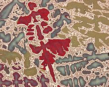
An image of the microstructures of ductile cast iron
141:, plastography and, collectively, materialography.
625:). Measurement may also require application of
334:techniques are used in metallographic analysis.
185:. After preparation, it is often analyzed using
224:) will respond alike and thus require the same
799:Metallography Part II - Microscopic Techniques
645:of a phase or constituent, measurement of the
792:Metallography Part I - Macroscopic Techniques
479:Differential interference contrast microscopy
435:, for example, are more vivid in DF than BF.
8:
801:, Karlsruhe University of Applied Sciences.
794:, Karlsruhe University of Applied Sciences.
709:Jacquet, P. A.; van Effenterre, A. (1957).
423:Most LOM observations are conducted using
518:Spatially resolve acoustic spectroscopy (
80:Learn how and when to remove this message
43:This article includes a list of general
701:
366:Design, resolution, and image contrast
7:
770:, 2nd Ed., ASM International, 1999.
503:DIC has largely replaced the older
548:wavelength-dispersive spectrometer
485:differential interference contrast
145:Preparing metallographic specimens
49:it lacks sufficient corresponding
25:
419:Bright- and dark-field microscopy
352:transmission electron microscopes
34:
27:Study of metals using microscopy
483:Another useful imaging mode is
532:energy dispersive spectrometer
1:
237:phenolic thermosetting resins
566:X-ray diffraction techniques
552:electron microprobe analyzer
348:scanning electron microscope
239:have been used, but modern
100:of bronze revealing a cast
836:
727:10.1051/metal/195754020107
591:Quantitative metallography
439:Polarized light microscopy
259:A macro etched copper disc
672:equiaxed grain structures
299:After polishing, certain
340:Light optical microscopy
797:Video on metallography
790:Video on metallography
787:, Cambridge University.
657:Standards organizations
561:An x-ray diffractometer
64:more precise citations.
768:Metallographic Etching
638:
562:
452:hexagonal close-packed
415:
407:
399:
362:
260:
210:grinding and polishing
170:
162:
154:
113:
105:
636:
560:
429:Dark-field microscopy
413:
405:
397:
360:
320:Pierre Armand Jacquet
258:
168:
160:
152:
111:
95:
715:Revue de Métallurgie
603:and non-metallic or
505:oblique illumination
499:Oblique illumination
450:(mainly metals with
322:and others in 1957.
228:during preparation.
685:Henry Clifton Sorby
605:composite materials
326:Analysis techniques
233:thermosetting resin
199:material properties
191:electron microscopy
661:ASTM International
639:
563:
448:crystal structures
416:
408:
400:
363:
261:
171:
163:
155:
114:
106:
815:Materials testing
572:x-ray diffraction
90:
89:
82:
16:(Redirected from
827:
747:
746:
706:
690:Holger F. Struer
667:quantitatively.
489:Georges Nomarski
433:Grain boundaries
85:
78:
74:
71:
65:
60:this article by
51:inline citations
38:
37:
30:
21:
835:
834:
830:
829:
828:
826:
825:
824:
805:
804:
783:HKDH Bhadeshia
780:
751:
750:
708:
707:
703:
698:
681:
665:microstructures
651:polycrystalline
643:volume fraction
593:
568:
528:
516:
514:SRAS microscopy
510:
501:
493:Wollaston prism
481:
470:film) is grown
444:Polarized light
441:
421:
368:
330:Many different
328:
301:microstructural
265:Silicon carbide
235:. In the past,
147:
86:
75:
69:
66:
56:Please help to
55:
39:
35:
28:
23:
22:
15:
12:
11:
5:
833:
831:
823:
822:
817:
807:
806:
803:
802:
795:
788:
779:
778:External links
776:
775:
774:
771:
764:
761:
758:
755:
749:
748:
721:(2): 107–125.
700:
699:
697:
694:
693:
692:
687:
680:
677:
592:
589:
584:hardened steel
567:
564:
527:
524:
515:
512:
500:
497:
480:
477:
440:
437:
420:
417:
367:
364:
327:
324:
146:
143:
88:
87:
42:
40:
33:
26:
24:
18:Metallographic
14:
13:
10:
9:
6:
4:
3:
2:
832:
821:
818:
816:
813:
812:
810:
800:
796:
793:
789:
786:
782:
781:
777:
772:
769:
765:
762:
759:
756:
753:
752:
744:
740:
736:
732:
728:
724:
720:
716:
712:
705:
702:
695:
691:
688:
686:
683:
682:
678:
676:
673:
668:
666:
662:
658:
654:
652:
648:
644:
635:
631:
628:
624:
620:
617:
613:
608:
606:
602:
598:
590:
588:
585:
581:
577:
573:
565:
559:
555:
553:
549:
545:
541:
537:
533:
525:
523:
521:
513:
511:
508:
506:
498:
496:
494:
490:
486:
478:
476:
473:
469:
466:or elemental
465:
461:
457:
453:
449:
445:
438:
436:
434:
430:
426:
418:
412:
404:
396:
392:
390:
386:
381:
379:
374:
365:
359:
355:
353:
350:(SEM), while
349:
343:
341:
335:
333:
325:
323:
321:
316:
314:
310:
306:
302:
297:
295:
291:
287:
283:
279:
275:
271:
266:
257:
253:
251:
246:
242:
238:
234:
229:
227:
223:
219:
213:
211:
207:
202:
200:
196:
192:
188:
184:
180:
176:
167:
159:
151:
144:
142:
140:
136:
132:
128:
126:
122:
118:
117:Metallography
110:
103:
99:
94:
84:
81:
73:
63:
59:
53:
52:
46:
41:
32:
31:
19:
767:
718:
714:
704:
669:
659:, including
655:
640:
623:ductile iron
609:
594:
569:
529:
517:
509:
502:
482:
442:
425:bright-field
422:
382:
369:
344:
336:
329:
317:
313:electrolytic
298:
262:
230:
214:
203:
197:and predict
172:
139:ceramography
129:
116:
115:
76:
67:
48:
766:G. Petzow,
472:epitaxially
373:diffraction
270:micrometres
226:consumables
123:, by using
70:August 2008
62:introducing
820:Metallurgy
809:Categories
696:References
647:grain size
627:stereology
616:spheroidal
332:microscopy
125:microscopy
98:micrograph
45:references
743:114572864
735:0035-1563
612:metrology
580:austenite
460:molybdate
378:dendrites
315:etchant.
250:ergonomic
222:ductility
179:polishing
135:polymeric
104:structure
102:dendritic
679:See also
619:graphite
544:nitrogen
468:selenium
464:chromate
385:contrast
274:porosity
218:hardness
206:abrasive
175:grinding
456:sulfide
294:napless
290:diamond
282:alumina
187:optical
183:etching
131:Ceramic
58:improve
741:
733:
601:alloys
597:metals
576:phases
542:, and
540:oxygen
536:carbon
286:silica
278:slurry
245:curing
195:alloys
181:, and
121:metals
47:, but
739:S2CID
582:in a
389:glare
292:on a
288:, or
241:epoxy
731:ISSN
599:and
520:SRAS
220:and
133:and
723:doi
649:in
621:in
307:or
280:of
189:or
811::
737:.
729:.
719:54
717:.
713:.
607:.
538:,
462:,
458:,
380:.
309:Zr
305:Ti
284:,
201:.
177:,
127:.
96:A
745:.
725::
216:(
83:)
77:(
72:)
68:(
54:.
20:)
Text is available under the Creative Commons Attribution-ShareAlike License. Additional terms may apply.