160:, followed in 1984 by the first paper that used visible radiation for near field scanning. The near-field optical (NFO) microscope involved a sub-wavelength aperture at the apex of a metal coated sharply pointed transparent tip, and a feedback mechanism to maintain a constant distance of a few nanometers between the sample and the probe. Lewis et al. were also aware of the potential of an NFO microscope at this time. They reported first results in 1986 confirming super-resolution. In both experiments, details below 50 nm (about λ
40:
2055:
127:. His original idea, proposed in 1928, was based upon the usage of intense nearly planar light from an arc under pressure behind a thin, opaque metal film with a small orifice of about 100 nm. The orifice was to remain within 100 nm of the surface, and information was to be collected by point-by-point scanning. He foresaw the illumination and the detector movement being the biggest technical difficulties.
2156:
433:(SERS). This technique can be used in an apertureless shear-force NSOM setup, or by using an AFM tip coated with gold or silver. The Raman signal is found to be significantly enhanced under the AFM tip. This technique has been used to give local variations in the Raman spectra under a single-walled nanotube. A highly sensitive optoacoustic spectrometer must be used for the detection of the Raman signal.
296:
1713:
288:
2168:
308:, which has a square pyramid shape with two facets coated with a metal. Such a probe has a high signal collection efficiency (>90%) and no frequency cutoff. Another alternative is "active tip" schemes, where the tip is functionalized with active light sources such as a fluorescent dye or even a light emitting diode that enables fluorescence excitation.
28:
373:
272:
934:
480:. It is normally limited to surface studies; however, it can be applied for subsurface investigations within the corresponding depth of field. In shear force mode and other contact operation it is not conducive for studying soft materials. It has long scan times for large sample areas for high resolution imaging.
250:
and have intensities that drop off exponentially with distance from the object. Because of this, the detector must be placed very close to the sample in the near field zone, typically a few nanometers. As a result, near field microscopy remains primarily a surface inspection technique. The detector is then
422:
Direct local Raman NSOM is based on Raman spectroscopy. Aperture Raman NSOM is limited by very hot and blunt tips, and by long collection times. However, apertureless NSOM can be used to achieve high Raman scattering efficiency factors (around 40). Topological artifacts make it hard to implement this
93:
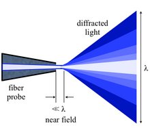
light is focused through an aperture with a diameter smaller than the excitation wavelength, resulting in an evanescent field (or near-field) on the far side of the aperture. When the sample is scanned at a small distance below the aperture, the optical resolution of transmitted or reflected light is
455:
The nanofocusing technique can create a nanometer-scale "white" light source at the tip apex, which can be used to illuminate a sample at near-field for spectroscopic analysis. The interband optical transitions in individual single-walled carbon nanotubes are imaged and a spatial resolution around 6
451:
method is a broadband nanoscale spectroscopy that combines apertureless NSOM with broadband illumination and FTIR detection to obtain a complete infrared spectrum at every spatial location. Sensitivity to a single molecular complex and nanoscale resolution up to 10 nm has been demonstrated with
418:
As the name implies, information is collected by spectroscopic means instead of imaging in the near field regime. Through near field spectroscopy (NFS), one can probe spectroscopically with sub-wavelength resolution. Raman SNOM and fluorescence SNOM are two of the most popular NFS techniques as they
172:
According to Abbe's theory of image formation, developed in 1873, the resolving capability of an optical component is ultimately limited by the spreading out of each image point due to diffraction. Unless the aperture of the optical component is large enough to collect all the diffracted light, the
311:
The merits of aperture and apertureless NSOM configurations can be merged in a hybrid probe design, which contains a metallic tip attached to the side of a tapered optical fiber. At visible range (400 nm to 900 nm), about 50% of the incident light can be focused to the tip apex, which is
249:
This treatment takes into account only the light diffracted into the far-field that propagates without any restrictions. NSOM makes use of evanescent or non propagating fields that exist only near the surface of the object. These fields carry the high frequency spatial information about the object
283:
Though there are many issues associated with the apertured tips (heating, artifacts, contrast, sensitivity, topology and interference among others), aperture mode remains more popular. This is primarily because apertureless mode is even more complex to set up and operate, and is not understood as
436:
Fluorescence NSOM is a highly popular and sensitive technique which makes use of fluorescence for near field imaging, and is especially suited for biological applications. The technique of choice here is apertureless back to the fiber emission in constant shear force mode. This technique uses
393:
from the returning reflected light. The scanning tip, depending upon the operation mode, is usually a pulled or stretched optical fiber coated with metal except at the tip or just a standard AFM cantilever with a hole in the center of the pyramidal tip. Standard optical detectors, such as
464:
NSOM can be vulnerable to artifacts that are not from the intended contrast mode. The most common root for artifacts in NSOM are tip breakage during scanning, striped contrast, displaced optical contrast, local far field light concentration, and topographic artifacts.
324:
Feedback mechanisms are usually used to achieve high resolution and artifact free images since the tip must be positioned within a few nanometers of the surfaces. Some of these mechanisms are constant force feedback and shear force feedback
384:
The primary components of an NSOM setup are the light source, feedback mechanism, the scanning tip, the detector and the piezoelectric sample stage. The light source is usually a laser focused into an optical fiber through a
335:
In shear force feedback mode, a tuning fork is mounted alongside the tip and made to oscillate at its resonance frequency. The amplitude is closely related to the tip-surface distance, and thus used as a feedback mechanism.
131:
also developed similar theories in 1956. He thought the moving of the pinhole or the detector when it is so close to the sample would be the most likely issue that could prevent the realization of such an instrument. It was
279:
There exist NSOM which can be operated in so-called aperture mode and NSOM for operation in a non-aperture mode. As illustrated, the tips used in the apertureless mode are very sharp and do not have a metal coating.
299:
Apertureless modes of operation: a) photon tunneling (PSTM) by a sharp transparent tip, b) PSTM by sharp opaque tip on smooth surface, and c) scanning interferometric apertureless microscopy with double
495:
utilize in-plane polarimetry to study physical properties inaccessible to near-field scanning optical microscopes including the spatial dependence of intramolecular vibrations in anisotropic molecules.
229:
1203:
Bao W, Melli M, Caselli N, Riboli F, Wiersma DS, Staffaroni M, et al. (December 2012). "Mapping local charge recombination heterogeneity by multidimensional nanospectroscopic imaging".
989:
Lewis AM, Isaacson M, Harootunian A, Muray A (1984). "Development of a 500 Å spatial resolution light microscope. I. Light is efficiently transmitted through λ/16 diameter apertures".
173:
finer aspects of the image will not correspond exactly to the object. The minimum resolution (d) for the optical component is thus limited by its aperture size, and expressed by the
441:-based dyes embedded in an appropriate resin. Edge filters are used for removal of all primary laser light. Resolution as low as 10 nm can be achieved using this technique.
1406:
Huth F, Govyadinov A, Amarie S, Nuansing W, Keilmann F, Hillenbrand R (August 2012). "Nano-FTIR absorption spectroscopy of molecular fingerprints at 20 nm spatial resolution".
1904:
1640:
344:
It is possible to take advantage of the various contrast techniques available to optical microscopy through NSOM but with much higher resolution. By using the change in the
2098:
284:
well. There are five primary modes of apertured NSOM operation and four primary modes of apertureless NSOM operation. The major ones are illustrated in the next figure.
2009:
1695:
468:
In apertureless NSOM, also known as scattering-type SNOM or s-SNOM, many of these artifacts are eliminated or can be avoided by proper technique application.
1937:
1700:
444:
Near field infrared spectrometry and near-field dielectric microscopy use near-field probes to combine sub-micron microscopy with localized IR spectroscopy.
94:
limited only by the diameter of the aperture. In particular, lateral resolution of 6 nm and vertical resolution of 2–5 nm have been demonstrated.
1391:
Pollock HM, Smith DA (2002). "The use of near-field probes for vibrational spectroscopy and photothermal imaging". In
Chalmers JM, Griffiths PR (eds.).
242:
for the optical component (maximum 1.3–1.4 for modern objectives with a very high magnification factor). Thus, the resolution limit is usually around λ
348:
of light or the intensity of the light as a function of the incident wavelength, it is possible to make use of contrast enhancing techniques such as
364:. It is also possible to provide contrast using the change in refractive index, reflectivity, local stress and magnetic properties amongst others.
1683:
1633:
492:
1508:"6 nm super-resolution optical transmission and scattering spectroscopic imaging of carbon nanotubes using a nanometer-scale white light source"
638:"6 nm super-resolution optical transmission and scattering spectroscopic imaging of carbon nanotubes using a nanometer-scale white light source"
491:
of the scanning tip. Metallic scanning tips naturally orient the polarization state perpendicular to the sample surface. Other techniques, like
316:(TERS) at tip apex, and collect the Raman signals through the same fiber. The lens-free fiber-in-fiber-out STM-NSOM-TERS has been demonstrated.
2136:
2004:
1730:
1305:
Hoshino K, Gopal A, Glaz MS, Vanden Bout DA, Zhang X (2012). "Nanoscale fluorescence imaging with quantum dot near-field electroluminescence".
1170:
1117:
419:
allow for the identification of nanosized features with chemical contrast. Some of the common near-field spectroscopic techniques are below.
864:
1340:
Kim S, Yu N, Ma X, Zhu Y, Liu Q, Liu M, Yan R (2019). "High external-efficiency nanofocusing for lens-free near-field optical nanoscopy".
2103:
1812:
1807:
1792:
1755:
1678:
430:
2194:
2039:
2019:
1668:
1626:
376:
Block diagram of an apertureless reflection-back-to-the-fiber NSOM setup with shear-force distance control and cross-polarization; 1:
361:
291:
Apertured modes of operation: a) illumination, b) collection, c) illumination collection, d) reflection and e) reflection collection.
932:, Pohl DW, "optical near field scanning microscope", published 1987-04-22, issued 1982-12-27, assigned to IBM.
556:"Visualizing nanoscale excitonic relaxation properties of disordered edges and grain boundaries in monolayer molybdenum disulfide"
2083:
2079:
1930:
535:
1822:
1787:
426:
313:
128:
101:, chemical structure and local stress. Dynamic properties can also be studied at a sub-wavelength scale using this technique.
2108:
1802:
1782:
1777:
1740:
1073:
Harootunian A, Betzig E, Isaacson M, Lewis A (1986). "Super-resolution fluorescence near-field scanning optical microscopy".
119:
is given credit for conceiving and developing the idea for an imaging instrument that would image by exciting and collecting
1163:
Atomic Force
Microscopy, Scanning Nearfield Optical Microscopy and Nanoscratching: Application to Rough and Natural Surfaces
183:
2204:
1745:
1690:
1608:
2172:
1563:
Ocelic N, Huber A, Hillenbrand R (2006-09-04). "Pseudoheterodyne detection for background-free near-field spectroscopy".
2214:
2160:
1923:
1842:
1837:
1254:
Michaelis J, Hettich C, Mlynek J, Sandoghdar V (May 2000). "Optical microscopy using a single-molecule light source".
258:
stage. The scanning can either be done at a constant height or with regulated height by using a feedback mechanism.
2126:
1858:
1772:
1712:
145:
79:
1999:
1868:
1832:
1827:
1750:
1735:
1649:
784:
Synge EH (1928). "A suggested method for extending the microscopic resolution into the ultramicroscopic region".
505:
357:
137:
124:
105:
1797:
1138:
97:
As in optical microscopy, the contrast mechanism can be easily adapted to study different properties, such as
35:, with the diffraction of light coming from NSOM fiber probe, showing wavelength of light and the near-field.
2087:
2069:
1984:
1863:
1663:
329:
312:
around 5 nm in radius. This hybrid probe can deliver the excitation light through the fiber to realize
157:
148:
using microwave radiation with a wavelength of 3 cm. A line grating was resolved with a resolution of λ
116:
1989:
484:
2199:
1994:
1817:
403:
87:
39:
929:
1572:
1519:
1462:
1415:
1349:
1314:
1263:
1212:
1082:
1029:
961:
887:
847:
756:
714:
659:
567:
395:
345:
44:
2209:
2074:
2014:
52:
1965:
1373:
1287:
1236:
911:
649:
407:
239:
174:
742:"Observation of nanostructure by scanning near-field optical microscope with small sphere probe"
1588:
1545:
1488:
1451:"Structural analysis and mapping of individual protein complexes by infrared nanospectroscopy"
1431:
1365:
1279:
1228:
1166:
1113:
1055:
903:
687:
593:
515:
488:
32:
1449:
Amenabar I, Poly S, Nuansing W, Hubrich EH, Govyadinov AA, Huth F, et al. (2013-12-04).
332:(AFM). Experiments can be performed in contact, intermittent contact, and non-contact modes.
2054:
2044:
1873:
1580:
1535:
1527:
1478:
1470:
1423:
1357:
1322:
1271:
1220:
1090:
1045:
1037:
998:
969:
895:
820:
793:
764:
722:
677:
667:
583:
575:
255:
98:
83:
389:, a beam splitter and a coupler. The polarizer and the beam splitter would serve to remove
2131:
1612:
399:
305:
48:
1188:
1018:"Near Field Scanning Optical Microscopy (NSOM): Development and Biophysical Applications"
1576:
1523:
1466:
1419:
1353:
1318:
1267:
1216:
1086:
1033:
965:
891:
851:
760:
718:
663:
571:
2024:
1889:
1540:
1507:
1483:
1450:
1050:
1017:
682:
637:
588:
555:
477:
1041:
2188:
2093:
1377:
1002:
380:
and crossed polarizers; 2: shear-force arrangement; 3: sample mount on a piezo stage.
377:
1240:
1291:
915:
353:
878:
Ash EA, Nicholls G (June 1972). "Super-resolution aperture scanning microscope".
510:
438:
390:
295:
251:
120:
1531:
769:
741:
672:
27:
2029:
1958:
1946:
1899:
1894:
1361:
824:
797:
141:
75:
1592:
1369:
705:
Dürig U, Pohl DW, Rohner F (1986). "Near-field optical scanning microscopy".
1765:
1618:
1224:
448:
386:
287:
1549:
1492:
1435:
1283:
1232:
1059:
1016:
Betzig E, Lewis A, Harootunian A, Isaacson M, Kratschmer E (January 1986).
907:
691:
597:
328:
Constant force feedback mode is similar to the feedback mechanism used in
554:
Bao W, Borys NJ, Ko C, Suh J, Fan W, Thron A, et al. (August 2015).
349:
133:
740:
Oshikane Y, Kataoka T, Okuda M, Hara S, Inoue H, Nakano M (April 2007).
1474:
1192:
The Optics
Laboratory, North Carolina State University. 12 October 2007
611:
579:
372:
1584:
1427:
1326:
476:
One limitation is a very short working distance and extremely shallow
271:
2034:
1915:
1673:
1275:
899:
811:
Synge EH (1932). "An application of piezoelectricity to microscopy".
726:
1506:
Ma X, Liu Q, Yu N, Xu D, Kim S, Liu Z, et al. (November 2021).
1094:
974:
949:
636:
Ma X, Liu Q, Yu N, Xu D, Kim S, Liu Z, et al. (November 2021).
78:
technique for nanostructure investigation that breaks the far field
17:
654:
537:
Optical
Spectroscopy of Colloidal CdSe Semiconductor Nanostructures
410:
NSOM for example, have much more stringent detector requirements.
371:
294:
286:
270:
90:
38:
26:
275:
Sketch of a) typical metal-coated tip, and b) sharp uncoated tip.
1919:
1622:
483:
An additional limitation is the predominant orientation of the
865:"Brief History and Simple Description of NSOM/SNOM Technology"
1711:
950:"Optical stethoscopy: Image recording with resolution λ/20"
2099:
Total internal reflection fluorescence microscopy (TIRF)
616:
224:{\displaystyle d=0.61{\frac {\lambda _{0}}{N\!A}}\;\!}
186:
43:
Comparison of photoluminescence maps recorded from a
2137:
Photo-activated localization microscopy (PALM/STORM)
2117:
2062:
1975:
1882:
1851:
1723:
1656:
406:, can be used. Highly specialized NSOM techniques,
223:
220:
212:
2040:Interference reflection microscopy (IRM/RICM)
1931:
1634:
1184:
1182:
8:
1133:
1131:
1129:
838:O'Keefe JA (1956). "Letters to the Editor".
749:Science and Technology of Advanced Materials
1938:
1924:
1916:
1641:
1627:
1619:
1156:
1154:
1152:
1150:
1148:
219:
1539:
1482:
1049:
973:
768:
681:
671:
653:
587:
543:(Ph.D. thesis). University of Notre Dame.
202:
196:
185:
2010:Differential interference contrast (DIC)
487:state of the interrogating light in the
246:/2 for conventional optical microscopy.
1139:Near-Field Scanning Optical Microscopy.
526:
493:anisotropic terahertz microspectroscopy
304:Some types of NSOM operation utilize a
238:is the wavelength in vacuum; NA is the
2005:Quantitative phase-contrast microscopy
1716:Typical atomic force microscopy set-up
68:scanning near-field optical microscopy
60:Near-field scanning optical microscopy
1142:Olympus America Inc. 12 October 2007.
7:
2167:
2132:Stimulated emission depletion (STED)
1393:Handbook of vibrational spectroscopy
152:/60. A decade later, a patent on an
431:surface enhanced Raman spectroscopy
267:Aperture and apertureless operation
156:near-field microscope was filed by
368:Instrumentation and standard setup
362:differential interference contrast
164:/10) in size could be recognized.
25:
2104:Lightsheet microscopy (LSFM/SPIM)
1112:. San Francisco: Addison Wesley.
2166:
2155:
2154:
2053:
1395:. Vol. 2. pp. 1472–92.
948:Pohl DW, Denk W, Lanz M (1984).
867:. Nanonics Inc. 12 October 2007.
82:by exploiting the properties of
1823:Scanning quantum dot microscopy
1615: (archived October 2, 2008)
427:Tip-enhanced Raman spectroscopy
314:tip-enhanced Raman spectroscopy
2109:Lattice light-sheet microscopy
2020:Second harmonic imaging (SHIM)
1778:Photothermal microspectroscopy
423:technique for rough surfaces.
140:who, in 1972, first broke the
1:
1042:10.1016/s0006-3495(86)83640-2
1003:10.1016/0304-3991(84)90201-8
1761:Near-field scanning optical
1731:Ballistic electron emission
55:(bottom). Scale bars: 1 μm.
2231:
1859:Scanning probe lithography
1532:10.1038/s41467-021-27216-5
770:10.1016/j.stam.2007.02.013
707:Journal of Applied Physics
673:10.1038/s41467-021-27216-5
254:across the sample using a
2195:Scanning probe microscopy
2150:
2051:
1953:
1869:Feature-oriented scanning
1833:Scanning SQUID microscopy
1828:Scanning SQUID microscope
1709:
1650:Scanning probe microscopy
1362:10.1038/s41566-019-0456-9
825:10.1080/14786443209461931
798:10.1080/14786440808564615
506:Fluorescence spectroscopy
429:(TERS) is an offshoot of
138:University College London
106:scanning probe microscopy
1813:Scanning joule expansion
1808:Scanning ion-conductance
1793:Scanning electrochemical
1756:Magnetic resonance force
1165:. Heidelberg: Springer.
47:flake using NSOM with a
2070:Fluorescence microscopy
2030:Structured illumination
1985:Bright-field microscopy
1864:Dip-pen nanolithography
1609:SNOM Scan Image Gallery
1565:Applied Physics Letters
1307:Applied Physics Letters
1225:10.1126/science.1227977
1075:Applied Physics Letters
954:Applied Physics Letters
456:nm has been reported.
414:Near-field spectroscopy
330:atomic force microscopy
117:Edward Hutchinson Synge
104:NSOM/SNOM is a form of
51:(top) and conventional
2142:Near-field (NSOM/SNOM)
2080:Multiphoton microscopy
1717:
381:
301:
292:
276:
225:
56:
36:
1995:Dark-field microscopy
1818:Scanning Kelvin probe
1715:
1512:Nature Communications
1455:Nature Communications
1190:Introduction to NSOM.
930:EP patent 0112401
642:Nature Communications
560:Nature Communications
375:
298:
290:
274:
226:
42:
31:Diagram illustrating
30:
2205:Laboratory equipment
2063:Fluorescence methods
1905:Vibrational analysis
1788:Scanning capacitance
396:avalanche photodiode
184:
45:molybdenum disulfide
2094:Image deconvolution
2075:Confocal microscopy
2015:Dispersion staining
1990:Köhler illumination
1803:Scanning Hall probe
1783:Piezoresponse force
1741:Electrostatic force
1577:2006ApPhL..89j1124O
1524:2021NatCo..12.6868M
1467:2013NatCo...4.2890A
1420:2012NanoL..12.3973H
1354:2019NaPho..13..636K
1319:2012ApPhL.101d3118H
1268:2000Natur.405..325M
1217:2012Sci...338.1317B
1211:(6112): 1317–1321.
1087:1986ApPhL..49..674H
1034:1986BpJ....49..269B
1022:Biophysical Journal
966:1984ApPhL..44..651P
892:1972Natur.237..510A
852:1956JOSA...46..359.
761:2007STAdM...8..181O
719:1986JAP....59.3318D
664:2021NatCo..12.6868M
572:2015NatCo...6.7993B
320:Feedback mechanisms
53:confocal microscopy
2215:Optical microscopy
1966:Optical microscopy
1947:Optical microscopy
1746:Kelvin probe force
1718:
1691:Scanning tunneling
1475:10.1038/ncomms3890
580:10.1038/ncomms8993
534:Herzog JB (2011).
382:
302:
293:
277:
262:Modes of operation
240:numerical aperture
221:
175:Rayleigh criterion
57:
37:
2182:
2181:
2127:Diffraction limit
1913:
1912:
1585:10.1063/1.2348781
1428:10.1021/nl301159v
1327:10.1063/1.4739235
1262:(6784): 325–328.
1172:978-3-540-28405-5
1119:978-0-19-510818-7
886:(5357): 510–512.
516:Near-field optics
217:
146:diffraction limit
33:near-field optics
16:(Redirected from
2222:
2170:
2169:
2158:
2157:
2120:limit techniques
2057:
1978:contrast methods
1976:Illumination and
1940:
1933:
1926:
1917:
1874:Millipede memory
1843:Scanning voltage
1838:Scanning thermal
1643:
1636:
1629:
1620:
1597:
1596:
1560:
1554:
1553:
1543:
1503:
1497:
1496:
1486:
1446:
1440:
1439:
1414:(8): 3973–3978.
1403:
1397:
1396:
1388:
1382:
1381:
1342:Nature Photonics
1337:
1331:
1330:
1302:
1296:
1295:
1276:10.1038/35012545
1251:
1245:
1244:
1200:
1194:
1186:
1177:
1176:
1161:Kaupp G (2006).
1158:
1143:
1135:
1124:
1123:
1108:Hecht E (2002).
1105:
1099:
1098:
1070:
1064:
1063:
1053:
1013:
1007:
1006:
986:
980:
979:
977:
945:
939:
938:
937:
933:
926:
920:
919:
900:10.1038/237510a0
875:
869:
868:
861:
855:
854:
835:
829:
828:
808:
802:
801:
781:
775:
774:
772:
746:
737:
731:
730:
727:10.1063/1.336848
702:
696:
695:
685:
675:
657:
633:
627:
626:
624:
623:
608:
602:
601:
591:
551:
545:
544:
542:
531:
230:
228:
227:
222:
218:
216:
207:
206:
197:
136:and Nicholls at
99:refractive index
84:evanescent waves
80:resolution limit
21:
2230:
2229:
2225:
2224:
2223:
2221:
2220:
2219:
2185:
2184:
2183:
2178:
2146:
2119:
2118:Sub-diffraction
2113:
2058:
2049:
1977:
1971:
1949:
1944:
1914:
1909:
1878:
1847:
1773:Photon scanning
1719:
1707:
1696:Electrochemical
1684:Photoconductive
1652:
1647:
1613:Wayback Machine
1605:
1600:
1562:
1561:
1557:
1505:
1504:
1500:
1448:
1447:
1443:
1405:
1404:
1400:
1390:
1389:
1385:
1339:
1338:
1334:
1304:
1303:
1299:
1253:
1252:
1248:
1202:
1201:
1197:
1187:
1180:
1173:
1160:
1159:
1146:
1136:
1127:
1120:
1107:
1106:
1102:
1095:10.1063/1.97565
1072:
1071:
1067:
1015:
1014:
1010:
991:Ultramicroscopy
988:
987:
983:
975:10.1063/1.94865
947:
946:
942:
935:
928:
927:
923:
877:
876:
872:
863:
862:
858:
840:J. Opt. Soc. Am
837:
836:
832:
810:
809:
805:
783:
782:
778:
744:
739:
738:
734:
704:
703:
699:
635:
634:
630:
621:
619:
612:"SNOM || WITec"
610:
609:
605:
553:
552:
548:
540:
533:
532:
528:
524:
502:
474:
462:
416:
400:photomultiplier
370:
342:
322:
306:campanile probe
269:
264:
245:
237:
208:
198:
182:
181:
170:
163:
151:
129:John A. O'Keefe
114:
86:. In SNOM, the
49:campanile probe
23:
22:
15:
12:
11:
5:
2228:
2226:
2218:
2217:
2212:
2207:
2202:
2197:
2187:
2186:
2180:
2179:
2177:
2176:
2164:
2151:
2148:
2147:
2145:
2144:
2139:
2134:
2129:
2123:
2121:
2115:
2114:
2112:
2111:
2106:
2101:
2096:
2091:
2077:
2072:
2066:
2064:
2060:
2059:
2052:
2050:
2048:
2047:
2042:
2037:
2032:
2027:
2025:4Pi microscope
2022:
2017:
2012:
2007:
2002:
2000:Phase contrast
1997:
1992:
1987:
1981:
1979:
1973:
1972:
1970:
1969:
1962:
1954:
1951:
1950:
1945:
1943:
1942:
1935:
1928:
1920:
1911:
1910:
1908:
1907:
1902:
1897:
1892:
1890:Nanotechnology
1886:
1884:
1880:
1879:
1877:
1876:
1871:
1866:
1861:
1855:
1853:
1849:
1848:
1846:
1845:
1840:
1835:
1830:
1825:
1820:
1815:
1810:
1805:
1800:
1795:
1790:
1785:
1780:
1775:
1770:
1769:
1768:
1758:
1753:
1751:Magnetic force
1748:
1743:
1738:
1736:Chemical force
1733:
1727:
1725:
1721:
1720:
1710:
1708:
1706:
1705:
1704:
1703:
1701:Spin polarized
1698:
1688:
1687:
1686:
1681:
1676:
1671:
1660:
1658:
1654:
1653:
1648:
1646:
1645:
1638:
1631:
1623:
1617:
1616:
1604:
1603:External links
1601:
1599:
1598:
1571:(10): 101124.
1555:
1498:
1441:
1398:
1383:
1348:(9): 636–643.
1332:
1297:
1246:
1195:
1178:
1171:
1144:
1125:
1118:
1100:
1065:
1028:(1): 269–279.
1008:
981:
940:
921:
870:
856:
830:
803:
776:
732:
697:
628:
603:
546:
525:
523:
520:
519:
518:
513:
508:
501:
498:
478:depth of field
473:
470:
461:
458:
415:
412:
402:tube (PMT) or
369:
366:
358:phase contrast
341:
338:
321:
318:
268:
265:
263:
260:
243:
235:
232:
231:
215:
211:
205:
201:
195:
192:
189:
169:
166:
161:
149:
113:
110:
24:
14:
13:
10:
9:
6:
4:
3:
2:
2227:
2216:
2213:
2211:
2208:
2206:
2203:
2201:
2198:
2196:
2193:
2192:
2190:
2175:
2174:
2165:
2163:
2162:
2153:
2152:
2149:
2143:
2140:
2138:
2135:
2133:
2130:
2128:
2125:
2124:
2122:
2116:
2110:
2107:
2105:
2102:
2100:
2097:
2095:
2092:
2089:
2085:
2081:
2078:
2076:
2073:
2071:
2068:
2067:
2065:
2061:
2056:
2046:
2043:
2041:
2038:
2036:
2033:
2031:
2028:
2026:
2023:
2021:
2018:
2016:
2013:
2011:
2008:
2006:
2003:
2001:
1998:
1996:
1993:
1991:
1988:
1986:
1983:
1982:
1980:
1974:
1968:
1967:
1963:
1961:
1960:
1956:
1955:
1952:
1948:
1941:
1936:
1934:
1929:
1927:
1922:
1921:
1918:
1906:
1903:
1901:
1898:
1896:
1893:
1891:
1888:
1887:
1885:
1881:
1875:
1872:
1870:
1867:
1865:
1862:
1860:
1857:
1856:
1854:
1850:
1844:
1841:
1839:
1836:
1834:
1831:
1829:
1826:
1824:
1821:
1819:
1816:
1814:
1811:
1809:
1806:
1804:
1801:
1799:
1798:Scanning gate
1796:
1794:
1791:
1789:
1786:
1784:
1781:
1779:
1776:
1774:
1771:
1767:
1764:
1763:
1762:
1759:
1757:
1754:
1752:
1749:
1747:
1744:
1742:
1739:
1737:
1734:
1732:
1729:
1728:
1726:
1722:
1714:
1702:
1699:
1697:
1694:
1693:
1692:
1689:
1685:
1682:
1680:
1677:
1675:
1672:
1670:
1667:
1666:
1665:
1662:
1661:
1659:
1655:
1651:
1644:
1639:
1637:
1632:
1630:
1625:
1624:
1621:
1614:
1610:
1607:
1606:
1602:
1594:
1590:
1586:
1582:
1578:
1574:
1570:
1566:
1559:
1556:
1551:
1547:
1542:
1537:
1533:
1529:
1525:
1521:
1517:
1513:
1509:
1502:
1499:
1494:
1490:
1485:
1480:
1476:
1472:
1468:
1464:
1460:
1456:
1452:
1445:
1442:
1437:
1433:
1429:
1425:
1421:
1417:
1413:
1409:
1402:
1399:
1394:
1387:
1384:
1379:
1375:
1371:
1367:
1363:
1359:
1355:
1351:
1347:
1343:
1336:
1333:
1328:
1324:
1320:
1316:
1313:(4): 043118.
1312:
1308:
1301:
1298:
1293:
1289:
1285:
1281:
1277:
1273:
1269:
1265:
1261:
1257:
1250:
1247:
1242:
1238:
1234:
1230:
1226:
1222:
1218:
1214:
1210:
1206:
1199:
1196:
1193:
1191:
1185:
1183:
1179:
1174:
1168:
1164:
1157:
1155:
1153:
1151:
1149:
1145:
1141:
1140:
1134:
1132:
1130:
1126:
1121:
1115:
1111:
1104:
1101:
1096:
1092:
1088:
1084:
1080:
1076:
1069:
1066:
1061:
1057:
1052:
1047:
1043:
1039:
1035:
1031:
1027:
1023:
1019:
1012:
1009:
1004:
1000:
996:
992:
985:
982:
976:
971:
967:
963:
959:
955:
951:
944:
941:
931:
925:
922:
917:
913:
909:
905:
901:
897:
893:
889:
885:
881:
874:
871:
866:
860:
857:
853:
849:
845:
841:
834:
831:
826:
822:
818:
814:
807:
804:
799:
795:
791:
787:
780:
777:
771:
766:
762:
758:
754:
750:
745:(free access)
743:
736:
733:
728:
724:
720:
716:
712:
708:
701:
698:
693:
689:
684:
679:
674:
669:
665:
661:
656:
651:
647:
643:
639:
632:
629:
618:. Ulm Germany
617:
613:
607:
604:
599:
595:
590:
585:
581:
577:
573:
569:
565:
561:
557:
550:
547:
539:
538:
530:
527:
521:
517:
514:
512:
509:
507:
504:
503:
499:
497:
494:
490:
486:
481:
479:
471:
469:
466:
459:
457:
453:
450:
445:
442:
440:
434:
432:
428:
424:
420:
413:
411:
409:
405:
401:
397:
392:
388:
379:
378:beam splitter
374:
367:
365:
363:
359:
355:
351:
347:
339:
337:
333:
331:
326:
319:
317:
315:
309:
307:
297:
289:
285:
281:
273:
266:
261:
259:
257:
256:piezoelectric
253:
247:
241:
213:
209:
203:
199:
193:
190:
187:
180:
179:
178:
176:
167:
165:
159:
155:
147:
143:
139:
135:
130:
126:
122:
118:
111:
109:
107:
102:
100:
95:
92:
89:
85:
81:
77:
73:
69:
65:
61:
54:
50:
46:
41:
34:
29:
19:
2200:Cell imaging
2171:
2159:
2141:
2088:Three-photon
1964:
1957:
1852:Applications
1760:
1664:Atomic force
1568:
1564:
1558:
1515:
1511:
1501:
1458:
1454:
1444:
1411:
1408:Nano Letters
1407:
1401:
1392:
1386:
1345:
1341:
1335:
1310:
1306:
1300:
1259:
1255:
1249:
1208:
1204:
1198:
1189:
1162:
1137:
1109:
1103:
1078:
1074:
1068:
1025:
1021:
1011:
994:
990:
984:
957:
953:
943:
924:
883:
879:
873:
859:
843:
839:
833:
816:
812:
806:
789:
785:
779:
752:
748:
735:
713:(10): 3318.
710:
706:
700:
645:
641:
631:
620:. Retrieved
615:
606:
563:
559:
549:
536:
529:
485:polarization
482:
475:
467:
463:
454:
446:
443:
435:
425:
421:
417:
383:
354:fluorescence
346:polarization
343:
334:
327:
323:
310:
303:
282:
278:
248:
233:
171:
153:
115:
103:
96:
71:
67:
63:
59:
58:
1679:Non-contact
1518:(1): 6868.
1081:(11): 674.
819:(83): 297.
792:(35): 356.
648:(1): 6868.
511:Nano-optics
472:Limitations
452:nano-FTIR.
439:merocyanine
391:stray light
300:modulation.
158:Dieter Pohl
121:diffraction
2210:Microscopy
2189:Categories
2084:Two-photon
1959:Microscope
1900:Microscopy
1895:Microscope
1669:Conductive
997:(3): 227.
960:(7): 651.
846:(5): 359.
755:(3): 181.
655:2006.04903
622:2017-04-06
522:References
489:near-field
125:near field
88:excitation
76:microscopy
1766:Nano-FTIR
1593:0003-6951
1378:256704795
1370:1749-4893
813:Phil. Mag
786:Phil. Mag
460:Artifacts
449:nano-FTIR
387:polarizer
200:λ
2161:Category
1883:See also
1674:Infrared
1550:34824270
1493:24301518
1461:: 2890.
1436:22703339
1284:10830956
1241:12220003
1233:23224550
1060:19431633
908:12635200
692:34824270
598:26269394
566:: 7993.
500:See also
350:staining
340:Contrast
252:rastered
2173:Commons
1611:at the
1573:Bibcode
1541:8617169
1520:Bibcode
1484:3863900
1463:Bibcode
1416:Bibcode
1350:Bibcode
1315:Bibcode
1292:1350535
1264:Bibcode
1213:Bibcode
1205:Science
1083:Bibcode
1051:1329633
1030:Bibcode
962:Bibcode
916:4144680
888:Bibcode
848:Bibcode
757:Bibcode
715:Bibcode
683:8617169
660:Bibcode
589:4557266
568:Bibcode
234:Here, λ
154:optical
123:in the
112:History
74:) is a
2035:Sarfus
1657:Common
1591:
1548:
1538:
1491:
1481:
1434:
1376:
1368:
1290:
1282:
1256:Nature
1239:
1231:
1169:
1116:
1110:Optics
1058:
1048:
936:
914:
906:
880:Nature
690:
680:
596:
586:
168:Theory
2045:Raman
1724:Other
1374:S2CID
1288:S2CID
1237:S2CID
912:S2CID
650:arXiv
541:(PDF)
408:Raman
91:laser
66:) or
1589:ISSN
1546:PMID
1489:PMID
1432:PMID
1366:ISSN
1280:PMID
1229:PMID
1167:ISBN
1114:ISBN
1056:PMID
904:PMID
688:PMID
594:PMID
447:The
360:and
194:0.61
142:Abbe
72:SNOM
64:NSOM
18:NSOM
1581:doi
1536:PMC
1528:doi
1479:PMC
1471:doi
1424:doi
1358:doi
1323:doi
1311:101
1272:doi
1260:405
1221:doi
1209:338
1091:doi
1046:PMC
1038:doi
999:doi
970:doi
896:doi
884:237
821:doi
794:doi
765:doi
723:doi
678:PMC
668:doi
584:PMC
576:doi
404:CCD
144:'s
134:Ash
2191::
2086:,
1587:.
1579:.
1569:89
1567:.
1544:.
1534:.
1526:.
1516:12
1514:.
1510:.
1487:.
1477:.
1469:.
1457:.
1453:.
1430:.
1422:.
1412:12
1410:.
1372:.
1364:.
1356:.
1346:13
1344:.
1321:.
1309:.
1286:.
1278:.
1270:.
1258:.
1235:.
1227:.
1219:.
1207:.
1181:^
1147:^
1128:^
1089:.
1079:49
1077:.
1054:.
1044:.
1036:.
1026:49
1024:.
1020:.
995:13
993:.
968:.
958:44
956:.
952:.
910:.
902:.
894:.
882:.
844:46
842:.
817:13
815:.
788:.
763:.
751:.
747:.
721:.
711:59
709:.
686:.
676:.
666:.
658:.
646:12
644:.
640:.
614:.
592:.
582:.
574:.
562:.
558:.
398:,
356:,
352:,
177::
108:.
2090:)
2082:(
1939:e
1932:t
1925:v
1642:e
1635:t
1628:v
1595:.
1583::
1575::
1552:.
1530::
1522::
1495:.
1473::
1465::
1459:4
1438:.
1426::
1418::
1380:.
1360::
1352::
1329:.
1325::
1317::
1294:.
1274::
1266::
1243:.
1223::
1215::
1175:.
1122:.
1097:.
1093::
1085::
1062:.
1040::
1032::
1005:.
1001::
978:.
972::
964::
918:.
898::
890::
850::
827:.
823::
800:.
796::
790:6
773:.
767::
759::
753:8
729:.
725::
717::
694:.
670::
662::
652::
625:.
600:.
578::
570::
564:6
244:0
236:0
214:A
210:N
204:0
191:=
188:d
162:0
150:0
70:(
62:(
20:)
Text is available under the Creative Commons Attribution-ShareAlike License. Additional terms may apply.