749:
617:
663:
347:
2198:
2174:
2162:
2186:
52:
780:, the distance between the lateral aspect of the medial malleolus and the medial border of the talus at the level of the talar dome, with a measurement greater than 4 mm being abnormal. Loss of any of these normal anatomic spaces can indirectly reflect ligamentous injury or occult fracture, and can be followed by MRI or CT.
40:
583:
In 2011, a relationship between proprioception of the ankle and balance performance was seen in the CNS. This was done by using a fMRI machine in order to see the changes in brain activity when the receptors of the ankle are stimulated. This implicates the ankle directly with the ability to balance.
571:
Mechanoreceptors of the ankle send proprioceptive sensory input to the central nervous system (CNS). Muscle spindles are thought to be the main type of mechanoreceptor responsible for proprioceptive attributes from the ankle. The muscle spindle gives feedback to the CNS system on the current length
791:
or talipes equinovarus, which occurs in one to two of every 1,000 live births, involves multiple abnormalities of the foot. Equinus refers to the downard deflection of the ankle, and is named for the walking on the toes in the manner of a horse. This does not occur because it is accompanied by an
756:
Varus or valgus deformity, if suspected, can be measured with the frontal tibiotalar surface angle (TTS), formed by the mid-longitudinal tibial axis (such as through a line bisecting the tibia at 8 and 13 cm above the tibial plafond) and the talar surface. An angle of less than 84 degrees is
724:
Treatment depends on the fracture type. Ankle stability largely dictates non-operative vs. operative treatment. Non-operative treatment includes splinting or casting while operative treatment includes fixing the fracture with metal implants through an open reduction internal fixation
562:
along the lateral aspect of the ankle region. The superior fibular retinaculum extends from the deep transverse fascia of the leg and lateral malleolus to calcaneus. The inferior fibular retinaculum is a continuous extension from the inferior extensor retinaculum to the calcaneus.
579:
relative to other muscular receptors that cross at the ankle joint. However, due to the multi-planar range of motion at the ankle joint there is not one group of muscles that is responsible for this. This helps to explain the relationship between the ankle and balance.
507:
is a Y-shaped structure. Its lateral attachment is on the calcaneus, and the band travels towards the anterior tibia where it is attached and blends with the superior extensor retinaculum. Along with that course, the band divides and another segment attaches to the
731:
Ankle fractures are common, occurring in over 1.8 per 1000 adults and 1 per 1000 children per year. In North
America this figure increases to more than 14 in ever 10,000 patients admitted to the Emergency Room. They occur most commonly in young males and older
799:
Ankle joint equinus, normally in adults, relates to restricted ankle joint range of motion(ROM). Calf muscle stretching exercises are normally helpful to increase the ankle joint dorsiflexion and used to manage clinical symptoms resulting from ankle equinus.
293:
Because the motion of the subtalar joint provides a significant contribution to positioning the foot, some authors will describe it as the lower ankle joint, and call the talocrural joint the upper ankle joint. Dorsiflexion and
Plantarflexion are the
216:
that connects the distal ends of the tibia and fibula in the lower limb with the proximal end of the talus. The articulation between the tibia and the talus bears more weight than that between the smaller fibula and the talus.
193:
of the foot. In common usage, the term ankle refers exclusively to the ankle region. In medical terminology, "ankle" (without qualifiers) can refer broadly to the region or specifically to the talocrural joint.
305:" (or talar mortise). The mortise is a rectangular socket. The ankle is composed of three joints: the talocrural joint (also called talotibial joint, tibiotalar joint, talar mortise, talar joint), the
776:, the horizontal distance between the medial border of the fibula and the lateral border of the anterior tibial prominence, with less than 10 mm being abnormal. The final measurement is the
1295:
Gatt, A. and
Chockalingam, N., 2011. Clinical Assessment of Ankle Joint DorsiflexionA Review of Measurement Techniques. Journal of the American Podiatric Medical Association, 101(1), pp.59-69.
1666:"Subluxation of the posterior tibial tendon into the tibiofibular syndesmosis secondary to high-level ankle fracture or dislocation: Surgical reduction technique guide and case study"
3399:
772:, the horizontal distance from the lateral border of the posterior tibial malleolus to the medial border of the fibula, with greater than 5 mm being abnormal. The second is
298:
that take place in the ankle joint. When the foot is plantar flexed, the ankle joint also allows some movements of side to side gliding, rotation, adduction, and abduction.
3376:
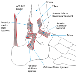
428:, i.e. the articulation between the medial aspect of the distal fibula and the lateral aspect of the distal tibia. An isolated injury to this ligament is often called a
2278:
512:. The tendons which pass through the superior extensor retinaculum are all sheathed along their paths through the inferior extensor retinaculum and the tendon of the
666:
Fracture of both sides of the ankle with dislocation as seen on anteroposterior X-ray. (1) fibula, (2) tibia, (arrow) medial malleolus, (arrowhead) lateral malleolus
2728:
1707:"The reliability and validity of radiographic measurements for determining the three-dimensional position of the talus in varus and valgus osteoarthritic ankles"
1237:
Imai, Kan; Ikoma, Kazuya; Kido, Masamitsu; Maki, Masahiro; Fujiwara, Hiroyoshi; Arai, Yuji; Oda, Ryo; Tokunaga, Daisaku; Inoue, Nozomu; Kubo, Toshikazu (2015).
424:
Though it does not span the ankle joint itself, the syndesmotic ligament makes an important contribution to the stability of the ankle. This ligament spans the
125:
2973:
2968:
2027:
523:
extends from the medial malleolus to the medical process of the calcaneus, and the following structures in order from medial to lateral: the tendon of the
443:
is more likely to occur when the ankle is plantar-flexed, as ligamentous support is more important in this position. The classic ankle sprain involves the
3065:
630:
1981:
Ono, K.; Nakamura, M.; Kurata, Y.; Hiroshima, K. (September 1984). "Ball-and-socket ankle joint: Anatomical and kinematic analysis of the hindfoot".
471:) allow the tendons to exert force across the angle between the leg and foot without lifting away from the angle, a process called bowstringing. The
629:
Of all major joints, the ankle is the most commonly injured. If the outside surface of the foot is twisted under the leg during weight bearing, the
3281:
1944:"The effect of calf muscle stretching exercises on ankle joint dorsiflexion and dynamic foot pressures, force and related temporal parameters"
3276:
2143:
2124:
2105:
1891:
1202:
1172:
1143:
1113:
1085:
1037:
1005:
973:
933:
904:
504:
472:
101:
2271:
3394:
3118:
2828:
2721:
3243:
3150:
721:
can help determine the need for X-rays. Special X-ray views called stress views help determine whether an ankle fracture is unstable.
3371:
852:
It has been suggested that dexterous control of toes has been lost in favour of a more precise voluntary control of the ankle joint.
3343:
3310:
3248:
2838:
1775:
3315:
3266:
3220:
3104:
2833:
492:
3271:
3225:
3183:
3100:
2996:
2264:
1165:
Rockwood and Green's
Fractures in Adults: Two Volumes Plus Integrated Content Website (Rockwood, Green, and Wilkins' Fractures)
488:
1392:
Ribot-Ciscar, E; Bergenheim, M; Albert, F; Roll, J. P. (2003). "Proprioceptive population coding of limb position in humans".
3386:
3353:
3348:
3173:
3109:
3075:
2991:
2786:
2714:
2197:
2173:
2161:
528:
363:
796:), which untreated, results in walking on the sides of the feet. Treatment may involve manipulation and casting or surgery.
258:
The talocrural joint is the only mortise and tenon joint in the human body, the term likening the skeletal structure to the
2185:
3145:
3113:
3070:
634:
544:
444:
359:
120:
3333:
3001:
748:
475:
extends between the anterior (forward) surfaces of the tibia and fibula near their lower (distal) ends. It contains the
247:
239:
3466:
3338:
3137:
2983:
2960:
2911:
2887:
2869:
2241:
310:
186:
729:). Significant recovery generally occurs within four months while completely recovery usually takes up to one year.
242:) from the narrowest point of the lower leg and includes the parts of the foot closer to the body (proximal) to the
3363:
2882:
2864:
2854:
2849:
2216:
1554:
520:
500:
391:
295:
2781:
575:
It was hypothesized that muscle spindle feedback from the ankle dorsiflexors played the most substantial role in
524:
3080:
1437:"Brain Activity during Ankle Proprioceptive Stimulation Predicts Balance Performance in Young and Older Adults"
742:
695:
513:
484:
452:
367:
84:
616:
286:
is a bony process extending distally off the medial tibia. The distal-most aspect of the fibula is called the
3235:
3128:
642:
532:
448:
290:. Together, the malleoli, along with their supporting ligaments, stabilize the talus underneath the tibia.
3325:
3302:
2916:
2775:
2083:
476:
132:
108:
96:
3258:
3178:
2939:
1907:
Gatt, Alfred; Chockalingam, Nachiappan (2011-01-01). "Clinical
Assessment of Ankle Joint Dorsiflexion".
1435:
Goble, D. J.; Coxon, J. P.; Van Impe, A.; Geurts, M.; Doumas, M.; Wenderoth, N.; Swinnen, S. P. (2011).
714:
536:
3212:
3165:
2796:
2770:
2765:
710:
687:
480:
395:
701:
Ankle fractures may result from excessive stress on the joint such as from rolling an ankle or from
3188:
3155:
551:
509:
496:
1219:
1160:
3426:
3421:
3413:
3203:
2894:
2067:
1417:
1374:
816:
1349:
Lephart, S. M.; Pincivero, D. M.; Rozzi, S. L. (1998). "Proprioception of the ankle and knee".
2791:
2139:
2120:
2101:
2059:
1998:
1963:
1924:
1887:
1856:
1822:
1771:
1762:
Chapter 5 - Radiological morphology of peritalar instability in varus and valgus tilted ankles
1744:
1726:
1687:
1646:
1597:
1523:
1505:
1466:
1409:
1366:
1331:
1278:
1260:
1198:
1192:
1168:
1139:
1109:
1081:
1033:
1025:
1001:
993:
969:
953:
929:
900:
892:
718:
683:
662:
597:
429:
410:
302:
287:
259:
2042:
Brouwer, B.; Ashby, P. (1992). "Corticospinal projections to lower limb motoneurons in man".
1133:
1073:
965:
958:
921:
301:
The bony arch formed by the tibial plafond and the two malleoli is referred to as the ankle "
3055:
3050:
3045:
3040:
3035:
2904:
2801:
2478:
2409:
2051:
1990:
1955:
1916:
1879:
1812:
1804:
1734:
1718:
1677:
1636:
1628:
1513:
1497:
1456:
1448:
1401:
1358:
1321:
1268:
1250:
559:
555:
379:
355:
283:
3461:
2934:
2899:
793:
279:
2427:
584:
Further research is needed in order to see to what extent does the ankle affect balance.
2706:
3451:
3092:
2684:
2594:
2542:
2399:
1943:
1817:
1792:
1739:
1706:
1641:
1616:
1518:
1485:
1461:
1436:
1273:
1238:
829:
820:
682:, and an inability to walk on the injured leg. Complications may include an associated
671:
655:
605:
576:
440:
335:
306:
210:
190:
182:
1761:
346:
3456:
3445:
2619:
2609:
2021:
2016:
1994:
1362:
463:
A number of tendons pass through the ankle region. Bands of connective tissue called
2071:
1421:
1378:
2604:
1808:
1632:
1452:
1326:
1309:
768:
For ligamentous injury, there are three main landmarks on X-rays: The first is the
702:
601:
540:
436:
604:, but how much energy is used in leg swing as opposed to advancing the whole-body
262:
of the same name. The bony architecture of the ankle consists of three bones: the
1883:
1705:
Nosewicz, Tomasz L.; Knupp, Markus; Bolliger, Lilianna; Hintermann, Beat (2012).
1682:
1665:
420:
is attached at the lateral malleolus and to the lateral surface of the calcaneus.
89:
2689:
2614:
2515:
2377:
2362:
675:
425:
383:
213:
1959:
2659:
2532:
2483:
2357:
2309:
2301:
2295:
2287:
1722:
1405:
1255:
451:
sprains. Another ligament that can be injured in a severe ankle sprain is the
271:
198:
1730:
1691:
1509:
1264:
2745:
2631:
2510:
2347:
2220:
1589:
706:
593:
387:
314:
231:
113:
2237:
1967:
1928:
1860:
1844:
1826:
1748:
1650:
1601:
1527:
1470:
1413:
1282:
1019:
1017:
17:
2240:. Your orthopaedic connection (American Academy of Orthopaedic Surgeons).
2063:
2002:
1370:
1335:
886:
884:
882:
2741:
2639:
2559:
2394:
2327:
2256:
788:
691:
679:
1942:
Macklin, Katriona; Healy, Aoife; Chockalingam, Nachiappan (March 2012).
592:
Historically, the role of the ankle in locomotion has been discussed by
313:. The joint surface of all bones in the ankle is covered with articular
2574:
2520:
2466:
2456:
2342:
2055:
1501:
2031:. Vol. 2 (11th ed.). Cambridge University Press. p. 58.
1054:
138:
2589:
2564:
2505:
2498:
2493:
2488:
2432:
2372:
2352:
2115:
McKinley, Michael P.; Martini, Frederic; Timmons, Michael J. (2000).
899:(7th ed.). Lippincott Williams & Wilkins. pp. 508–669.
638:
600:. There is no question that ankle push-off is a significant force in
572:
of the muscle it innervates and to any change in length that occurs.
267:
206:
1920:
51:
459:
Retinacula, tendons and their synovial sheaths, vessels, and nerves
2737:
2649:
2599:
2579:
2569:
2525:
2471:
2461:
2447:
2404:
2332:
1296:
747:
741:
The initial evaluation of suspected ankle pathology is usually by
661:
615:
345:
263:
202:
170:
72:
3015:
2926:
2820:
2811:
2674:
2664:
2654:
2584:
2437:
2419:
2337:
2322:
2317:
1135:
Gray's
Anatomy E-Book: The Anatomical Basis of Clinical Practice
861:
726:
447:(ATFL), which is also the most commonly injured ligament during
243:
235:
162:
39:
2710:
2260:
1239:"Joint space width of the tibiotalar joint in the healthy foot"
274:. The articular surface of the tibia may be referred to as the
2753:
2679:
2644:
2551:
2389:
2384:
2367:
866:
378:
supports the medial side of the joint, and is attached at the
166:
1194:
Kinesiology - E-Book: The
Skeletal System and Muscle Function
987:
985:
320:
The distances between the bones in the ankle are as follows:
2020:
189:. The movements produced at this joint are dorsiflexion and
891:
Moore, Keith L.; Dalley, Arthur F.; Agur, A. M. R. (2013).
435:
The bony architecture of the ankle joint is most stable in
1770:. University of Amsterdam, Faculty of Medicine (AMC-UvA).
1583:
1581:
1579:
1577:
1575:
1486:"A unified perspective on ankle push-off in human walking"
641:) as it is weaker than the medial ligament and it resists
413:
of the fibula to the dorsal and ventral ends of the talus.
1670:
Foot & Ankle
Surgery: Techniques, Reports & Cases
1167:. Lippincott Williams & Wilkins. pp. 1928–1971.
1617:"Interventions for treating ankle fractures in children"
1549:
1547:
1545:
1543:
1541:
1539:
1537:
815:
The word ankle or ancle is common, in various forms, to
674:
is a break of one or more of the bones that make up the
2096:
Anderson, Stephen A.; Calais-Germain, Blandine (1993).
1080:(3rd ed.). Houghton Mifflin Harcourt. p. 22.
928:(3rd ed.). Houghton Mifflin Harcourt. p. 22.
620:
A diagram illustrating varying severity of ankle sprain
487:
within its tendon sheath and the unsheathed tendons of
761:, and an angle of more than 94 degrees is regarded as
2215:
Ardizzone, Remy; Valmassy, Ronald L. (October 2005).
1909:
Journal of the
American Podiatric Medical Association
1314:
The
Journal of Bone and Joint Surgery. British Volume
1615:
Yeung DE, Jia X, Miller CA, Barker SL (April 2016).
996:. In Kolt, Gregory S.; Snyder-Mackler, Lynn (eds.).
3412:
3385:
3362:
3324:
3301:
3294:
3257:
3234:
3211:
3201:
3164:
3136:
3127:
3091:
3023:
3014:
2982:
2959:
2952:
2925:
2819:
2810:
2752:
2630:
2550:
2541:
2446:
2418:
2308:
2294:
1555:"Ankle Fractures (Broken Ankle) - OrthoInfo - AAOS"
956:. In Gay, Spencer B.; Woodcock, Richard J. (eds.).
324:
Talus - medial malleolus : 1.70 ± 0.13 mm
119:
107:
95:
83:
71:
66:
61:
32:
957:
230:The ankle region is found at the junction of the
1793:"Radiological evaluation of a high ankle sprain"
1186:
1184:
1838:
1836:
1797:Proceedings (Baylor University. Medical Center)
1596:. Treasure Island (FL): StatPearls Publishing.
1127:
1125:
1099:
1097:
409:support the lateral side of the joint from the
382:of the tibia and connect in four places to the
1876:Epidemiology of Human Congenital Malformations
1197:. Elsevier Health Sciences. pp. 284–292.
992:Williams, D. S. Blaise; Taunton, Jack (2007).
964:. Lippincott Williams & Wilkins. pp.
330:Talus - lateral malleolus: 2.13 ± 0.20 mm
2722:
2272:
2203:Dorsum of Foot. Ankle joint. Deep dissection.
2179:Dorsum of Foot. Ankle joint. Deep dissection.
2167:Dorsum of Foot. Ankle joint. Deep dissection.
1032:. Elsevier Health Sciences. pp. 745–55.
1000:. Elsevier Health Sciences. pp. 420–39.
947:
945:
209:(both in the leg). The talocrural joint is a
8:
2191:Ankle joint. Deep dissection. Anterior view.
1768:Acute and chronic aspects of hindfoot trauma
1664:Ptak, Nathaniel A.; Rigby, Ryan B. (2024).
1621:The Cochrane Database of Systematic Reviews
1484:Zelik, Karl E.; Adamczyk, Peter G. (2016).
1310:"Mechanoreceptors in human ankle ligaments"
832:
823:
705:. Types of ankle fractures include lateral
327:Talus - tibial plafond: 2.04 ± 0.29 mm
197:The main bones of the ankle region are the
3298:
3208:
3133:
3020:
2956:
2816:
2729:
2715:
2707:
2547:
2279:
2265:
2257:
2136:Essentials of Human Anatomy and Physiology
516:is also contained within the retinaculum.
50:
38:
1874:Källén, Bengt (2014). "Pes Equinovarus".
1816:
1738:
1681:
1640:
1517:
1460:
1325:
1272:
1254:
709:, medial malleolus, posterior malleolus,
398:, and to the medial surface of the talus.
2217:"How To Diagnose Lateral Ankle Injuries"
2119:. Englewood Cliffs, N.J: Prentice Hall.
1791:Evans, JM; Schucany, WG (October 2006).
1055:"Talocrural Articulation or Ankle-joint"
998:Physical Therapies in Sport and Exercise
819:, probably connected in origin with the
807:and fusion of the talo-navicular joint.
2157:
878:
678:. Symptoms may include pain, swelling,
499:passes under the retinaculum while the
354:The ankle joint is bound by the strong
1308:Michelson, J. D.; Hutchins, C (1995).
1191:Joseph E. Muscolino (21 August 2016).
1078:Webster's New World Medical Dictionary
926:Webster's New World Medical Dictionary
136:
29:
27:Region where the foot and the leg meet
505:inferior extensor retinaculum of foot
473:superior extensor retinaculum of foot
309:(also called talocalcaneal), and the
7:
2138:. San Francisco: Benjamin Cummings.
2974:posterior of the head of the fibula
1490:The Journal of Experimental Biology
3036:medial of talocrural joint/deltoid
2969:anterior of the head of the fibula
2244:from the original on 23 March 2010
1243:Journal of Foot and Ankle Research
25:
3066:lateral collateral of ankle joint
2839:Posterior meniscofemoral ligament
1132:Susan Standring (7 August 2015).
803:Occasionally a human ankle has a
358:and three lateral ligaments: the
161:(informal) is the area where the
3151:plantar calcaneonavicular/spring
2834:Anterior meniscofemoral ligament
2219:. Podiatry Today. Archived from
2196:
2184:
2172:
2160:
1995:10.1097/01241398-198409000-00007
1983:Journal of Pediatric Orthopedics
1363:10.2165/00007256-199825030-00002
1159:David P. Barei (29 March 2012).
1108:. Philadelphia: Wolters Kluwer.
654:This section is an excerpt from
1588:Wire J, Slane VH (9 May 2019).
1297:https://doi.org/10.7547/1010059
407:posterior talofibular ligaments
169:meet. The ankle includes three
56:Lateral view of the human ankle
3156:bifurcated (calcaneonavicular)
2134:Marieb, Elaine Nicpon (2000).
1809:10.1080/08998280.2006.11928206
1633:10.1002/14651858.CD010836.pub2
1453:10.1523/JNEUROSCI.4159-11.2011
1327:10.1302/0301-620X.77B2.7706334
1218:Joseph H Volker (2018-08-08).
1163:. In Robert W. Bucholz (ed.).
752:Tibiotalar surface angle (TTS)
529:flexor digitorum longus muscle
364:posterior talofibular ligament
1:
792:inward rotation of the foot (
545:flexor hallucis longus muscle
445:anterior talofibular ligament
360:anterior talofibular ligament
334:Decreased distances indicate
3282:interosseous cuneometatarsal
3002:Interosseous membrane of leg
1884:10.1007/978-3-319-01472-2_22
1843:Gore AI, Spencer JP (2004).
1766:T.L. Nosewicz (2018-09-25).
1683:10.1016/j.fastrc.2023.100357
1138:. Elsevier Health Sciences.
1028:. In Adams, James G. (ed.).
1024:del Castillo, Jorge (2012).
635:anterior talofibular portion
3277:interosseous intercuneiform
3189:bifurcated (calcaneocuboid)
2100:. Chicago: Eastland Press.
2044:Experimental Brain Research
1394:Experimental Brain Research
1106:Clinically oriented anatomy
994:"Foot, ankle and lower leg"
897:Clinically Oriented Anatomy
805:ball-and-socket ankle joint
637:, is subject to tearing (a
350:Ligaments of ankle and feet
311:Inferior tibiofibular joint
187:inferior tibiofibular joint
3483:
3326:Intermetatarsal/metatarsal
2081:
1960:10.1016/j.foot.2011.09.001
653:
521:flexor retinaculum of foot
501:superficial peroneal nerve
392:calcaneonavicular ligament
2236:Haddad, Steven L. (ed.).
1723:10.1007/s00256-012-1421-6
1406:10.1007/s00221-003-1384-x
1256:10.1186/s13047-015-0086-5
1059:Anatomy of the Human Body
1026:"Foot and Ankle Injuries"
954:"Musculoskeletal Imaging"
952:Milner, Brent K. (1999).
833:
645:of the talocrural joint.
525:tibialis posterior muscle
493:extensor digitorum longus
131:
49:
37:
3303:Tarsometatarsal/Lisfranc
770:tibiofibular clear space
743:projectional radiography
696:post-traumatic arthritis
554:hold the tendons of the
543:, and the tendon of the
514:fibularis tertius muscle
489:extensor hallucis longus
485:tibialis anterior muscle
453:calcaneofibular ligament
418:calcaneofibular ligament
368:calcaneofibular ligament
238:. It extends downwards (
2028:Encyclopædia Britannica
1441:Journal of Neuroscience
533:posterior tibial artery
483:and the tendons of the
3349:superficial transverse
3184:plantar calcaneocuboid
3093:Subtalar/talocalcaneal
2997:Posterior tibiofibular
2288:Human regional anatomy
2084:anatomical terminology
824:
753:
715:trimalleolar fractures
667:
621:
503:is outside of it. The
477:anterior tibial artery
351:
133:Anatomical terminology
3174:dorsal calcaneocuboid
3138:Talocalcaneonavicular
3076:posterior talofibular
2992:Anterior tibiofibular
2984:Inferior tibiofibular
2961:Superior tibiofibular
2940:Infrapatellar fat pad
2787:transverse acetabular
1161:"56. Pilon Fractures"
1104:Moore, Keith (2018).
751:
665:
619:
612:Clinical significance
349:
3146:dorsal talonavicular
3071:anterior talofibular
3046:posterior tibiotalar
3025:Talocrural and ankle
1878:. pp. 111–113.
1053:Gray, Henry (1918).
774:tibiofibular overlap
688:compartment syndrome
527:, the tendon of the
396:navicular tuberosity
282:for "ceiling"). The
3364:Metatarsophalangeal
3041:anterior tibiotalar
2098:Anatomy of Movement
510:plantar aponeurosis
497:deep peroneal nerve
246:and upper surface (
201:(in the foot), the
3467:Lower limb anatomy
2238:"Foot & Ankle"
2223:on January 4, 2010
2082:This article uses
2056:10.1007/bf00229889
1845:"The newborn foot"
1711:Skeletal Radiology
1502:10.1242/jeb.140376
1030:Emergency Medicine
817:Germanic languages
778:medial clear space
754:
668:
622:
552:fibular retinacula
352:
175:ankle joint proper
3439:
3438:
3435:
3434:
3408:
3407:
3290:
3289:
3236:Cuboideonavicular
3197:
3196:
3129:Transverse tarsal
3010:
3009:
2948:
2947:
2792:acetabular labrum
2704:
2703:
2700:
2699:
2154:Additional images
2145:978-0-8053-4940-5
2126:978-0-13-010011-5
2107:978-0-939616-17-6
1893:978-3-319-01471-5
1717:(12): 1567–1573.
1590:"Ankle Fractures"
1559:www.orthoinfo.org
1496:(23): 3676–3683.
1204:978-0-323-39935-7
1174:978-1-4511-6144-1
1145:978-0-7020-6851-5
1115:978-1-4963-4721-3
1087:978-0-544-18897-6
1039:978-1-4557-3394-1
1007:978-0-443-10351-3
975:978-0-683-30663-7
935:978-0-544-18897-6
906:978-1-4511-1945-9
719:Ottawa ankle rule
684:high ankle sprain
633:, especially the
598:Leonardo da Vinci
430:high ankle sprain
411:lateral malleolus
288:lateral malleolus
260:woodworking joint
155:talocrural region
147:
146:
142:
16:(Redirected from
3474:
3299:
3209:
3134:
3021:
2957:
2817:
2802:zona orbicularis
2731:
2724:
2717:
2708:
2548:
2479:Vertebral column
2281:
2274:
2267:
2258:
2253:
2251:
2249:
2232:
2230:
2228:
2200:
2188:
2176:
2164:
2149:
2130:
2111:
2076:
2075:
2039:
2033:
2032:
2024:
2013:
2007:
2006:
1978:
1972:
1971:
1939:
1933:
1932:
1904:
1898:
1897:
1871:
1865:
1864:
1849:Am Fam Physician
1840:
1831:
1830:
1820:
1788:
1782:
1781:
1759:
1753:
1752:
1742:
1702:
1696:
1695:
1685:
1661:
1655:
1654:
1644:
1612:
1606:
1605:
1585:
1570:
1569:
1567:
1565:
1551:
1532:
1531:
1521:
1481:
1475:
1474:
1464:
1447:(45): 16344–52.
1432:
1426:
1425:
1389:
1383:
1382:
1346:
1340:
1339:
1329:
1305:
1299:
1293:
1287:
1286:
1276:
1258:
1234:
1228:
1227:
1215:
1209:
1208:
1188:
1179:
1178:
1156:
1150:
1149:
1129:
1120:
1119:
1101:
1092:
1091:
1069:
1063:
1062:
1050:
1044:
1043:
1021:
1012:
1011:
989:
980:
979:
963:
960:Radiology Recall
949:
940:
939:
917:
911:
910:
888:
837:, meaning bent.
836:
835:
827:
631:lateral ligament
625:Traumatic injury
567:Mechanoreceptors
560:fibularis brevis
556:fibularis longus
380:medial malleolus
376:deltoid ligament
356:deltoid ligament
284:medial malleolus
179:talocrural joint
139:edit on Wikidata
54:
42:
30:
21:
3482:
3481:
3477:
3476:
3475:
3473:
3472:
3471:
3442:
3441:
3440:
3431:
3404:
3387:Interphalangeal
3381:
3358:
3354:deep transverse
3320:
3286:
3253:
3230:
3193:
3160:
3123:
3087:
3081:calcaneofibular
3006:
2978:
2944:
2935:Patellar tendon
2921:
2870:fibular/lateral
2806:
2748:
2735:
2705:
2696:
2626:
2537:
2442:
2414:
2304:
2290:
2285:
2247:
2245:
2235:
2226:
2224:
2214:
2211:
2204:
2201:
2192:
2189:
2180:
2177:
2168:
2165:
2156:
2146:
2133:
2127:
2114:
2108:
2095:
2092:
2087:
2080:
2079:
2041:
2040:
2036:
2015:
2014:
2010:
1980:
1979:
1975:
1941:
1940:
1936:
1921:10.7547/1010059
1906:
1905:
1901:
1894:
1873:
1872:
1868:
1842:
1841:
1834:
1790:
1789:
1785:
1778:
1765:
1760:
1756:
1704:
1703:
1699:
1663:
1662:
1658:
1627:(4): CD010836.
1614:
1613:
1609:
1587:
1586:
1573:
1563:
1561:
1553:
1552:
1535:
1483:
1482:
1478:
1434:
1433:
1429:
1391:
1390:
1386:
1351:Sports Medicine
1348:
1347:
1343:
1307:
1306:
1302:
1294:
1290:
1236:
1235:
1231:
1217:
1216:
1212:
1205:
1190:
1189:
1182:
1175:
1158:
1157:
1153:
1146:
1131:
1130:
1123:
1116:
1103:
1102:
1095:
1088:
1071:
1070:
1066:
1052:
1051:
1047:
1040:
1023:
1022:
1015:
1008:
991:
990:
983:
976:
951:
950:
943:
936:
919:
918:
914:
907:
890:
889:
880:
875:
858:
850:
843:
813:
794:varus deformity
786:
739:
734:
733:
659:
651:
643:inward rotation
627:
614:
590:
569:
461:
344:
256:
250:) of the foot.
228:
223:
143:
57:
45:
28:
23:
22:
15:
12:
11:
5:
3480:
3478:
3470:
3469:
3464:
3459:
3454:
3444:
3443:
3437:
3436:
3433:
3432:
3430:
3429:
3424:
3418:
3416:
3410:
3409:
3406:
3405:
3403:
3402:
3397:
3391:
3389:
3383:
3382:
3380:
3379:
3374:
3368:
3366:
3360:
3359:
3357:
3356:
3351:
3346:
3341:
3336:
3330:
3328:
3322:
3321:
3319:
3318:
3313:
3307:
3305:
3296:
3292:
3291:
3288:
3287:
3285:
3284:
3279:
3274:
3269:
3263:
3261:
3259:Intercuneiform
3255:
3254:
3252:
3251:
3246:
3240:
3238:
3232:
3231:
3229:
3228:
3223:
3217:
3215:
3213:Cuneonavicular
3206:
3199:
3198:
3195:
3194:
3192:
3191:
3186:
3181:
3176:
3170:
3168:
3166:Calcaneocuboid
3162:
3161:
3159:
3158:
3153:
3148:
3142:
3140:
3131:
3125:
3124:
3122:
3121:
3116:
3107:
3097:
3095:
3089:
3088:
3086:
3085:
3084:
3083:
3078:
3073:
3060:
3059:
3058:
3056:tibionavicular
3053:
3051:tibiocalcaneal
3048:
3043:
3029:
3027:
3018:
3012:
3011:
3008:
3007:
3005:
3004:
2999:
2994:
2988:
2986:
2980:
2979:
2977:
2976:
2971:
2965:
2963:
2954:
2950:
2949:
2946:
2945:
2943:
2942:
2937:
2931:
2929:
2927:Patellofemoral
2923:
2922:
2920:
2919:
2914:
2909:
2908:
2907:
2902:
2892:
2891:
2890:
2885:
2877:intracapsular:
2874:
2873:
2872:
2867:
2859:
2858:
2857:
2852:
2844:extracapsular:
2841:
2836:
2831:
2825:
2823:
2814:
2808:
2807:
2805:
2804:
2799:
2794:
2789:
2784:
2779:
2773:
2768:
2758:
2756:
2750:
2749:
2736:
2734:
2733:
2726:
2719:
2711:
2702:
2701:
2698:
2697:
2695:
2694:
2693:
2692:
2687:
2682:
2677:
2672:
2662:
2657:
2652:
2647:
2642:
2636:
2634:
2628:
2627:
2625:
2624:
2623:
2622:
2617:
2612:
2607:
2602:
2597:
2592:
2582:
2577:
2572:
2567:
2562:
2556:
2554:
2545:
2539:
2538:
2536:
2535:
2530:
2529:
2528:
2523:
2518:
2508:
2503:
2502:
2501:
2496:
2486:
2481:
2476:
2475:
2474:
2469:
2464:
2453:
2451:
2444:
2443:
2441:
2440:
2435:
2430:
2424:
2422:
2416:
2415:
2413:
2412:
2407:
2402:
2397:
2392:
2387:
2382:
2381:
2380:
2375:
2370:
2365:
2360:
2355:
2350:
2345:
2340:
2335:
2330:
2320:
2314:
2312:
2306:
2305:
2300:
2298:
2292:
2291:
2286:
2284:
2283:
2276:
2269:
2261:
2255:
2254:
2233:
2210:
2209:External links
2207:
2206:
2205:
2202:
2195:
2193:
2190:
2183:
2181:
2178:
2171:
2169:
2166:
2159:
2155:
2152:
2151:
2150:
2144:
2131:
2125:
2112:
2106:
2091:
2088:
2078:
2077:
2034:
2019:, ed. (1911).
2017:Chisholm, Hugh
2008:
1989:(5): 564–568.
1973:
1934:
1899:
1892:
1866:
1832:
1783:
1776:
1754:
1697:
1656:
1607:
1571:
1533:
1476:
1427:
1384:
1341:
1300:
1288:
1229:
1210:
1203:
1180:
1173:
1151:
1144:
1121:
1114:
1093:
1086:
1072:WebMD (2009).
1064:
1045:
1038:
1013:
1006:
981:
974:
941:
934:
920:WebMD (2009).
912:
905:
877:
876:
874:
871:
870:
869:
864:
857:
854:
849:
846:
842:
839:
812:
809:
785:
782:
763:talipes valgus
738:
735:
672:ankle fracture
660:
656:Ankle fracture
652:
650:
647:
626:
623:
613:
610:
608:is not clear.
606:center of mass
589:
586:
577:proprioception
568:
565:
460:
457:
441:sprained ankle
422:
421:
414:
399:
343:
340:
336:osteoarthritis
332:
331:
328:
325:
307:subtalar joint
255:
252:
227:
224:
222:
219:
191:plantarflexion
183:subtalar joint
145:
144:
135:
129:
128:
123:
117:
116:
111:
105:
104:
99:
93:
92:
87:
81:
80:
75:
69:
68:
64:
63:
59:
58:
55:
47:
46:
43:
35:
34:
26:
24:
14:
13:
10:
9:
6:
4:
3:
2:
3479:
3468:
3465:
3463:
3460:
3458:
3455:
3453:
3450:
3449:
3447:
3428:
3425:
3423:
3420:
3419:
3417:
3415:
3411:
3401:
3398:
3396:
3393:
3392:
3390:
3388:
3384:
3378:
3375:
3373:
3370:
3369:
3367:
3365:
3361:
3355:
3352:
3350:
3347:
3345:
3342:
3340:
3337:
3335:
3332:
3331:
3329:
3327:
3323:
3317:
3314:
3312:
3309:
3308:
3306:
3304:
3300:
3297:
3293:
3283:
3280:
3278:
3275:
3273:
3270:
3268:
3265:
3264:
3262:
3260:
3256:
3250:
3247:
3245:
3242:
3241:
3239:
3237:
3233:
3227:
3224:
3222:
3219:
3218:
3216:
3214:
3210:
3207:
3205:
3200:
3190:
3187:
3185:
3182:
3180:
3177:
3175:
3172:
3171:
3169:
3167:
3163:
3157:
3154:
3152:
3149:
3147:
3144:
3143:
3141:
3139:
3135:
3132:
3130:
3126:
3120:
3117:
3115:
3111:
3108:
3106:
3102:
3099:
3098:
3096:
3094:
3090:
3082:
3079:
3077:
3074:
3072:
3069:
3068:
3067:
3064:
3061:
3057:
3054:
3052:
3049:
3047:
3044:
3042:
3039:
3038:
3037:
3034:
3031:
3030:
3028:
3026:
3022:
3019:
3017:
3013:
3003:
3000:
2998:
2995:
2993:
2990:
2989:
2987:
2985:
2981:
2975:
2972:
2970:
2967:
2966:
2964:
2962:
2958:
2955:
2951:
2941:
2938:
2936:
2933:
2932:
2930:
2928:
2924:
2918:
2917:anterolateral
2915:
2913:
2910:
2906:
2903:
2901:
2898:
2897:
2896:
2893:
2889:
2886:
2884:
2881:
2880:
2878:
2875:
2871:
2868:
2866:
2865:medial/tibial
2863:
2862:
2860:
2856:
2853:
2851:
2848:
2847:
2845:
2842:
2840:
2837:
2835:
2832:
2830:
2827:
2826:
2824:
2822:
2818:
2815:
2813:
2809:
2803:
2800:
2798:
2795:
2793:
2790:
2788:
2785:
2783:
2782:head of femur
2780:
2777:
2776:ischiofemoral
2774:
2772:
2769:
2767:
2763:
2760:
2759:
2757:
2755:
2751:
2747:
2743:
2739:
2732:
2727:
2725:
2720:
2718:
2713:
2712:
2709:
2691:
2688:
2686:
2683:
2681:
2678:
2676:
2673:
2671:
2668:
2667:
2666:
2663:
2661:
2658:
2656:
2653:
2651:
2648:
2646:
2643:
2641:
2638:
2637:
2635:
2633:
2629:
2621:
2618:
2616:
2613:
2611:
2608:
2606:
2603:
2601:
2598:
2596:
2593:
2591:
2588:
2587:
2586:
2583:
2581:
2578:
2576:
2573:
2571:
2568:
2566:
2563:
2561:
2558:
2557:
2555:
2553:
2549:
2546:
2544:
2540:
2534:
2531:
2527:
2524:
2522:
2519:
2517:
2514:
2513:
2512:
2509:
2507:
2504:
2500:
2497:
2495:
2492:
2491:
2490:
2487:
2485:
2482:
2480:
2477:
2473:
2470:
2468:
2465:
2463:
2460:
2459:
2458:
2455:
2454:
2452:
2449:
2445:
2439:
2436:
2434:
2431:
2429:
2426:
2425:
2423:
2421:
2417:
2411:
2408:
2406:
2403:
2401:
2398:
2396:
2393:
2391:
2388:
2386:
2383:
2379:
2376:
2374:
2371:
2369:
2366:
2364:
2361:
2359:
2356:
2354:
2351:
2349:
2346:
2344:
2341:
2339:
2336:
2334:
2331:
2329:
2326:
2325:
2324:
2321:
2319:
2316:
2315:
2313:
2311:
2307:
2303:
2299:
2297:
2293:
2289:
2282:
2277:
2275:
2270:
2268:
2263:
2262:
2259:
2248:September 21,
2243:
2239:
2234:
2227:September 21,
2222:
2218:
2213:
2212:
2208:
2199:
2194:
2187:
2182:
2175:
2170:
2163:
2158:
2153:
2147:
2141:
2137:
2132:
2128:
2122:
2118:
2117:Human Anatomy
2113:
2109:
2103:
2099:
2094:
2093:
2089:
2085:
2073:
2069:
2065:
2061:
2057:
2053:
2050:(3): 649–54.
2049:
2045:
2038:
2035:
2030:
2029:
2023:
2022:"Ankle"
2018:
2012:
2009:
2004:
2000:
1996:
1992:
1988:
1984:
1977:
1974:
1969:
1965:
1961:
1957:
1953:
1949:
1945:
1938:
1935:
1930:
1926:
1922:
1918:
1914:
1910:
1903:
1900:
1895:
1889:
1885:
1881:
1877:
1870:
1867:
1862:
1858:
1855:(4): 865–72.
1854:
1850:
1846:
1839:
1837:
1833:
1828:
1824:
1819:
1814:
1810:
1806:
1802:
1798:
1794:
1787:
1784:
1779:
1777:9789463750479
1773:
1769:
1763:
1758:
1755:
1750:
1746:
1741:
1736:
1732:
1728:
1724:
1720:
1716:
1712:
1708:
1701:
1698:
1693:
1689:
1684:
1679:
1676:(1): 100357.
1675:
1671:
1667:
1660:
1657:
1652:
1648:
1643:
1638:
1634:
1630:
1626:
1622:
1618:
1611:
1608:
1603:
1599:
1595:
1591:
1584:
1582:
1580:
1578:
1576:
1572:
1560:
1556:
1550:
1548:
1546:
1544:
1542:
1540:
1538:
1534:
1529:
1525:
1520:
1515:
1511:
1507:
1503:
1499:
1495:
1491:
1487:
1480:
1477:
1472:
1468:
1463:
1458:
1454:
1450:
1446:
1442:
1438:
1431:
1428:
1423:
1419:
1415:
1411:
1407:
1403:
1399:
1395:
1388:
1385:
1380:
1376:
1372:
1368:
1364:
1360:
1357:(3): 149–55.
1356:
1352:
1345:
1342:
1337:
1333:
1328:
1323:
1320:(2): 219–24.
1319:
1315:
1311:
1304:
1301:
1298:
1292:
1289:
1284:
1280:
1275:
1270:
1266:
1262:
1257:
1252:
1248:
1244:
1240:
1233:
1230:
1225:
1221:
1220:"Ankle Joint"
1214:
1211:
1206:
1200:
1196:
1195:
1187:
1185:
1181:
1176:
1170:
1166:
1162:
1155:
1152:
1147:
1141:
1137:
1136:
1128:
1126:
1122:
1117:
1111:
1107:
1100:
1098:
1094:
1089:
1083:
1079:
1075:
1074:"ankle joint"
1068:
1065:
1060:
1056:
1049:
1046:
1041:
1035:
1031:
1027:
1020:
1018:
1014:
1009:
1003:
999:
995:
988:
986:
982:
977:
971:
967:
962:
961:
955:
948:
946:
942:
937:
931:
927:
923:
916:
913:
908:
902:
898:
894:
887:
885:
883:
879:
872:
868:
865:
863:
860:
859:
855:
853:
847:
845:
841:Other animals
840:
838:
831:
826:
822:
818:
810:
808:
806:
801:
797:
795:
790:
784:Abnormalities
783:
781:
779:
775:
771:
766:
764:
760:
759:talipes varus
750:
746:
744:
736:
730:
728:
722:
720:
716:
712:
708:
704:
699:
697:
693:
690:, stiffness,
689:
685:
681:
677:
673:
664:
657:
648:
646:
644:
640:
636:
632:
624:
618:
611:
609:
607:
603:
599:
595:
587:
585:
581:
578:
573:
566:
564:
561:
557:
553:
548:
546:
542:
538:
534:
530:
526:
522:
517:
515:
511:
506:
502:
498:
495:muscles. The
494:
490:
486:
482:
478:
474:
470:
466:
458:
456:
454:
450:
446:
442:
438:
433:
431:
427:
419:
415:
412:
408:
404:
400:
397:
393:
389:
385:
381:
377:
373:
372:
371:
369:
365:
361:
357:
348:
341:
339:
337:
329:
326:
323:
322:
321:
318:
316:
312:
308:
304:
299:
297:
291:
289:
285:
281:
277:
273:
269:
265:
261:
253:
251:
249:
245:
241:
237:
233:
225:
220:
218:
215:
212:
208:
204:
200:
195:
192:
188:
184:
180:
176:
172:
168:
164:
160:
156:
152:
140:
134:
130:
127:
124:
122:
118:
115:
112:
110:
106:
103:
100:
98:
94:
91:
88:
86:
82:
79:
76:
74:
70:
65:
60:
53:
48:
41:
36:
31:
19:
3422:Longitudinal
3344:interosseous
3179:long plantar
3119:interosseous
3062:
3032:
3024:
2953:Tibiofibular
2876:
2843:
2821:Tibiofemoral
2761:
2669:
2428:Adam's apple
2246:. Retrieved
2225:. Retrieved
2221:the original
2135:
2116:
2097:
2047:
2043:
2037:
2026:
2011:
1986:
1982:
1976:
1954:(1): 10–17.
1951:
1947:
1937:
1915:(1): 59–69.
1912:
1908:
1902:
1875:
1869:
1852:
1848:
1803:(4): 402–5.
1800:
1796:
1786:
1767:
1757:
1714:
1710:
1700:
1673:
1669:
1659:
1624:
1620:
1610:
1593:
1562:. Retrieved
1558:
1493:
1489:
1479:
1444:
1440:
1430:
1400:(4): 512–9.
1397:
1393:
1387:
1354:
1350:
1344:
1317:
1313:
1303:
1291:
1246:
1242:
1232:
1223:
1213:
1193:
1164:
1154:
1134:
1105:
1077:
1067:
1058:
1048:
1029:
997:
959:
925:
915:
896:
893:"Lower Limb"
851:
844:
814:
804:
802:
798:
787:
777:
773:
769:
767:
762:
758:
757:regarded as
755:
740:
723:
703:blunt trauma
700:
669:
628:
591:
582:
574:
570:
549:
541:tibial nerve
518:
468:
464:
462:
437:dorsiflexion
434:
423:
417:
406:
402:
375:
353:
333:
319:
300:
292:
275:
257:
229:
196:
178:
174:
159:jumping bone
158:
154:
150:
148:
102:A01.1.00.041
77:
3204:intertarsal
2861:collateral
2771:pubofemoral
2766:iliofemoral
1224:Earth's Lab
745:("X-ray").
711:bimalleolar
676:ankle joint
469:retinaculum
467:(singular:
426:syndesmosis
384:talar shelf
254:Ankle joint
214:hinge joint
67:Identifiers
44:Human ankle
18:Ankle-joint
3446:Categories
3427:Transverse
3400:collateral
3377:collateral
2912:transverse
2846:popliteal
2595:Fingernail
2090:References
1594:StatPearls
602:human gait
465:retinacula
439:. Thus, a
366:, and the
270:, and the
185:, and the
3105:posterior
2888:posterior
2879:cruciate
2746:human leg
2742:ligaments
2511:Genitalia
1731:0364-2348
1692:2667-3967
1510:0022-0949
1265:1757-1146
1249:(1): 26.
873:Footnotes
848:Evolution
707:malleolus
649:Fractures
594:Aristotle
449:inversion
388:calcaneus
342:Ligaments
315:cartilage
296:movements
221:Structure
3101:anterior
3063:lateral:
2883:anterior
2640:Buttocks
2560:Shoulder
2395:Mandible
2328:Forehead
2242:Archived
2072:24650165
1968:21944945
1948:The Foot
1929:21242472
1861:14989573
1827:17106502
1749:22609967
1651:27033333
1602:31194464
1528:27903626
1471:22072686
1422:14626459
1414:12677332
1379:13099542
1283:26146520
856:See also
789:Clubfoot
732:females.
692:malunion
680:bruising
588:Function
403:anterior
240:distally
234:and the
211:synovial
165:and the
3395:plantar
3372:plantar
3334:plantar
3311:plantar
3267:plantar
3244:plantar
3221:plantar
3202:Distal
3110:lateral
3033:medial:
2905:lateral
2895:menisci
2855:arcuate
2850:oblique
2829:Capsule
2797:capsule
2762:femoral
2744:of the
2685:Toenail
2575:Forearm
2521:Scrotum
2467:Midriff
2457:Abdomen
2450:(Trunk)
2400:Occiput
2343:Eyebrow
2064:1644127
2003:6490876
1818:1618742
1740:3478506
1642:7111433
1564:20 June
1519:5201006
1462:6633212
1371:9554026
1336:7706334
1274:4490633
966:258–383
922:"ankle"
834:αγκυλος
825:angulus
811:History
737:Imaging
386:of the
303:mortise
276:plafond
157:or the
90:D000842
62:Details
3462:Joints
3414:Arches
3339:dorsal
3316:dorsal
3272:dorsal
3249:dorsal
3226:dorsal
3114:medial
2900:medial
2738:Joints
2620:Little
2610:Middle
2590:Finger
2565:Axilla
2506:Pelvis
2499:Nipple
2494:Breast
2489:Thorax
2433:Throat
2410:Temple
2373:Tongue
2353:Eyelid
2142:
2123:
2104:
2070:
2062:
2001:
1966:
1927:
1890:
1859:
1825:
1815:
1774:
1764:, in:
1747:
1737:
1729:
1690:
1649:
1639:
1600:
1526:
1516:
1508:
1469:
1459:
1420:
1412:
1377:
1369:
1334:
1281:
1271:
1263:
1201:
1171:
1142:
1112:
1084:
1036:
1004:
972:
932:
903:
717:. The
713:, and
694:, and
639:sprain
539:, the
531:, the
394:, the
362:, the
280:French
268:fibula
266:, the
248:dorsum
226:Region
207:fibula
205:, and
181:, the
173:: the
171:joints
153:, the
78:tarsus
3452:Ankle
3295:Other
2670:Ankle
2650:Thigh
2605:Index
2600:Thumb
2580:Wrist
2570:Elbow
2543:Limbs
2526:Vulva
2516:Penis
2472:Navel
2462:Waist
2448:Torso
2405:Scalp
2378:Tooth
2363:Mouth
2333:Cheek
2068:S2CID
1418:S2CID
1375:S2CID
830:Greek
828:, or
821:Latin
272:talus
264:tibia
203:tibia
199:talus
151:ankle
137:[
73:Latin
33:Ankle
3457:Foot
3016:Foot
2812:Knee
2740:and
2690:Sole
2675:Heel
2665:Foot
2660:Calf
2655:Knee
2615:Ring
2585:Hand
2533:Anus
2484:Back
2438:Nape
2420:Neck
2358:Nose
2338:Chin
2323:Face
2318:Hair
2310:Head
2302:Skin
2296:Body
2250:2017
2229:2017
2140:ISBN
2121:ISBN
2102:ISBN
2060:PMID
1999:PMID
1964:PMID
1925:PMID
1888:ISBN
1857:PMID
1823:PMID
1772:ISBN
1745:PMID
1727:ISSN
1688:ISSN
1647:PMID
1625:2016
1598:PMID
1566:2019
1524:PMID
1506:ISSN
1467:PMID
1410:PMID
1367:PMID
1332:PMID
1279:PMID
1261:ISSN
1199:ISBN
1169:ISBN
1140:ISBN
1110:ISBN
1082:ISBN
1034:ISBN
1002:ISBN
970:ISBN
930:ISBN
901:ISBN
862:Foot
727:ORIF
596:and
558:and
550:The
537:vein
535:and
519:The
491:and
481:vein
479:and
416:The
405:and
401:The
374:The
244:heel
236:foot
163:foot
149:The
126:9665
97:TA98
85:MeSH
2754:Hip
2680:Toe
2645:Hip
2632:Leg
2552:Arm
2390:Jaw
2385:Ear
2368:Lip
2348:Eye
2052:doi
1991:doi
1956:doi
1917:doi
1913:101
1880:doi
1813:PMC
1805:doi
1735:PMC
1719:doi
1678:doi
1637:PMC
1629:doi
1514:PMC
1498:doi
1494:219
1457:PMC
1449:doi
1402:doi
1398:149
1359:doi
1322:doi
1269:PMC
1251:doi
867:Leg
670:An
232:leg
177:or
167:leg
121:FMA
114:165
109:TA2
3448::
2066:.
2058:.
2048:89
2046:.
2025:.
1997:.
1985:.
1962:.
1952:22
1950:.
1946:.
1923:.
1911:.
1886:.
1853:69
1851:.
1847:.
1835:^
1821:.
1811:.
1801:19
1799:.
1795:.
1743:.
1733:.
1725:.
1715:41
1713:.
1709:.
1686:.
1672:.
1668:.
1645:.
1635:.
1623:.
1619:.
1592:.
1574:^
1557:.
1536:^
1522:.
1512:.
1504:.
1492:.
1488:.
1465:.
1455:.
1445:31
1443:.
1439:.
1416:.
1408:.
1396:.
1373:.
1365:.
1355:25
1353:.
1330:.
1318:77
1316:.
1312:.
1277:.
1267:.
1259:.
1245:.
1241:.
1222:.
1183:^
1124:^
1096:^
1076:.
1057:.
1016:^
984:^
968:.
944:^
924:.
895:.
881:^
765:.
698:.
686:,
547:.
455:.
432:.
390:,
370:.
338:.
317:.
3112:/
3103:/
2778:)
2764:(
2730:e
2723:t
2716:v
2280:e
2273:t
2266:v
2252:.
2231:.
2148:.
2129:.
2110:.
2086:.
2074:.
2054::
2005:.
1993::
1987:4
1970:.
1958::
1931:.
1919::
1896:.
1882::
1863:.
1829:.
1807::
1780:.
1751:.
1721::
1694:.
1680::
1674:4
1653:.
1631::
1604:.
1568:.
1530:.
1500::
1473:.
1451::
1424:.
1404::
1381:.
1361::
1338:.
1324::
1285:.
1253::
1247:8
1226:.
1207:.
1177:.
1148:.
1118:.
1090:.
1061:.
1042:.
1010:.
978:.
938:.
909:.
725:(
658:.
278:(
141:]
20:)
Text is available under the Creative Commons Attribution-ShareAlike License. Additional terms may apply.