264:, which enable them to locally depolarize adjacent cells. Gap junctions allow the passage of positive cations from the depolarization of the pacemaker cell to adjacent contractile cells. This starts the depolarization and eventual action potential in contractile cells. Having cardiomyocytes connected via gap junctions allow all contractile cells of the heart to act in a coordinated fashion and contract as a unit. All the while being in sync with the pacemaker cells; this is the property that allows the pacemaker cells to control contraction in all other cardiomyocytes.
129:
32:
478:
330:, and depolarizes the other potential pacemaker cells (AV node) to initiate action potentials before these other cells have had a chance to generate their own spontaneous action potential, thus they contract and propagate electrical impulses to the pace set by the cells of the SA node. This is the normal conduction of electrical activity in the heart.
385:. These two relative ion concentration changes slowly depolarize (make more positive) the inside membrane potential (voltage) of the cell, giving these cells their pacemaker potential. When the membrane potential gets depolarized to about -40mV it has reached threshold (cells enter phase 0), allowing an action potential to be generated.
401:
produces the rising phase of the action potential, which results in the reversal of membrane potential to a peak of about +10mV. It is important to note that intracellular calcium causes muscular contraction in contractile cells, and is the effector ion. In heart pacemaker cells, phase 0 depends on the activation of
322:, will also produce a spontaneous action potential at a rate of 30-40 beats per minute, so if the SA and AV node both fail to function, these cells can become pacemakers. These cells will be initiating action potentials and contraction at a much lower rate than the primary or secondary pacemaker cells.
418:
gets more negative). The calcium channels are also inactivated soon after they open. In addition, as sodium channels become inactivated, sodium permeability into the cell is decreased. These ion concentration changes slowly repolarize the cell to resting membrane potential (-60mV). Another important
531:
that generates electrical impulses delivered by electrodes to the chambers of the heart either the upper atria, or lower ventricles to cause the targeted chambers to contract and pump blood. By doing so, the artificial pacemaker takes over from the primary SA node pacemaker to regulate the function
400:
The SA and AV node do not have fast sodium channels like neurons, and the depolarization is mainly caused by a slow influx of calcium ions. (The funny current also increases). Calcium enters the cell via voltage-sensitive calcium channels that open when the threshold is reached. This calcium influx
215:
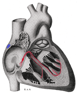
Schematic representation of the sinoatrial node and the atrioventricular bundle of His. The location of the SA node is shown in blue. The bundle, represented in red, originates near the orifice of the coronary sinus, undergoes slight enlargement to form the AV node. The AV node tapers down into the
431:
restores ion concentrations of sodium and potassium ions by pumping sodium out of the cell and pumping (exchanging) potassium into the cell. Restoring these ion concentrations is vital because it enables the cell to reset itself and enables it to repeat the process of spontaneous depolarization
175:
that create these rhythmic impulses, setting the pace for blood pumping, are called pacemaker cells, and they directly control the heart rate. They make up the cardiac pacemaker, that is, the natural pacemaker of the heart. In most humans, the highest concentration of pacemaker cells is in the
405:
instead of the activation of voltage-gated fast sodium channels, which are responsible for initiating action potentials in contractile (non-pacemaker) cells. For this reason, the pacemaker action potential rising phase slope is more gradual than that of the contractile cell (image 2).
492:
also known as an ectopic focus or ectopic foci, is an excitable group of cells that causes a premature heart beat outside the normally functioning SA node of the heart. It is thus a cardiac pacemaker that is ectopic, producing an ectopic beat. If chronic this can result in
325:
The SA node controls the rate of contraction for the entire heart muscle because its cells have the quickest rate of spontaneous depolarization, thus they initiate action potentials the quickest. The action potential generated by the SA node passes down the
212:
376:
that surrounds the cells. However, in pacemaker cells, this potassium permeability (efflux) decreases as time goes on, causing a slow depolarization. In addition, there is a slow, continuous inward flow of
216:
bundle of His, which passes into the ventricular septum and divides into two bundle branches, the left and right bundles. The ultimate distribution cannot be completely shown in this diagram.
414:
The reversal of membrane potential triggers the opening of potassium leak channels, resulting in the rapid loss of potassium ions from the inside of the cell, causing repolarization (V
449:
before it travels down the electrical conduction system, a group of cells further down the heart will become its pacemaker. This center is typically represented by cells inside the
354:
in the body, these cells will slowly depolarize by themselves and do not need any outside innervation from the autonomic nervous system to fire action potentials.
1032:
327:
250:
188:
137:
647:
527:
An artificial cardiac pacemaker (or artificial pacemaker, so as not to be confused with the natural cardiac pacemaker) or just pacemaker is an
338:
There are 3 main stages in the generation of an action potential in a pacemaker cell. Since the stages are analogous to contraction of
299:
which acts as the secondary pacemaker. The cells of the AV node normally discharge at about 40-60 beats per minute, and are called the
1185:
271:, ultimately resulting in contraction, approximately 100 times per minute. This native rate is constantly modified by the activity of
49:
708:
596:
203:(or simply "pacemaker") may be used after damage to the body's intrinsic conduction system to produce these impulses synthetically.
115:
703:
96:
68:
276:
53:
75:
522:
342:, they have the same naming system. This can lead to some confusion. There is no phase 1 or 2, just phases 0, 3, and 4.
199:, in which the contractions lose their rhythm. In humans, and sometimes in other animals, a mechanical device called an
20:
393:
Though much faster than the depolarization of phase 4, the upstroke in a pacemaker cell is slow compared to that in an
1110:
887:
640:
128:
272:
82:
42:
19:
This article is about the natural pacemaker in the heart. For the medical device that simulates the function, see
911:
369:
1180:
506:
420:
280:
246:
164:
64:
891:
804:
772:
760:
229:(SA node) is the primary pacemaker of the heart. It is a region of cardiac muscle on the wall of the upper
1129:
907:
879:
871:
633:
402:
1052:
957:
925:
799:
718:
450:
428:
296:
419:
note at this phase is that ionic pumps restore ion concentrations to pre-action potential status. The
1154:
1047:
983:
903:
875:
822:
723:
510:
200:
177:
1105:
688:
424:
315:
1149:
1117:
777:
750:
698:
528:
311:
254:
234:
192:
144:
253:. Only one percent of the heart muscle cells are conductive, the rest of the cardiomyocytes are
588:
167:. The rate at which these impulses fire controls the rate of cardiac contraction, that is, the
89:
1159:
1087:
883:
782:
767:
755:
683:
678:
592:
554:
489:
458:
382:
358:
921:
817:
787:
580:
446:
160:
1067:
1062:
1042:
915:
899:
895:
693:
466:
319:
226:
612:
1024:
965:
947:
861:
745:
454:
268:
172:
148:
1174:
1057:
581:
482:
462:
373:
307:
238:
973:
969:
961:
867:
261:
230:
187:
Sometimes a secondary pacemaker sets the pace, if the SA node is damaged or if the
181:
1097:
1079:
1014:
1006:
953:
857:
847:
794:
550:
502:
498:
366:
196:
31:
1122:
937:
284:
168:
617:
494:
362:
558:
477:
469:
are occasionally capable of acting as the default or "escape" pacemaker.
350:
The key to the rhythmic firing of pacemaker cells is that, unlike other
211:
625:
260:
The pacemaker cells are connected to neighboring contractile cells via
133:
378:
351:
156:
587:(5th ed.). San Francisco: Pearson/Benjamin Cummings. pp.
180:, the natural and primary pacemaker, and the resultant rhythm is a
979:
657:
476:
445:
If the SA node does not function, or the impulse generated in the
306:
Further down the electrical conducting system of the heart is the
210:
152:
127:
394:
361:(-60mV to -70mV) is caused by a continuous outflow or "leak" of
629:
25:
237:
entrance. The cells that make up the SA node are specialized
16:
Network of cells that facilitate rhythmic heart contraction
1142:
1096:
1078:
1023:
1005:
998:
936:
846:
839:
734:
671:
664:
249:. These signals are propagated through the heart's
56:. Unsourced material may be challenged and removed.
549:Kashou AH, Basit H, Chhabra L (January 2020).
432:leading to activation of an action potential.
287:in adult humans is about 70 beats per minute.
641:
532:of the heart's electrical conduction system.
159:is initiated by electrical impulses known as
8:
423:ionic pump works to pump calcium out of the
1002:
843:
738:
668:
648:
634:
626:
427:, thus effectively relaxing the cell. The
583:Biology : concepts & connections
328:electrical conduction system of the heart
291:Secondary (AV junction and Bundle of His)
189:electrical conduction system of the heart
138:electrical conduction system of the heart
116:Learn how and when to remove this message
453:(AV node), which is an area between the
551:"Physiology, Sinoatrial Node (SA Node)"
541:
295:Impulses from the sinus node reach the
132:Image showing the cardiac pacemaker or
908:moderator band/septomarginal trabecula
579:Neil A. Campbell; et al. (2006).
7:
54:adding citations to reliable sources
267:Cells in the SA node spontaneously
136:, the primary pacemaker within the
14:
245:that can spontaneously generate
30:
334:Generation of action potentials
163:that in the heart are known as
41:needs additional citations for
465:. If the AV node also fails,
283:, so that the average resting
1:
513:may be used to counter this.
346:Phase 4 - Pacemaker potential
523:Artificial cardiac pacemaker
251:electrical conduction system
21:Artificial cardiac pacemaker
1111:sternopericardial ligaments
888:valve of inferior vena cava
1202:
709:posterior interventricular
520:
481:Illustration depicting an
18:
1186:Cardiac electrophysiology
1123:epicardium/visceral layer
912:crista supraventricularis
813:
741:
704:anterior interventricular
247:cardiac action potentials
165:cardiac action potentials
529:implanted medical device
507:ventricular fibrillation
421:sodium-calcium exchanger
410:Phase 3 - Repolarization
381:, called the "funny" or
357:In all other cells, the
281:autonomic nervous system
892:valve of coronary sinus
805:atrioventricular septum
773:interventricular septum
403:L-type calcium channels
1130:fold of left vena cava
880:limbus of fossa ovalis
485:
217:
140:
926:pulmonary circulation
517:Artificial pacemakers
480:
451:atrioventricular node
441:Damage to the SA node
436:Clinical significance
429:sodium/potassium pump
316:right bundle branches
297:atrioventricular node
279:nerve fibers via the
214:
131:
1155:Coronary circulation
984:systemic circulation
823:intervenous tubercle
511:artificial pacemaker
340:cardiac muscle cells
201:artificial pacemaker
178:sinoatrial (SA) node
50:improve this article
1106:fibrous pericardium
425:intracellular space
301:secondary pacemaker
193:Cardiac arrhythmias
65:"Cardiac pacemaker"
1150:Circulatory system
1118:serous pericardium
1080:Pericardial cavity
778:trabeculae carneae
751:interatrial septum
486:
447:SA node is blocked
389:Phase 0 - Upstroke
235:superior vena cava
218:
141:
1168:
1167:
1160:Coronary arteries
1138:
1137:
1088:pericardial sinus
1048:Bachmann's bundle
1038:cardiac pacemaker
1033:Conduction system
994:
993:
884:crista terminalis
835:
834:
831:
830:
783:chordae tendineae
756:pectinate muscles
613:Junctional Rhythm
490:ectopic pacemaker
473:Ectopic pacemaker
383:pacemaker current
359:resting potential
221:Primary pacemaker
161:action potentials
126:
125:
118:
100:
1193:
1003:
958:atrial appendage
922:pulmonary artery
872:atrial appendage
844:
818:cardiac skeleton
788:papillary muscle
739:
669:
650:
643:
636:
627:
620:
609:
603:
602:
586:
576:
570:
569:
567:
565:
546:
121:
114:
110:
107:
101:
99:
58:
34:
26:
1201:
1200:
1196:
1195:
1194:
1192:
1191:
1190:
1181:Cardiac anatomy
1171:
1170:
1169:
1164:
1134:
1092:
1074:
1068:Purkinje fibers
1063:bundle branches
1019:
990:
948:pulmonary veins
932:
916:pulmonary valve
900:right ventricle
896:tricuspid valve
827:
809:
761:terminal sulcus
730:
660:
656:Anatomy of the
654:
624:
623:
610:
606:
599:
578:
577:
573:
563:
561:
548:
547:
543:
538:
525:
519:
475:
467:Purkinje fibers
443:
438:
417:
412:
391:
348:
336:
320:Purkinje fibers
293:
277:parasympathetic
243:pacemaker cells
227:sinoatrial node
223:
209:
155:muscle) in all
122:
111:
105:
102:
59:
57:
47:
35:
24:
17:
12:
11:
5:
1199:
1197:
1189:
1188:
1183:
1173:
1172:
1166:
1165:
1163:
1162:
1157:
1152:
1146:
1144:
1140:
1139:
1136:
1135:
1133:
1132:
1127:
1126:
1125:
1115:
1114:
1113:
1102:
1100:
1094:
1093:
1091:
1090:
1084:
1082:
1076:
1075:
1073:
1072:
1071:
1070:
1065:
1060:
1055:
1050:
1045:
1040:
1029:
1027:
1021:
1020:
1018:
1017:
1011:
1009:
1000:
996:
995:
992:
991:
989:
988:
966:left ventricle
942:
940:
934:
933:
931:
930:
862:coronary sinus
852:
850:
841:
837:
836:
833:
832:
829:
828:
826:
825:
820:
814:
811:
810:
808:
807:
802:
797:
792:
791:
790:
785:
780:
775:
765:
764:
763:
758:
753:
742:
736:
732:
731:
729:
728:
727:
726:
721:
713:
712:
711:
706:
701:
696:
686:
681:
675:
673:
666:
662:
661:
655:
653:
652:
645:
638:
630:
622:
621:
604:
597:
571:
553:. StatPearls.
540:
539:
537:
534:
521:Main article:
518:
515:
474:
471:
442:
439:
437:
434:
415:
411:
408:
390:
387:
347:
344:
335:
332:
292:
289:
239:cardiomyocytes
222:
219:
208:
205:
191:has problems.
149:cardiac muscle
124:
123:
38:
36:
29:
15:
13:
10:
9:
6:
4:
3:
2:
1198:
1187:
1184:
1182:
1179:
1178:
1176:
1161:
1158:
1156:
1153:
1151:
1148:
1147:
1145:
1141:
1131:
1128:
1124:
1121:
1120:
1119:
1116:
1112:
1109:
1108:
1107:
1104:
1103:
1101:
1099:
1095:
1089:
1086:
1085:
1083:
1081:
1077:
1069:
1066:
1064:
1061:
1059:
1058:bundle of His
1056:
1054:
1051:
1049:
1046:
1044:
1041:
1039:
1036:
1035:
1034:
1031:
1030:
1028:
1026:
1022:
1016:
1013:
1012:
1010:
1008:
1004:
1001:
997:
987:
985:
981:
975:
971:
967:
963:
959:
955:
951:
949:
944:
943:
941:
939:
935:
929:
927:
923:
917:
913:
909:
905:
901:
897:
893:
889:
885:
881:
877:
873:
869:
865:
863:
859:
854:
853:
851:
849:
845:
842:
838:
824:
821:
819:
816:
815:
812:
806:
803:
801:
798:
796:
793:
789:
786:
784:
781:
779:
776:
774:
771:
770:
769:
766:
762:
759:
757:
754:
752:
749:
748:
747:
744:
743:
740:
737:
733:
725:
722:
720:
717:
716:
714:
710:
707:
705:
702:
700:
697:
695:
692:
691:
690:
687:
685:
682:
680:
677:
676:
674:
670:
667:
663:
659:
651:
646:
644:
639:
637:
632:
631:
628:
619:
615:
614:
608:
605:
600:
598:0-13-193480-5
594:
590:
585:
584:
575:
572:
560:
556:
552:
545:
542:
535:
533:
530:
524:
516:
514:
512:
508:
504:
500:
496:
491:
484:
483:ectopic focus
479:
472:
470:
468:
464:
463:atrial septum
461:, within the
460:
456:
452:
448:
440:
435:
433:
430:
426:
422:
409:
407:
404:
398:
396:
388:
386:
384:
380:
375:
371:
368:
365:ions through
364:
360:
355:
353:
345:
343:
341:
333:
331:
329:
323:
321:
317:
313:
309:
308:Bundle of His
304:
302:
298:
290:
288:
286:
282:
278:
274:
270:
265:
263:
262:gap junctions
258:
256:
252:
248:
244:
240:
236:
232:
228:
220:
213:
206:
204:
202:
198:
194:
190:
185:
183:
179:
174:
170:
166:
162:
158:
154:
150:
146:
139:
135:
130:
120:
117:
109:
106:December 2009
98:
95:
91:
88:
84:
81:
77:
74:
70:
67: –
66:
62:
61:Find sources:
55:
51:
45:
44:
39:This article
37:
33:
28:
27:
22:
1143:Blood supply
1037:
1015:heart valves
977:
974:aortic sinus
970:aortic valve
962:mitral valve
945:
919:
904:infundibulum
876:fossa ovalis
868:right atrium
855:
611:
607:
582:
574:
562:. Retrieved
544:
526:
487:
444:
413:
399:
392:
356:
349:
339:
337:
324:
305:
300:
294:
266:
259:
242:
233:near to the
231:right atrium
224:
186:
182:sinus rhythm
142:
112:
103:
93:
86:
79:
72:
60:
48:Please help
43:verification
40:
1098:Pericardium
1007:Endocardium
954:left atrium
858:venae cavae
848:Right heart
699:interatrial
503:bradycardia
499:tachycardia
367:ion channel
273:sympathetic
255:contractile
197:heart block
145:contraction
1175:Categories
1025:Myocardium
938:Left heart
768:ventricles
536:References
495:arhythmias
459:ventricles
318:, and the
285:heart rate
269:depolarize
195:can cause
169:heart rate
76:newspapers
618:eMedicine
363:potassium
241:known as
840:Chambers
735:Internal
715:borders
694:coronary
559:29083608
497:such as
374:membrane
370:proteins
1053:AV node
1043:SA node
672:Surface
665:General
372:in the
352:neurons
207:Control
157:animals
134:SA node
90:scholar
999:Layers
914:), →
795:valves
595:
564:10 May
557:
379:sodium
310:. The
171:. The
92:
85:
78:
71:
63:
980:aorta
960:) →
800:cusps
746:atria
719:right
689:sulci
658:heart
509:. An
505:, or
455:atria
173:cells
153:heart
97:JSTOR
83:books
982:and
976:) →
924:and
894:) →
724:left
684:apex
679:base
593:ISBN
566:2020
555:PMID
457:and
395:axon
314:and
312:left
275:and
225:The
143:The
69:news
616:at
589:473
488:An
303:.
147:of
52:by
1177::
968:→
964:→
952:→
918:→
910:,
906:,
898:→
890:,
886:,
882:,
878:,
874:,
866:→
860:,
591:.
501:,
397:.
257:.
184:.
986:)
978:(
972:(
956:(
950:)
946:(
928:)
920:(
902:(
870:(
864:)
856:(
649:e
642:t
635:v
601:.
568:.
416:m
151:(
119:)
113:(
108:)
104:(
94:·
87:·
80:·
73:·
46:.
23:.
Text is available under the Creative Commons Attribution-ShareAlike License. Additional terms may apply.