103:
126:
51:
to assess blood flow through the major arteries in the brain, and can scan through bone. It is not usual for this technique to be referred to simply as "cranial ultrasound". Additionally, cranial ultrasound can be used for intra-operative imaging in adults undergoing neurosurgery once the skull has
68:(PVL), which tends to occur later on. One of the main purposes of routine cranial ultrasound scanning in neonatal units is to identify these problems as they develop. If severe intraventricular haemorrhage is noted then the baby will need to be scanned more frequently in case post-hemorrhagic
110:
A 5 to 7.5 MHz probe is used to scan deeper structures in the brain. A 7 to 12 MHz probe is used for scanning superficial structures for detecting lesions between the brain and the skull,
94:. A typical regimen might involve performing a scan on the first, third and seventh day of a premature baby's life, and then at regular intervals until the baby reaches term.
87:, and signs and symptoms that suggests central nervous system disorder such as seizures, microcephaly, macrocephaly, hypotonia, and unexplained poor feeding at term.
197:
Other refinements of cranial ultrasound technique include serial measurement of the width of the lateral ventricles ("ventricular index") to monitor suspected
102:
144:
A standard cranial ultrasound examination usually involves recording of approximately 11 views of the brain from different angles, six in the
137:, to aid conduction of ultrasound waves. Ideally scans are performed during sleep or when the infant is calm. The operator then uses an
178:
is the most commonly used acoustic window for cranial ultrasounds, more advanced operators may gain additional views, especially of
90:
Most neonatal units in the developed world routinely perform serial cranial ultrasound scans on babies who are born significantly
552:
39:
is a technique for scanning the brain using high-frequency sound waves. It is used almost exclusively in babies because their
506:
van Wezel-Meijler G, Steggerda SJ, Leijser LM (February 2010). "Cranial ultrasonography in neonates: role and limitations".
425:
65:
262:
scan when the infant is near term, as well as routine cranial ultrasound, to avoid missing more subtle abnormalities.
61:
152:
and parasaggital planes. This allows all parts of the ventricles and most of the rest of the brain to be visualised.
125:
43:(the soft spot on the skull) provides an "acoustic window". A different form of ultrasound-based brain scanning,
547:
225:
221:
Operator dependency: the quality of images obtained relies on the skill of the person performing sonography.
179:
141:
probe to examine the baby's brain, viewing the images on a computer screen and recording them as necessary.
111:
84:
155:
Who performs the scans varies between different health systems. In many hospitals in the United
Kingdom
187:
44:
60:
Premature babies are especially vulnerable to certain conditions involving the brain. These include
557:
213:
Cranial ultrasound is a very safe technique as it is non-invasive and does not involve any kind of
175:
134:
77:
496:
van Wezel-Meijler, G, "Neonatal
Cranial Ultrasonography" pp85-90, Springer Berlin Heidelberg, 2007
562:
464:
444:
407:
387:
214:
202:
183:
160:
138:
73:
48:
523:
368:
515:
456:
399:
358:
348:
198:
91:
315:
239:
is very small, for example is post-mature infants, scanning may be technically difficult.
363:
336:
156:
541:
191:
145:
69:
468:
411:
168:
164:
519:
303:
298:
280:
251:
460:
403:
243:
236:
229:
40:
353:
320:
247:
106:
Coronal anterior section through anterior fontanelle of a one-year-old girl.
527:
372:
149:
129:
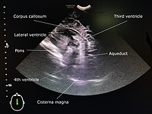
Mid sagittal section through anterior fontanelle of a one-year-old girl.
159:
or neonatologists usually perform cranial ultrasound; in other systems
83:
Other indications include babies that requires ventilatory support,
52:
been opened, for example to help identify the margins of a tumour.
124:
115:
101:
119:
445:"Practical guide to neonatal cranial ultrasound (CrUS): basics"
388:"Practical guide to neonatal cranial ultrasound (CrUS): basics"
337:"Neonatal cranial sonography: A concise review for clinicians"
259:
114:
thrombosis, cerebral oedema, and evaluating the structures of
335:
Gupta P, Sodhi KS, Saxena AK, Khandelwal N, Singhi P (2016).
133:
A water-based gel is applied to the infant's head, over the
64:(IVH), which often occurs during the first few days, and
258:
Therefore, many neonatal services prefer to perform an
482:
Lissauer T, Fanaroff AA, Miall L, Fanaroff J (2015).
224:
Some brain structures are poorly visualised, notably
16:
Brain scan technique using high-frequency sound waves
26:
21:
217:. However, it is subject to certain limitations.
279:Dumain T (4 February 2022). Melinosky C (ed.).
330:
328:
8:
304:Intraventricular hemorrhage of the newborn
47:, can be used in any age group. This uses
362:
352:
232:if only the anterior fontanelle is used.
271:
316:Pediatric Periventricular Leukomalacia
18:
486:. John Wiley & Sons. p. 187.
80:is blocked by blood-clots) develops.
7:
443:James, Anitha C. (September 2018).
386:James, Anitha C. (September 2018).
341:Journal of Pediatric Neurosciences
14:
242:Damage to the brain soft tissue (
281:"What Is a Cranial Ultrasound?"
426:"Cranial Ultrasound guideline"
246:), for example resulting from
1:
520:10.1053/j.semperi.2009.10.002
449:Paediatrics and Child Health
392:Paediatrics and Child Health
161:advanced nurse practitioners
66:periventricular leukomalacia
76:as the natural flow of the
62:intraventricular hemorrhage
579:
461:10.1016/j.paed.2018.07.003
404:10.1016/j.paed.2018.07.003
182:structures, by using the
508:Seminars in Perinatology
354:10.4103/1817-1745.181261
299:MedlinePlus Encyclopedia
171:may perform most scans.
553:Medical ultrasonography
484:Neonatology at a Glance
228:structures such as the
112:superior sagittal sinus
85:neonatal encephalopathy
30:technique to scan brain
205:to assess blood flow.
199:ventricular dilatation
148:plane and five in the
130:
107:
254:, may be hard to see.
128:
105:
188:posterior fontanelle
45:transcranial Doppler
432:. 12 February 2016.
176:anterior fontanelle
135:anterior fontanelle
78:cerebrospinal fluid
215:ionising radiation
184:mastoid fontanelle
131:
108:
49:Doppler ultrasound
37:Cranial ultrasound
22:Cranial ultrasound
72:(swelling of the
34:
33:
570:
532:
531:
503:
497:
494:
488:
487:
479:
473:
472:
440:
434:
433:
430:NHS Forth Valley
422:
416:
415:
383:
377:
376:
366:
356:
332:
323:
312:
306:
295:
289:
288:
276:
19:
578:
577:
573:
572:
571:
569:
568:
567:
548:Medical imaging
538:
537:
536:
535:
505:
504:
500:
495:
491:
481:
480:
476:
442:
441:
437:
424:
423:
419:
385:
384:
380:
334:
333:
326:
313:
309:
296:
292:
278:
277:
273:
268:
226:posterior fossa
211:
180:posterior fossa
100:
58:
17:
12:
11:
5:
576:
574:
566:
565:
560:
555:
550:
540:
539:
534:
533:
498:
489:
474:
455:(9): 424–430.
435:
417:
398:(9): 424–430.
378:
324:
307:
290:
270:
269:
267:
264:
256:
255:
240:
233:
222:
210:
207:
157:paediatricians
99:
96:
57:
54:
32:
31:
28:
24:
23:
15:
13:
10:
9:
6:
4:
3:
2:
575:
564:
561:
559:
556:
554:
551:
549:
546:
545:
543:
529:
525:
521:
517:
513:
509:
502:
499:
493:
490:
485:
478:
475:
470:
466:
462:
458:
454:
450:
446:
439:
436:
431:
427:
421:
418:
413:
409:
405:
401:
397:
393:
389:
382:
379:
374:
370:
365:
360:
355:
350:
346:
342:
338:
331:
329:
325:
322:
318:
317:
311:
308:
305:
301:
300:
294:
291:
286:
282:
275:
272:
265:
263:
261:
253:
249:
245:
241:
238:
234:
231:
227:
223:
220:
219:
218:
216:
208:
206:
204:
200:
195:
193:
189:
185:
181:
177:
172:
170:
166:
162:
158:
153:
151:
147:
142:
140:
136:
127:
123:
121:
117:
113:
104:
97:
95:
93:
88:
86:
81:
79:
75:
71:
70:hydrocephalus
67:
63:
55:
53:
50:
46:
42:
38:
29:
25:
20:
514:(1): 28–38.
511:
507:
501:
492:
483:
477:
452:
448:
438:
429:
420:
395:
391:
381:
344:
340:
314:
310:
297:
293:
284:
274:
257:
250:or abnormal
212:
196:
173:
169:sonographers
165:radiologists
154:
143:
132:
109:
89:
82:
59:
36:
35:
347:(1): 7–13.
252:myelination
209:Limitations
201:and colour
190:and/or the
558:Pediatrics
542:Categories
266:References
244:parenchyma
237:fontanelle
230:cerebellum
174:While the
139:ultrasound
74:ventricles
41:fontanelle
563:Neurology
321:eMedicine
248:ischaemia
98:Technique
92:premature
528:20109970
469:56987166
412:56987166
373:27195026
194:window.
192:temporal
150:sagittal
364:4862295
235:If the
203:Doppler
146:coronal
27:Purpose
526:
467:
410:
371:
361:
186:, the
465:S2CID
408:S2CID
285:WedMD
116:sulci
524:PMID
369:PMID
120:gyri
118:and
56:Uses
516:doi
457:doi
400:doi
359:PMC
349:doi
319:at
260:MRI
167:or
544::
522:.
512:34
510:.
463:.
453:28
451:.
447:.
428:.
406:.
396:28
394:.
390:.
367:.
357:.
345:11
343:.
339:.
327:^
302::
283:.
163:,
122:.
530:.
518::
471:.
459::
414:.
402::
375:.
351::
287:.
Text is available under the Creative Commons Attribution-ShareAlike License. Additional terms may apply.