718:, from the inner enamel epithelium. Changes will also start occurring in the adjacent dental papilla, very quickly after reversed polarity of the cells of the inner enamel epithelium. To contain increasing amounts of protein-synthesizing organelles, odontoblasts as their cytoplasm (the liquid inside a cell but outside the nucleus). increases in volume after the ectomesenchymal cells beside the acellular zone rapidly enlarge and elongate to become preodontoblasts. When the odontoblasts differentiate and increase in size to occupy the acellular zone between the dental papilla and the inner enamel epithelium, the zone slowly is removed. With their nuclei positioned away from the inner enamel epithelium, these newly differentiated cells are distinguished by being highly polarized.
615:(upper jaw) The probable need for a rich blood supply would seem to show that the cell mass will soon be highly productive in the formation of dental tissues. Therefore, when the late bell stage of the tooth germ development has been reached, most of the cells would have been differentiated to an apparent endpoint where the cells will now begin their formative role when the first three stages of the tissue development are almost completed, and the tissues can now start to begin secreting.
147:
29:
135:
632:
Blood vessels going into the dental papilla are formed into groups that coincides with the positions of where the roots will develop in future. As time passes, the viability of the tissue is affected as the blood supply becomes steadily reduced in stages and the volume of pulpal tissue starts decreasing too.
380:. The dental lamina is a band of epithelial tissue which connects the developing tooth bud to the oral epithelium. The dental lamina eventually disintegrates into small clusters of epithelium and is reabsorbed. The dental lamina is first evidence of tooth development and begins at the sixth week in utero.
447:, the epithelial tooth germ forms a bell-shaped structure in the labio-lingual section and is characterised by the formation of the dental sac. The peripheral cells of the dental papilla undergo differentiation, growing larger in size and taking a columnar (uni-layered) form and are now referred to as
398:
A section of the ectomesenchyme (a group of tissue made up of neurocrest cells which are present in the initial development of an embryo. This forms the hard and soft tissues of the neck and skull), condenses into a mass within the concavity of the cap of the enamel organ. This mass is now considered
713:
Growth factors in the cells of the inner enamel epithelium and expressions of signaling molecules bring about the differentiation of odontoblast through normal development of the dental papilla. Exhibiting a central nucleus and few organelles, the dental papilla cells are small and undifferentiated.
689:
are among the few which have been studied during the tooth development process. Of which, the verve-related signaling molecules seems to show a trend that suggest an early implication of innervation of tooth development. Similar to how many molecules are able to stimulate axonal growth or migration,
606:
When all of the individual components of the tooth germ have become developed, the entire cell mass would have appeared to have migrated deeper into the underlying connective tissues. This phenomenon, which will continue throughout the whole life of the teeth, is most possibly due to the cell mass
631:
Clusters of blood vessels are found branching out around the tooth germ in the dental follicle and going into the dental papilla during the cap stage. In the dental papilla the number of blood vessels increase and the matrix deposition will begin once the maximum is reached during the bell stage.
661:
starts. The timing is not similar to the establishment of the neural supply and the papillary vascular supply even though a feasible relationship has been assumed between the developing nerve and blood supplies. Furthermore, histo-chemistry studies have shown that in the makeup of pioneer nerve
426:
in future development. The periodontium is the tissue that surrounds and supports the teeth. It includes the connective tissue and overlying keratinised membrane lining the oral cavity that surrounds the teeth, the periodontal ligament, cementum which provides a protective covering for the root
518:
are secreted in successive layers. The mesenchymal tissue of dental papilla and dental sac and the ectodermal tissue of enamel undergo induction. The outer cells of dental papilla are induced by preameloblasts (cells within the enamel from which a cell that takes part in forming dental enamel
451:(the outer part of the dental pulp). This differentiation begins at the apex of the dental papilla, gradually extending downwards. This differentiation occurs to supplement the development of the dental sac which is responsible for cementum, periodontal ligament and the
368:. Histodifferentiation is the differentiation of different tissue types during the development of an embryo/ undifferentiated group of cells. Furthermore, morphogenesis is a predominant physiological process during the cap stage. This is due to formation of
543:
Ectomesenchymal cells will multiply continuously in a localized area such that when the bell stage of development is reached, both the epithelial component and the ectomesenchymal component will seem to have been surrounded by something that presents as a
527:
and result in formation of the dentin matrix/pre-dentin (the innermost section of the dentin, which is not mineralized and located adjacent to pulp tissues in the crown area and root area). The central cells of dental papilla form the
348:
The cap stage is the second stage of tooth development and occurs during the ninth or tenth week of prenatal development. Unequal proliferation of the tooth bud forms a three-dimensional cap shape. Overlying this cap structure is the
657:. The plexus of Raschkow is a network of nerves immediately beneath the odontoblast layer of the dentine, first described by J. Raschkow in 1835. However, the nerve fibers will only begin entering the dental papilla (pulp) when
372:
of the tooth. The primordium contains each of the primordial tissue types, essential for the development of successive teeth. These primordial tissues together form the enamel organ, dental papilla and dental sac.
652:
is the clear target of these dental nerve fibers. The dental follicle is a fibrous sac that surrounds the odontogenic organ and developing tooth. The plexus is a system of connections of blood vessels, nerves, or
324:
of a tooth and its supporting structures form from these distinct cellular aggregations. Similar to dental follicle, the dental papilla has a very rich blood supply and provides nutrition to the
662:
fibers heading towards the tooth germ, automatic nerve fibers are not present. Therefore, the starting innervation of the developing teeth is involved with the sensory innovation of the future
484:
Contain very big nuclei and have small quantities of the intra-cellular organelles involved in protein synthesis. The cells contact each other through desmosomes and gap junctions
827:
226:
1504:
1097:
934:
905:
202:
399:
the dental papilla. Note that dental papilla is originally derived from ectomesenchyme. Ectomesenchyme (type of mesenchyme) is derived from
395:. It gives rise to the nervous system, sense organs, outer layer of the skin, teeth and the membrane lining the oral cavity (mouth).
1589:
1069:
1038:
973:
853:
803:
770:
548:
sac. Therefore, among a complicated mass of highly differentiated cells, it would appear to have three major components, which are:
112:
1383:
706:
cells allows comprehension and explanation of normal development and affects their recruitment when needed to start repairing the
1497:
46:
255:
93:
50:
690:
various molecules are also within the bounds of possibility of being involved in the initial innervation of the tooth germ.
734:
of the developing tooth germ but do not describe the significant functional changes that occur during development, such as
65:
221:
214:
1090:
72:
418:
The existing ectomesenchyme around the outside of the cap of the enamel organ then condenses into the dental sac. A
1490:
1314:
1288:
1283:
1125:
415:. The dentinoenamel junction is the surface at which the enamel and the dentin of the crown of a tooth are joined.
39:
1309:
1227:
1201:
1196:
209:
79:
1222:
727:
520:
391:
which is the outermost of the three germ layers of the forming embryo. The other two are: the mesoderm and the
361:
317:
275:
185:
1642:
1609:
1604:
1513:
1335:
444:
436:
376:
Also during the cap stage is the formation of a depression within the deepest part of each tooth bud of the
337:
61:
1584:
1431:
1421:
1330:
1248:
1083:
412:
233:
197:
139:
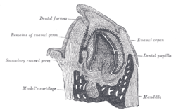
Vertical section of the mandible of an early human fetus. × 25. (Dental papilla labeled at center right.)
1243:
1120:
365:
731:
663:
470:
Separated from the peripheral cells of the dental papilla by a basement membrane and a cell free zone
440:
387:
organ. It is important to note that enamel is an ectodermal product as it is originally derived from
1619:
1454:
17:
798:(4th ed.). Maryland Heights: Elsevier/Saunders. pp. 51, 52, 58, 59, 60, 61, 62, 63, 66.
1614:
821:
532:
of the pulp during root development. These cells then become surrounded by newly formed dentin..
498:
the transport of materials to and from the enamel-forming cells in the internal enamel epithelium
519:
develops) to differentiate into odontoblasts (dentin-secreting cells). The odontoblasts undergo
869:
1376:
1293:
1065:
1044:
1034:
969:
930:
901:
849:
809:
799:
776:
766:
419:
404:
1206:
966:
Master
Dentistry Volume 3 Oral Biology: Oral Anatomy, Histology, Physiology and Biochemistry
730:
proceeds into three stages: the bud, cap and bell stage. these terms are descriptive of the
654:
582:
452:
321:
86:
1467:
1170:
1143:
649:
554:
313:
263:
357:
tissue known as the dental papilla superiorly, and lies within the epithelial concavity.
279:
146:
1563:
1406:
1371:
1106:
924:
714:
At this stage, the cells are separated by an acellular zone, that consist of some fine
703:
658:
524:
350:
644:
head towards the developing tooth. The nerve fibers will branch out and create a rich
1636:
1594:
1363:
1163:
735:
377:
1599:
1537:
1426:
1396:
1340:
1253:
1153:
682:
671:
667:
558:→ The ectomesenchymal cells which are part of the fibrous sac that have been formed
423:
408:
400:
384:
325:
309:
298:
283:
190:
1527:
1354:
1135:
699:
641:
607:
moving towards a rich blood supply that can be found in the deeper parts of the
448:
267:
28:
1532:
1401:
990:
686:
529:
369:
251:
1048:
813:
780:
1553:
287:
271:
574:
The tissues which have been derived from each of the three components are:
364:
occur at this stage; such as cytodifferentiation, histodifferentiation and
481:
Involved in the maintenance of the shape of the enamel and the environment
422:
separate the enamel organ and the dental sac. The dental sac produces the
1558:
1267:
1158:
715:
608:
515:
392:
388:
354:
581:→ will develop to become the periodontal ligament, the cementum and the
239:
1180:
1148:
612:
591:
1436:
707:
645:
294:
290:
1482:
995:(6th ed.). Sinauer Associates – via www.ncbi.nlm.nih.gov.
564:→ The ectomesenchymal cells which are lying deep to the enamel organ
134:
545:
302:
173:
1075:
756:
754:
752:
678:
1486:
1079:
1031:
Ten Cate's oral histology: development, structure, and function
989:
Gilbert, Scott F. (May 11, 2000). "Induction and
Competence".
640:
During the bud to cap stage of tooth development, the pioneer
22:
603:
Till this point, no dental tissues have been created yet.
794:
J., Fehrenbach, Margaret; Popowics, Tracy (2015-02-02).
411:
and dental papilla which will be the site of the future
336:
Formation of dental papilla occurs in the cap stage of
439:
which occurs between the eleventh and twelfth week of
383:
This is responsible for the cap-like structure of the
900:. United States of America: Oxford University Press.
796:
Illustrated dental embryology, histology, and anatomy
1062:
Oral
Histology: development, structure, and function
514:
During the apposition stage, the enamel, dentin and
1572:
1546:
1520:
1362:
1353:
1323:
1302:
1276:
1266:
1236:
1215:
1189:
1179:
1134:
1113:
473:
Rich in RNA but do not contain alkaline phosphatase
220:
208:
196:
184:
172:
167:
162:
127:
53:. Unsourced material may be challenged and removed.
619:Nerve and vascular supply during early development
590:→ will develop to become the dental pulp and the
286:. The dental papilla appears after 8–10 weeks of
848:. Tokyo: Ishiyaku Publishers, Inc. p. 41.
648:around the tooth germ in that structure as the
891:
889:
887:
885:
883:
215:papilla_by_E4.0.3.3.1.0.12 E4.0.3.3.1.0.12
1498:
1091:
8:
870:"Medical Definition of histodifferentiation"
826:: CS1 maint: multiple names: authors list (
677:Nerve-related signaling molecules, such as
293:life. The dental papilla gives rise to the
1505:
1491:
1483:
1359:
1273:
1186:
1098:
1084:
1076:
1033:(8th ed.). St. Louis, Mo.: Elsevier.
599:→ will develop to create the enamel solely
145:
133:
113:Learn how and when to remove this message
1029:Nanci, Antonio; Ten Cate, A. R. (2013).
951:The American Heritage Medical Dictionary
320:. This is of importance because all the
919:
917:
846:Human Tooth and Dental Arch Development
761:Creanor, Stephen, ed. (February 2016).
748:
819:
435:The bell stage is the fourth stage of
427:surface and supporting alveolar bone.
237:
124:
929:(2nd ed.). Elsevier, Inc. 2008.
510:Apposition stage and maturation stage
151:Histologic slide showing a tooth bud.
7:
839:
837:
316:together forms one unit, called the
51:adding citations to reliable sources
765:. Chichester, West Sussex: Wiley.
14:
1590:Epithelial cell rests of Malassez
1016:Farlex Partner Medical Dictionary
1005:J Nat Sci Biol Med. 2015 Jul-Dec;
953:. Houghton Mifflin Company. 2004.
570:→ purely the epithelial component
670:. Nerve fibers never enters the
27:
964:Berkovitz, Barry K. B. (2010).
763:Essential clinical oral biology
38:needs additional citations for
898:Oxford dictionary of Dentistry
501:the concentration of materials
1:
968:. Elsevier Health Sciences.
681:line-derived growth factor,
694:Odontoblast differentiation
353:, which is attached to the
312:organ, dental papilla, and
278:. It lies below a cellular
1659:
1126:Universal Numbering System
15:
1463:
1450:
1417:
1392:
926:Mosby's Dental Dictionary
738:and histodifferentiation.
495:the synthesis of proteins
232:
144:
132:
896:Ireland, Robert (2010).
16:Not to be confused with
1610:Inner enamel epithelium
1605:Outer enamel epithelium
443:. During this stage of
1585:Epithelial root sheath
1432:Dental-enamel junction
1422:Cementoenamel junction
1384:Zuckerkandl's tubercle
844:Ooë, Tadahiro (1981).
413:dentinoenamel junction
234:Anatomical terminology
1121:Glossary of dentistry
992:Developmental Biology
611:(lower jaw) and the
262:is a condensation of
664:periodontal ligament
441:prenatal development
366:morphodifferentation
256:prenatal development
47:improve this article
1620:Stratum intermedium
702:differentiate from
579:The dental follicle
492:Is concerned with:
407:exists between the
18:interdental papilla
1615:Stellate reticulum
698:Understanding how
588:The dental papilla
562:The dental papilla
157:C: dental follicle
1630:
1629:
1514:Tooth development
1480:
1479:
1476:
1475:
1377:Cusp of Carabelli
1349:
1348:
1262:
1261:
936:978-0-323-04963-4
907:978-0-19-953301-5
728:Tooth development
655:lymphatic vessels
459:Epithelium layers
437:tooth development
420:basement membrane
405:basement membrane
360:Various types of
248:
247:
243:
155:B: dental papilla
123:
122:
115:
97:
1650:
1507:
1500:
1493:
1484:
1360:
1274:
1187:
1100:
1093:
1086:
1077:
1064:. 5th ed. 1998.
1060:Cate, A.R. Ten.
1053:
1052:
1026:
1020:
1019:
1012:
1006:
1003:
997:
996:
986:
980:
979:
961:
955:
954:
947:
941:
940:
921:
912:
911:
893:
878:
877:
866:
860:
859:
841:
832:
831:
825:
817:
791:
785:
784:
758:
716:collagen fibrils
597:The enamel organ
568:The enamel organ
453:alveolar process
403:cells (NCCs). A
276:developing tooth
240:edit on Wikidata
149:
137:
125:
118:
111:
107:
104:
98:
96:
62:"Dental papilla"
55:
31:
23:
1658:
1657:
1653:
1652:
1651:
1649:
1648:
1647:
1633:
1632:
1631:
1626:
1568:
1542:
1521:Precursor cells
1516:
1511:
1481:
1472:
1468:Dental alveolus
1459:
1446:
1413:
1388:
1345:
1319:
1315:Second premolar
1298:
1289:Lateral incisor
1284:Central incisor
1258:
1232:
1228:Second premolar
1211:
1202:Lateral incisor
1197:Central incisor
1175:
1130:
1109:
1104:
1057:
1056:
1041:
1028:
1027:
1023:
1014:
1013:
1009:
1004:
1000:
988:
987:
983:
976:
963:
962:
958:
949:
948:
944:
937:
923:
922:
915:
908:
895:
894:
881:
874:Merriam-Webster
868:
867:
863:
856:
843:
842:
835:
818:
806:
793:
792:
788:
773:
760:
759:
750:
745:
724:
704:ectomesenchymal
696:
650:dental follicle
638:
629:
627:Vascular supply
621:
555:dental follicle
538:
536:Differentiation
521:differentiation
512:
461:
433:
362:differentiation
346:
334:
314:dental follicle
264:ectomesenchymal
244:
158:
156:
154:
153:A: enamel organ
152:
140:
119:
108:
102:
99:
56:
54:
44:
32:
21:
12:
11:
5:
1656:
1654:
1646:
1645:
1643:Parts of tooth
1635:
1634:
1628:
1627:
1625:
1624:
1623:
1622:
1617:
1612:
1607:
1597:
1592:
1587:
1582:
1580:Dental papilla
1576:
1574:
1570:
1569:
1567:
1566:
1564:Tooth eruption
1561:
1556:
1550:
1548:
1544:
1543:
1541:
1540:
1535:
1530:
1524:
1522:
1518:
1517:
1512:
1510:
1509:
1502:
1495:
1487:
1478:
1477:
1474:
1473:
1471:
1470:
1464:
1461:
1460:
1458:
1457:
1451:
1448:
1447:
1445:
1444:
1442:Dental papilla
1439:
1434:
1429:
1424:
1418:
1415:
1414:
1412:
1411:
1410:
1409:
1407:Apical foramen
1399:
1393:
1390:
1389:
1387:
1386:
1381:
1380:
1379:
1368:
1366:
1357:
1351:
1350:
1347:
1346:
1344:
1343:
1338:
1333:
1327:
1325:
1321:
1320:
1318:
1317:
1312:
1310:First premolar
1306:
1304:
1300:
1299:
1297:
1296:
1291:
1286:
1280:
1278:
1271:
1264:
1263:
1260:
1259:
1257:
1256:
1251:
1246:
1240:
1238:
1234:
1233:
1231:
1230:
1225:
1223:First premolar
1219:
1217:
1213:
1212:
1210:
1209:
1204:
1199:
1193:
1191:
1184:
1177:
1176:
1174:
1173:
1168:
1167:
1166:
1161:
1156:
1151:
1140:
1138:
1132:
1131:
1129:
1128:
1123:
1117:
1115:
1111:
1110:
1107:Dental anatomy
1105:
1103:
1102:
1095:
1088:
1080:
1074:
1073:
1055:
1054:
1039:
1021:
1007:
998:
981:
974:
956:
942:
935:
913:
906:
879:
861:
854:
833:
804:
786:
771:
747:
746:
744:
741:
740:
739:
723:
720:
695:
692:
659:dentinogenesis
637:
634:
628:
625:
620:
617:
601:
600:
594:
585:
572:
571:
565:
559:
537:
534:
525:repolarization
511:
508:
507:
506:
505:
504:
503:
502:
499:
496:
487:
486:
485:
482:
476:
475:
474:
471:
460:
457:
432:
429:
351:ectomesenchyme
345:
342:
333:
330:
274:sections of a
260:dental papilla
246:
245:
236:
230:
229:
224:
218:
217:
212:
206:
205:
200:
194:
193:
188:
182:
181:
179:papilla dentis
176:
170:
169:
165:
164:
160:
159:
150:
142:
141:
138:
130:
129:
128:Dental papilla
121:
120:
35:
33:
26:
13:
10:
9:
6:
4:
3:
2:
1655:
1644:
1641:
1640:
1638:
1621:
1618:
1616:
1613:
1611:
1608:
1606:
1603:
1602:
1601:
1598:
1596:
1595:Dental lamina
1593:
1591:
1588:
1586:
1583:
1581:
1578:
1577:
1575:
1571:
1565:
1562:
1560:
1557:
1555:
1552:
1551:
1549:
1545:
1539:
1536:
1534:
1531:
1529:
1526:
1525:
1523:
1519:
1515:
1508:
1503:
1501:
1496:
1494:
1489:
1488:
1485:
1469:
1466:
1465:
1462:
1456:
1453:
1452:
1449:
1443:
1440:
1438:
1435:
1433:
1430:
1428:
1425:
1423:
1420:
1419:
1416:
1408:
1405:
1404:
1403:
1400:
1398:
1395:
1394:
1391:
1385:
1382:
1378:
1375:
1374:
1373:
1370:
1369:
1367:
1365:
1361:
1358:
1356:
1352:
1342:
1339:
1337:
1334:
1332:
1329:
1328:
1326:
1322:
1316:
1313:
1311:
1308:
1307:
1305:
1301:
1295:
1292:
1290:
1287:
1285:
1282:
1281:
1279:
1275:
1272:
1269:
1265:
1255:
1252:
1250:
1247:
1245:
1242:
1241:
1239:
1235:
1229:
1226:
1224:
1221:
1220:
1218:
1214:
1208:
1205:
1203:
1200:
1198:
1195:
1194:
1192:
1188:
1185:
1182:
1178:
1172:
1169:
1165:
1162:
1160:
1157:
1155:
1152:
1150:
1147:
1146:
1145:
1142:
1141:
1139:
1137:
1133:
1127:
1124:
1122:
1119:
1118:
1116:
1112:
1108:
1101:
1096:
1094:
1089:
1087:
1082:
1081:
1078:
1071:
1070:0-8151-2952-1
1067:
1063:
1059:
1058:
1050:
1046:
1042:
1040:9780323078467
1036:
1032:
1025:
1022:
1017:
1011:
1008:
1002:
999:
994:
993:
985:
982:
977:
975:9780702044588
971:
967:
960:
957:
952:
946:
943:
938:
932:
928:
927:
920:
918:
914:
909:
903:
899:
892:
890:
888:
886:
884:
880:
875:
871:
865:
862:
857:
855:9780912791005
851:
847:
840:
838:
834:
829:
823:
815:
811:
807:
805:9781455776856
801:
797:
790:
787:
782:
778:
774:
772:9781118939666
768:
764:
757:
755:
753:
749:
742:
737:
736:morphogenesis
733:
729:
726:
725:
721:
719:
717:
711:
709:
705:
701:
693:
691:
688:
684:
680:
675:
673:
669:
665:
660:
656:
651:
647:
643:
635:
633:
626:
624:
618:
616:
614:
610:
604:
598:
595:
593:
589:
586:
584:
583:alveolar bone
580:
577:
576:
575:
569:
566:
563:
560:
557:
556:
551:
550:
549:
547:
541:
535:
533:
531:
526:
522:
517:
509:
500:
497:
494:
493:
491:
490:
488:
483:
480:
479:
477:
472:
469:
468:
466:
465:
464:
458:
456:
454:
450:
446:
445:odontogenesis
442:
438:
430:
428:
425:
421:
416:
414:
410:
406:
402:
396:
394:
390:
386:
381:
379:
378:dental lamina
374:
371:
367:
363:
358:
356:
352:
343:
341:
339:
338:odontogenesis
331:
329:
327:
323:
319:
315:
311:
306:
304:
300:
296:
292:
289:
285:
282:known as the
281:
277:
273:
269:
266:cells called
265:
261:
257:
253:
241:
235:
231:
228:
225:
223:
219:
216:
213:
211:
207:
204:
201:
199:
195:
192:
189:
187:
183:
180:
177:
175:
171:
166:
161:
148:
143:
136:
131:
126:
117:
114:
106:
95:
92:
88:
85:
81:
78:
74:
71:
67:
64: –
63:
59:
58:Find sources:
52:
48:
42:
41:
36:This article
34:
30:
25:
24:
19:
1600:Enamel organ
1579:
1538:Cementoblast
1441:
1336:Second molar
1249:Second molar
1114:Nomenclature
1061:
1030:
1024:
1015:
1010:
1001:
991:
984:
965:
959:
950:
945:
925:
897:
873:
864:
845:
795:
789:
762:
712:
697:
683:neurotrophin
676:
672:enamel organ
642:nerve fibers
639:
636:Nerve supply
630:
622:
605:
602:
596:
587:
578:
573:
567:
561:
552:
542:
539:
513:
462:
449:odontoblasts
434:
424:periodontium
417:
409:enamel organ
401:neural crest
397:
382:
375:
359:
347:
335:
326:enamel organ
307:
284:enamel organ
268:odontoblasts
259:
249:
203:A05.1.03.054
178:
109:
103:October 2018
100:
90:
83:
76:
69:
57:
45:Please help
40:verification
37:
1528:Odontoblast
1341:Third molar
1331:First molar
1254:Third molar
1244:First molar
700:odontoblast
280:aggregation
168:Identifiers
1533:Ameloblast
1402:Root canal
1268:Mandibular
743:References
732:morphology
687:semaphorin
679:glial cell
530:primordium
431:Bell stage
370:primordium
355:mesodermal
332:Embryology
318:tooth germ
272:histologic
270:, seen in
252:embryology
73:newspapers
1554:Dentition
1181:Maxillary
1171:Deciduous
1144:Permanent
1049:769803484
822:cite book
814:905370300
781:917888653
344:Cap stage
1637:Category
1559:Teething
1303:Premolar
1216:Premolar
1159:premolar
722:See also
623:Source:
609:mandible
540:Source:
516:cementum
489:Stratum
463:Source:
393:endoderm
389:ectoderm
1547:General
1455:Mamelon
1277:Incisor
1190:Incisor
1149:incisor
1018:. 2012.
613:maxilla
592:dentine
546:fibrous
322:tissues
191:D003771
163:Details
87:scholar
1437:Dentin
1427:Enamel
1294:Canine
1207:Canine
1154:canine
1068:
1047:
1037:
972:
933:
904:
852:
812:
802:
779:
769:
708:dentin
646:plexus
478:Outer
467:Inner
385:enamel
310:enamel
295:dentin
291:uteral
258:, the
89:
82:
75:
68:
60:
1573:Other
1364:Crown
1355:Parts
1324:Molar
1270:teeth
1237:Molar
1183:teeth
1164:molar
1136:Teeth
303:tooth
301:of a
288:intra
238:[
227:57662
174:Latin
94:JSTOR
80:books
1397:Pulp
1372:Cusp
1066:ISBN
1045:OCLC
1035:ISBN
970:ISBN
931:ISBN
902:ISBN
850:ISBN
828:link
810:OCLC
800:ISBN
777:OCLC
767:ISBN
685:and
668:pulp
666:and
553:The
523:and
308:The
299:pulp
297:and
254:and
198:TA98
186:MeSH
66:news
250:In
222:FMA
49:by
1639::
1043:.
916:^
882:^
872:.
836:^
824:}}
820:{{
808:.
775:.
751:^
710:.
674:.
455:.
340:.
328:.
305:.
210:TE
1506:e
1499:t
1492:v
1099:e
1092:t
1085:v
1072:.
1051:.
978:.
939:.
910:.
876:.
858:.
830:)
816:.
783:.
242:]
116:)
110:(
105:)
101:(
91:·
84:·
77:·
70:·
43:.
20:.
Text is available under the Creative Commons Attribution-ShareAlike License. Additional terms may apply.