195:
159:
171:
183:
38:
282:
288:
90:
541:
158:
257:"This is an open access article distributed under the Creative Commons Attribution License, which permits unrestricted use, distribution, and reproduction in any medium, provided the original work is properly cited."
170:
329:– You must give appropriate credit, provide a link to the license, and indicate if changes were made. You may do so in any reasonable manner, but not in any way that suggests the licensor endorses you or your use.
194:
56:
52:
48:
42:
101:
69:
144:
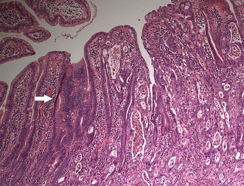
Light microscopy of small intestinal adenocarcinoma, showing small bowel infiltrated with adenocarcinoma cells. Tissue section stained with haematoxylin and eosin; magnification ×100.
590:
182:
164:
Gross pathology, serosal view, of small intestine with adenocarcinoma, showing cancer with infiltrative growth, causing stenosis.
452:
416:
540:
535:
60:
37:
400:
147:
472:
436:
383:
551:
233:
572:
568:
The following pages on the
English Knowledge (XXG) use this file (pages on other projects are not listed):
224:(2013). "Primary Small Bowel Tumour Presenting as Bowel Obstruction in a Patient with a Virgin Abdomen".
298:
295:
109:
176:
Luminal view, showing the stenotic infiltrative growth across the entire intestinal wall.
96:
212:
249:
238:
508:
Click on a date/time to view the file as it appeared at that time.
267:
Xilin Wu1, Hester Y. S. Cheung, Cliff C. C. Chung, Michael K. W. Li
243:
84:
600:
134:
Light microscopy of small intestinal adenocarcinoma.jpg
74:(1,327 × 1,013 pixels, file size: 559 KB, MIME type:
128:
108:
Commons is a freely licensed media file repository.
367:
Light microscopy of small intestinal adenocarcinoma
200:Immunohistochemistry with cytokeratin 7, 19 and 20
453:Creative Commons Attribution 4.0 International
89:
281:
8:
336:https://creativecommons.org/licenses/by/4.0
312:– to copy, distribute and transmit the work
510:
363:
226:International Journal of Clinical Medicine
583:The following other wikis use this file:
570:
486:
470:
450:
434:
414:
398:
381:
378:
359:
352:
154:
7:
558:User created page with UploadWizard
499:
287:
244:
242:
232:
372:
366:
303:
278:
138:
121:
67:
371:
342:Creative Commons Attribution 4.0
357:
323:Under the following conditions:
294:This file is licensed under the
286:
280:
193:
181:
169:
157:
88:
31:
21:
354:
139:
14:
353:
26:
1:
299:Attribution 4.0 International
379:Items portrayed in this file
619:
597:Usage on vi.wikipedia.org
587:Usage on sr.wikipedia.org
591:Злоћудни рак танког црева
500:
16:
356:
95:This is a file from the
557:
263:
239:10.4236/ijcm.2013.46050
218:
211:
208:
131:
99:. Information from its
573:Small intestine cancer
536:16:14, 15 January 2020
102:description page there
41:Size of this preview:
152:From the same case:
61:1,327 × 1,013 pixels
318:– to adapt the work
47:Other resolutions:
57:1,006 × 768 pixels
579:Global file usage
561:
437:copyright license
365:
271:
270:
117:
116:
97:Wikimedia Commons
32:Global file usage
610:
553:Mikael Häggström
548:
401:copyright status
349:
346:
343:
340:
337:
296:Creative Commons
290:
289:
284:
283:
253:
248:
246:
236:
214:
197:
188:Light microscopy
185:
173:
161:
150:
143:
135:
129:
113:
92:
91:
85:
79:
77:
64:
53:629 × 480 pixels
49:315 × 240 pixels
43:785 × 599 pixels
618:
617:
613:
612:
611:
609:
608:
607:
577:
569:
562:
554:
546:
502:
501:
498:
497:
496:
495:
494:
493:
492:
491:
489:
479:
478:
477:
475:
464:
463:
462:
461:
460:
459:
458:
457:
455:
443:
442:
441:
439:
428:
427:
426:
425:
424:
423:
422:
421:
419:
407:
406:
405:
403:
392:
391:
390:
389:
388:
386:
370:
369:
368:
351:
350:
347:
344:
341:
338:
335:
334:
302:
291:
277:
272:
231:(06): 287–290.
223:
204:
201:
198:
189:
186:
177:
174:
165:
162:
146:
133:
126:
119:
118:
107:
106:
105:is shown below.
81:
75:
73:
66:
65:
46:
12:
11:
5:
616:
614:
606:
605:
604:
603:
595:
594:
593:
581:
580:
576:
575:
567:
566:
565:
560:
559:
556:
552:
549:
545:1,327 × 1,013
543:
538:
533:
529:
528:
525:
522:
519:
516:
513:
506:
505:
490:
487:
485:
484:
483:
482:
481:
480:
476:
471:
469:
468:
467:
466:
465:
456:
451:
449:
448:
447:
446:
445:
444:
440:
435:
433:
432:
431:
430:
429:
420:
415:
413:
412:
411:
410:
409:
408:
404:
399:
397:
396:
395:
394:
393:
387:
382:
380:
377:
376:
375:
374:
373:
362:
361:
358:
355:
333:
332:
331:
330:
321:
320:
319:
313:
306:You are free:
293:
292:
279:
276:
273:
269:
268:
265:
261:
260:
259:
258:
220:
216:
215:
210:
206:
205:
203:
202:
199:
192:
190:
187:
180:
178:
175:
168:
166:
163:
156:
136:
127:
125:
122:
120:
115:
114:
93:
83:
82:
40:
36:
35:
34:
29:
24:
19:
13:
10:
9:
6:
4:
3:
2:
615:
602:
599:
598:
596:
592:
589:
588:
586:
585:
584:
578:
574:
571:
563:
555:
550:
544:
542:
539:
537:
534:
531:
530:
526:
523:
520:
517:
514:
512:
511:
509:
503:
474:
454:
438:
418:
402:
385:
328:
325:
324:
322:
317:
314:
311:
308:
307:
305:
304:
300:
297:
285:
274:
266:
262:
256:
255:
254:
251:
247:
240:
235:
230:
227:
221:
217:
207:
196:
191:
184:
179:
172:
167:
160:
155:
153:
151:
149:
142:
137:
130:
123:
111:
104:
103:
98:
94:
87:
86:
80:
71:
70:Original file
62:
58:
54:
50:
44:
39:
33:
30:
28:
25:
23:
20:
18:
15:
582:
507:
504:File history
326:
315:
309:
228:
225:
222:
145:
140:
110:You can help
100:
68:
22:File history
488:20 May 2013
417:copyrighted
327:attribution
213:20 May 2013
132:Description
564:File usage
521:Dimensions
339:CC BY 4.0
76:image/jpeg
27:File usage
518:Thumbnail
515:Date/Time
473:inception
275:Licensing
250:2158-284X
141:English:
547:(559 KB)
360:Captions
316:to remix
310:to share
301:license.
601:Ung thư
532:current
527:Comment
384:depicts
364:English
124:Summary
72:
264:Author
219:Source
524:User
348:true
345:true
245:ISSN
209:Date
148:edit
17:File
234:DOI
241:.
229:04
59:|
55:|
51:|
45:.
252:.
237::
112:.
78:)
63:.
Text is available under the Creative Commons Attribution-ShareAlike License. Additional terms may apply.


