109:, the cells that normally function as lipid store of the body, are well equipped to handle the excess lipids. Yet, too great of an excess will overburden these cells and cause a spillover into non-adipose cells, which do not have the necessary storage space. When the storage capacity of non-adipose cells is exceeded, cellular dysfunction and/or death result. The mechanism by which lipotoxicity causes death and dysfunction is not well understood. The cause of
20:
236:
hence were considered predictive for insulin resistance and causative in obesity-associated insulin resistance. However, endurance athletes also have high IMCL levels despite being highly insulin sensitive, which indicates that not the level of IMCL accumulation per se, but rather the characteristics of this intramyocellular fat determine whether it negatively affects insulin signaling. Intramyocellular lipids are mainly stored in
235:
The skeletal muscle accounts for more than 80 percent of the postprandial whole body glucose uptake and therefore plays an important role in glucose homeostasis. Skeletal muscle lipid levels – intramyocellular lipids (IMCL) – correlate negatively with insulin sensitivity in a sedentary population and
266:
The final strategy focuses on inhibiting the apoptotic pathways and signaling cascades. This is accomplished by using drugs that inhibit production of specific chemicals required for the pathways to be functional. While this may prove to the most effective protection against cell death, it will also
124:
has been named as the causative agent. The causative role of obesity in lipotoxicity is controversial. Some researchers claim that obesity has protective effects against lipotoxicity as it results in extra adipose tissue in which excess lipids can be stored. Others claim obesity is a risk factor for
251:
The first strategy focuses on decreasing the lipid content of non-adipose tissues. This can be accomplished by either increasing the oxidation of the lipids, or increasing their secretion and transport. Current treatments involve extreme weight loss and leptin treatment.
240:, the organelles for fat storage. Recent research indicates that creating intramyocellular neutral lipid storage capacity for example by increasing the abundance of lipid droplet coat proteins protects against obesity-associated insulin resistance in skeletal muscle.
82:
In normal cellular operations, there is a balance between the production of lipids, and their oxidation or transport. In lipotoxic cells, there is an imbalance between the amount of lipids produced and the amount used. Upon entrance of the cell,
350:
Bosma M, Kersten S, Hesselink MKC, and
Schrauwen P. Re-evaluating lipotoxic triggers in skeletal muscle: Relating intramyocellular lipid metabolism to insulin sensitivity. Prog Lipid Res 2012; 51: 36-49|doi=10.1016/j.plipres.2011.11.003
187:, the ratio of monounsaturated fatty acids and saturated fatty acids leads to apoptosis and liver damage. There are several potential mechanisms by which the excess fatty acids can cause cell death and damage. They may activate
182:
An excess of free fatty acids in liver cells plays a role in
Nonalcoholic Fatty Liver Disease (NAFLD). In the liver, it is the type of fatty acid, not the quantity, that determines the extent of the lipotoxic effects. In
215:
in the endoplasmic reticulum. Researchers are working on treatments that will increase the oxidation of these fatty acids within the heart in order to prevent the lipotoxic effects.
125:
lipotoxicity. Both sides accept that high fat diets put patients at increased risk for lipotoxic cells. Individuals with high numbers of lipotoxic cells usually experience both
120:
Currently, there is no universally accepted theory for why certain individuals are afflicted with lipotoxicity. Research is ongoing into a genetic cause, but no individual
549:
95:
molecule and is considered the most neutral and harmless type of intracellular lipid storage. Alternatively, fatty acids can be converted to lipid intermediates like
113:
and extent of cellular dysfunction is related to the type of cell affected, as well as the type and quantity of excess lipids. A theory has been put forward by
88:
117:
relating the development of lipotoxicity to the perturbation of membrane glycerophospholipid/sphingolipid homeostasis and their associated signalling events.
694:"Overexpression of PLIN5 in skeletal muscle promotes oxidative gene expression and intramyocellular lipid content without compromising insulin sensitivity"
693:
255:
Another strategy is focusing on diverting excess lipids away from non-adipose tissues, and towards adipose tissues. This is accomplished with
211:
Lipotoxicity in cardiac tissue is attributed to excess saturated fatty acids. The apoptosis that follows is believed to be caused by
227:, causing their dysfunction and death. The effects of the lipotoxicity is treated with leptin therapy and insulin sensitizers.
734:
Unger, Roger (January 2005). "Longevity, lipotoxicity and leptin: the adipocyte defense against feasting and famine".
103:
and fatty acyl-CoAs. These lipid intermediates can impair cellular function, which is referred to as lipotoxicity.
692:
Bosma, M.; Sparks, L. M.; Hooiveld, G.; Jorgensen, J.; Houten, S. M.; Schrauwen, P.; Hesselink, M. K. C. (2013).
212:
769:
Smith, U; Hammarstedt (March 2010). "Antagonistic effects of thiazolidinediones and cytokines in lipotoxicity".
192:
196:
303:
Garbarino, Jeanne; Stephen L. Sturley (2009). "Saturated with fat: new perspectives on lipotoxicity".
408:"Sphingolipids and glycerophospholipids - The "ying and yang" of lipotoxicity in metabolic diseases"
864:
388:
328:
130:
31:
166:
and death in severe cases. The current accepted treatments for lipotoxicity in renal cells are
859:
835:
786:
751:
716:
674:
625:
576:
525:
484:
435:
427:
380:
320:
267:
require the most research and development due to the specificity required of the medications.
256:
146:
Renal lipotoxicity occurs when excess long-chain nonesterified fatty acids are stored in the
825:
817:
778:
743:
708:
664:
656:
615:
607:
566:
558:
515:
474:
466:
419:
372:
312:
260:
278:
refers to the beneficial effects of lipids in a cell or a tissue, primarily lipid-mediated
869:
406:
Rodriguez-Cuenca, S.; Pellegrinelli, V.; Campbell, M.; Oresic, M.; Vidal-Puig, A. (2017).
171:
151:
59:
830:
805:
669:
644:
620:
595:
571:
544:
479:
454:
279:
237:
188:
163:
96:
43:
39:
853:
376:
200:
155:
63:
545:"Lipotoxicity in Nonalcoholic Fatty Liver Disease: Not All Lipids Are Created Equal"
392:
199:. These lipotoxic effects have been shown to be prevented by the presence of excess
332:
423:
248:
The methods to prevent and treat lipotoxicity are divided into three main groups.
782:
747:
712:
611:
316:
74:, and is estimated to affect approximately 25% of the adult American population.
184:
84:
23:
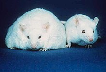
Two mice; the mouse on the left has more fat stores than the mouse on the right.
821:
470:
283:
224:
154:
cells. It is believed that these fatty acids are delivered to the kidneys via
106:
431:
407:
223:
Lipotoxicity affects the pancreas when excess free fatty acids are found in
110:
100:
839:
790:
755:
720:
678:
629:
580:
529:
488:
439:
384:
324:
771:
Biochimica et
Biophysica Acta (BBA) - Molecular and Cell Biology of Lipids
701:
Biochimica et
Biophysica Acta (BBA) - Molecular and Cell Biology of Lipids
600:
Biochimica et
Biophysica Acta (BBA) - Molecular and Cell Biology of Lipids
520:
503:
159:
92:
71:
806:"Lipoexpediency: de novo lipogenesis as a metabolic signal transmitter"
455:"Gluttony, Sloth and the Metabolic Syndrome: A Roadmap to Lipotoxicity"
167:
133:. However, no causative mechanism has been found for this correlation.
114:
67:
47:
660:
562:
147:
126:
19:
363:
Schaffer, Jean (June 2003). "Lipotoxicity: when tissues overeat".
55:
51:
35:
18:
121:
16:
Metabolic disorder in which lipids accumulate in non-fat tissue
158:. This condition leads to tubulointerstitial inflammation and
87:
can be converted to different types of lipids for storage.
305:
Current
Opinion in Clinical Nutrition and Metabolic Care
286:. The term was coined as an antonym to lipotoxicity.
282:
events, that may occur even in the setting of excess
543:Alkhouri, Naim; Dixon and Feldstein (August 2009).
550:Expert Review of Gastroenterology & Hepatology
645:"Lipotoxicity and Decreased Islet Graft Survival"
804:Lodhi IJ, Wei X, Semenkovich CF (January 2011).
346:
344:
342:
62:. Lipotoxicity is believed to have a role in
8:
191:, stimulate apoptotic pathways, or initiate
46:. The tissues normally affected include the
263:proteins responsible for lipid metabolism.
358:
356:
829:
668:
619:
570:
519:
478:
91:consists of three fatty acids bound to a
459:Trends in Endocrinology & Metabolism
295:
259:, a group of medications that activate
42:, leading to cellular dysfunction and
34:that results from the accumulation of
7:
14:
643:Leitão, Cristiane (March 2010).
377:10.1097/00041433-200306000-00008
1:
424:10.1016/j.plipres.2017.01.002
365:Current Opinion in Lipidology
783:10.1016/j.bbalip.2009.11.006
748:10.1016/j.biochi.2004.11.014
713:10.1016/j.bbalip.2013.01.007
612:10.1016/j.bbalip.2009.09.023
317:10.1097/mco.0b013e32832182ee
596:"Lipotoxicity in the Heart"
137:Effects in different organs
886:
594:Wende, Adam (March 2010).
453:Unger, Roger (June 2010).
412:Progress in Lipid Research
822:10.1016/j.tem.2010.09.002
471:10.1016/j.tem.2010.01.009
213:unfolded protein response
810:Trends Endocrinol. Metab
244:Prevention and treatment
203:within the hepatocytes.
193:cellular stress response
502:Weinberg, J.M (2006).
170:therapy and intensive
162:in mild cases, and to
24:
521:10.1038/sj.ki.5001834
197:endoplasmic reticulum
115:Cambridge researchers
38:intermediates in non-
22:
508:Kidney International
280:signal transmission
257:thiazolidinediones
131:insulin resistance
32:metabolic syndrome
25:
661:10.2337/dc09-1387
563:10.1586/egh.09.32
877:
844:
843:
833:
801:
795:
794:
766:
760:
759:
731:
725:
724:
698:
689:
683:
682:
672:
640:
634:
633:
623:
591:
585:
584:
574:
540:
534:
533:
523:
514:(9): 1560–1566.
499:
493:
492:
482:
450:
444:
443:
403:
397:
396:
360:
351:
348:
337:
336:
300:
261:nuclear receptor
885:
884:
880:
879:
878:
876:
875:
874:
850:
849:
848:
847:
803:
802:
798:
768:
767:
763:
733:
732:
728:
696:
691:
690:
686:
642:
641:
637:
593:
592:
588:
542:
541:
537:
501:
500:
496:
452:
451:
447:
405:
404:
400:
362:
361:
354:
349:
340:
302:
301:
297:
292:
273:
246:
233:
231:Skeletal muscle
221:
209:
189:death receptors
180:
172:insulin therapy
152:proximal tubule
144:
139:
89:Triacylglycerol
80:
60:skeletal muscle
17:
12:
11:
5:
883:
881:
873:
872:
867:
862:
852:
851:
846:
845:
796:
777:(3): 377–380.
761:
726:
684:
655:(3): 658–660.
635:
606:(3): 311–319.
586:
557:(4): 445–451.
535:
504:"Lipotoxicity"
494:
465:(6): 345–352.
445:
398:
371:(3): 281–287.
352:
338:
311:(2): 110–116.
294:
293:
291:
288:
276:Lipoexpediency
272:
271:Lipoexpediency
269:
245:
242:
238:lipid droplets
232:
229:
220:
217:
208:
205:
179:
176:
164:kidney failure
143:
140:
138:
135:
97:diacylglycerol
79:
76:
40:adipose tissue
15:
13:
10:
9:
6:
4:
3:
2:
882:
871:
868:
866:
863:
861:
858:
857:
855:
841:
837:
832:
827:
823:
819:
815:
811:
807:
800:
797:
792:
788:
784:
780:
776:
772:
765:
762:
757:
753:
749:
745:
741:
737:
730:
727:
722:
718:
714:
710:
707:(4): 844–52.
706:
702:
695:
688:
685:
680:
676:
671:
666:
662:
658:
654:
650:
649:Diabetes Care
646:
639:
636:
631:
627:
622:
617:
613:
609:
605:
601:
597:
590:
587:
582:
578:
573:
568:
564:
560:
556:
552:
551:
546:
539:
536:
531:
527:
522:
517:
513:
509:
505:
498:
495:
490:
486:
481:
476:
472:
468:
464:
460:
456:
449:
446:
441:
437:
433:
429:
425:
421:
417:
413:
409:
402:
399:
394:
390:
386:
382:
378:
374:
370:
366:
359:
357:
353:
347:
345:
343:
339:
334:
330:
326:
322:
318:
314:
310:
306:
299:
296:
289:
287:
285:
281:
277:
270:
268:
264:
262:
258:
253:
249:
243:
241:
239:
230:
228:
226:
218:
216:
214:
206:
204:
202:
201:triglycerides
198:
194:
190:
186:
177:
175:
173:
169:
165:
161:
157:
156:serum albumin
153:
149:
141:
136:
134:
132:
128:
123:
118:
116:
112:
108:
104:
102:
98:
94:
90:
86:
77:
75:
73:
69:
65:
64:heart failure
61:
57:
53:
49:
45:
41:
37:
33:
29:
21:
813:
809:
799:
774:
770:
764:
742:(1): 57–64.
739:
735:
729:
704:
700:
687:
652:
648:
638:
603:
599:
589:
554:
548:
538:
511:
507:
497:
462:
458:
448:
415:
411:
401:
368:
364:
308:
304:
298:
275:
274:
265:
254:
250:
247:
234:
222:
210:
181:
145:
119:
105:
81:
28:Lipotoxicity
27:
26:
284:fatty acids
185:hepatocytes
85:fatty acids
865:Metabolism
854:Categories
816:(1): 1–8.
290:References
225:beta cells
107:Adipocytes
736:Biochimie
432:1873-2194
418:: 14–29.
111:apoptosis
101:ceramides
860:Diabetes
840:20889351
791:19941972
756:15733738
721:23353597
679:20009097
630:19818871
581:19673631
530:16955100
489:20223680
440:28104532
393:23895380
385:12840659
325:19202381
219:Pancreas
160:fibrosis
93:glycerol
72:diabetes
831:3011046
670:2827526
621:2823976
572:2775708
480:2880185
333:7169311
195:in the
168:fibrate
142:Kidneys
68:obesity
48:kidneys
870:Lipids
838:
828:
789:
754:
719:
677:
667:
628:
618:
579:
569:
528:
487:
477:
438:
430:
391:
383:
331:
323:
148:kidney
127:leptin
70:, and
697:(PDF)
389:S2CID
329:S2CID
207:Heart
178:Liver
78:Cause
56:heart
52:liver
44:death
36:lipid
30:is a
836:PMID
787:PMID
775:1801
752:PMID
717:PMID
705:1831
675:PMID
626:PMID
604:1801
577:PMID
526:PMID
485:PMID
436:PMID
428:ISSN
381:PMID
321:PMID
150:and
129:and
122:gene
58:and
826:PMC
818:doi
779:doi
744:doi
709:doi
665:PMC
657:doi
616:PMC
608:doi
567:PMC
559:doi
516:doi
475:PMC
467:doi
420:doi
373:doi
313:doi
856::
834:.
824:.
814:22
812:.
808:.
785:.
773:.
750:.
740:87
738:.
715:.
703:.
699:.
673:.
663:.
653:33
651:.
647:.
624:.
614:.
602:.
598:.
575:.
565:.
553:.
547:.
524:.
512:70
510:.
506:.
483:.
473:.
463:21
461:.
457:.
434:.
426:.
416:66
414:.
410:.
387:.
379:.
369:14
367:.
355:^
341:^
327:.
319:.
309:12
307:.
174:.
99:,
66:,
54:,
50:,
842:.
820::
793:.
781::
758:.
746::
723:.
711::
681:.
659::
632:.
610::
583:.
561::
555:3
532:.
518::
491:.
469::
442:.
422::
395:.
375::
335:.
315::
Text is available under the Creative Commons Attribution-ShareAlike License. Additional terms may apply.