94:, the location of particular types of molecules can be determined. Natural lipids do not fluoresce, so it is always necessary to include a dye molecule in order to study lipid bilayers with fluorescence microscopy. To some extent, the addition of the dye molecule always changes the system, and in some cases it can be difficult to say whether the observed effect is due to the lipids, the dye or, most commonly, some combination of the two. The dye is usually attached either to a lipid or a molecule that closely resembles a lipid, but since the dye domain is relatively large it can alter the behavior of this other molecule. This is a particularly contentious issue when studying the
349:, allowing researchers to tune the experimental baseline by mixing water and deuterated water. Using reflectometry rather than scattering with neutrons or x-rays allow experimenters to probe supported bilayers or multilayer stacks. These measurements are more complicated to perform an analyze, but allow determination of cross sectional composition, including the location and concentration of water within the bilayer. In the case of both neutron and x-ray scattering measurements, the information provided is an ensemble average of the system and is therefore subject to uncertainty based on thermal fluctuations in these highly mobile structures.
245:(AFM) has been used in recent years to image and probe the physical properties of lipid bilayers. AFM is a promising technique because it has the potential to image with nanometer resolution at room temperature and even underwater, conditions necessary for natural bilayer behavior. These capabilities have allowed direct imaging of the subtle ripple phase transition in a supported bilayer. Another AFM experiment performed in a
231:
341:. One limitation of x-ray techniques is that x-rays are relatively insensitive to light elements such as hydrogen. This effect is a consequence of the fact that x-rays interact with matter by scattering off of electron density which decreases with decreasing atomic number. In contrast, neutrons scatter off of nuclear density and nuclear magnetic fields so sensitivity does not decrease monotonically with
285:
level, this higher resolution has been invaluable. In 1960, when the structure of the bilayer was still debated, it was electron microscopy that offered the first direct visualization of the two apposing leaflets. In conjunction with rapid freezing techniques, electron microscopy has also been used to study the mechanisms of inter- and intracellular transport, for instance in demonstrating that
333:
because each has different advantages and disadvantages. X-rays interact only weakly with water, so bulk samples can be probed with relatively easy sample preparation. This is one of the reasons that x-ray scattering was the technique first used to systematically study inter-bilayer spacing. X-ray scattering can also yield information on the average spacing between individual
147:
259:
important when studying metastable systems such as vesicles adsorbed on a substrate, since the AFM tip can induce rupture and other structural changes. Care must also be taken to choose an appropriate material and surface preparation for the AFM tip, as hydrophobic surfaces can interact strongly with lipids and disrupt the bilayer structure.
126:(FRET). In FRET, two dye molecules are chosen such that the emission spectrum of one overlaps the absorption spectrum of the other. This energy transfer is extremely distance dependent, so it is possible to tell with angstrom resolution how far apart the two dyes are. This can be used for instance to determine when two bilayers
113:
by exposure to an intense light source. This area is then monitored over time as the “dead” dye molecules diffuse out and are replaced by intact dye molecules from the surrounding bilayer. By fitting this recovery curve it is possible to calculate the diffusion coefficient of the bilayer. An argument
284:
interacts with the sample rather than a beam of light as in traditional microscopy. Electrons have a much shorter wavelength than light so electron microscopy has much higher resolution than light microscopy, potentially down to the atomic scale. Because lipid bilayers are arranged on the molecular
121:
and image processing this limit can be extended, but typically not much below 100 nanometers, which is much smaller than a typical cell but much larger than the thickness of a lipid bilayer. More recently, advanced microscopy methods have allowed much greater resolution under certain circumstances,
197:
of a bilayer. Because capacitance is inversely proportional to thickness and bilayers are very thin they typically have a very large capacitance, on the order of 2 μF/cm. Capacitance measurements are particularly useful when dealing with black lipid membranes, as they can be used to determine
182:
of the bilayer. This resistance is typically quite high for intact bilayers, often exceeding 100 GΩ since the hydrophobic core is impermeable to charged hydrated species. Because this resistance is so large, the presence of even a few nanometer-scale holes results in a dramatic increase in current
301:
conditions with the associated water frozen, or a metallic negative can be made from a frozen sample. It is also typically necessary to stain the bilayer with a heavy metal compound such as osmium tetroxide or uranyl acetate because the low atomic weight constituents of lipids (carbon, nitrogen,
332:
Both X-rays and high-energy neutrons are used to probe the structure and periodicity of biological structures including bilayers because they can be tuned to interact with matter at the relevant (angstrom-nm) length scales. Often, these two classes of experiment provide complementary information
254:
labeling of the lipids, as the probe tip interacts mechanically with the bilayer surface. Because of this, the same scan can reveal information about both the bilayer and any associated structures, even to the extent of resolving individual membrane proteins. In addition to imaging, AFM can also
249:
under aqueous buffer medium allowed (1) to determine the formation of transmembrane pores (holes) around nanoparticles of approximately 1.2 to 22 nm diameter via subtraction of AFM images from series recorded during the lipid bilayer formation and (2) to observe adsorption of single insulin
161:
Electrical measurements are the most straightforward way to characterize one of the more important functions of a bilayer, namely its ability to segregate and prevent the flow of ions in solution. Accordingly, electrical characterization was one of the first tools used to study the properties of
258:
Although AFM is a powerful and versatile tool for studying lipid bilayers, there are some practical limitations and difficulties. Because of the fragile nature of the bilayer, extremely low scanning forces (typically 50pN or less) must be used to avoid damage. This consideration is particularly
296:
The limitations of electron microscopy in the study of lipid structures deal primarily with sample preparation. Most electron microscopes require the sample to be under vacuum, which is incompatible with hydration at room temperature. To surmount this problem, samples can be imaged under
268:
192:
electrode since this reaction is stable, reversible, involves a single electron transfer and can produce large currents. In addition to simple DC current measurements it is also possible to perform AC electrical characterization to extract information about the capacitance and complex
29:
bonds and is irreversibly destroyed if removed from water. In spite of these limitations dozens of techniques have been developed over the last seventy years to allow investigations of the structure and function of bilayers. The first general approach was to utilize non-destructive
72:
221:
Hydrated bilayers show rich vibrational dynamics and are good media for efficient vibrational energy transfer. Vibrational properties of lipid monolayers and bilayers has been investigated by ultrafast spectroscopic techniques and recently developed computational methods.
114:
against the use of this technique is that what is actually being studied is the diffusion of the dye, not the lipid. While correct, this distinction is not always important, since the mobility of the dye is often dominated by the mobility of the bilayer.
24:
makes it a very difficult structure to study. An individual bilayer, since it is only a few nanometers thick, is invisible in traditional light microscopy. The bilayer is also a relatively fragile structure since it is held together entirely by
423:
K. C. Melikov, V. A. Frolov, A. Shcherbakov, A. V. Samsonov, Y. A. Chizmadzhev and L. V. Chernomordik."Voltage-Induced
Nonconductive Pre-Pores and Metastable Single Pores in Unmodified Planar Lipid Bilayer " Biophysical Journal. 80. (2001)
177:
Fundamentally, all electrical measurements of bilayers involve the placement of an electrode on either side of the membrane. By applying a bias across these electrodes and measuring the resulting current, it is possible to determine the
592:
G. Zaccai, J. K. Blasie and B. P. Schoenborn."Neutron diffraction studies on the location of water in lecithin bilayer model membranes." Proceedings of the
National Academy of Sciences of the United States of America. 72. (1975)
255:
probe the mechanical nature of small delicate structures such as lipid bilayers. One study demonstrated the possibility of measuring the elastic modulus of individual nano-scale membranes suspended over porous anodic alumina.
271:
Image from a
Transmission Electron Microscope of a lipid vesicle. The two dark bands around the edge are the two leaflets of the bilayer. Similar electron micrographs confirmed the bilayer nature of the cell membrane in the
19:
is the use of various optical, chemical and physical probing methods to study the properties of lipid bilayers. Many of these techniques are elaborate and require expensive equipment because the fundamental nature of the
187:
can be resolved. In such DC measurements, it is necessary to use electrochemically active electrodes to provide the necessary positive charges on one side and negative charges on the other. The most common system is the
485:
T. Kaasgaard, C. Leidy, J. H. Crowe, O. E. Mouritsen and K. Jorgensen."Temperature-Controlled
Structure and Kinetics of Ripple Phases in One- and Two-Component Supported Lipid Bilayers " Biophysical Journal. 85. (2003)
451:
Alireza
Mashaghi et al. Optical anisotropy of supported lipid structures probed by waveguide spectroscopy and its application to study of supported lipid bilayer formation kinetics Anal. Chem., 80 (10), 3666–3676
564:
J. E. Heuser, T. S. Reese, M. J. Dennis, Y. Jan, L. Jan and L. Evans."Synaptic vesicle exocytosis captured by quick freezing and correlated with quantal transmitter release." Journal of Cell
Biology. 81. (1979)
546:
J. Schneider, W. Barger and G. U. Lee."Nanometer scale surface properties of supported lipid bilayers measured with hydrophobic and hydrophilic atomic force microscope probes." Langmuir. 19. (2003) 1899-1907.
517:
S. Steltenkamp, M. M. Muller, M. Deserno, C. Hennesthal, C. Steinem and A. Janshoff."Mechanical properties of pore-spanning lipid bilayers probed by atomic force microscopy." Biophysical
Journal. 91. (2006)
536:
K. Dimitrievski, M. Zach, V. P. Zhadanov and B. Kasemo."Imaging and manipulation of adsorbed lipid vesicles by an AFM tip : Experiment and Monte Carlo simulations." Colloids and
Surfaces B. 47. (2006)
38:
and electrical resistance which measured bilayer properties but did not actually image the bilayer. Later, protocols were developed to modify the bilayer and allow its direct visualization at first in the
86:
is a technique whereby certain molecules can be excited with one wavelength of light and will emit another longer wavelength of light. Because each fluorescent molecule has a unique spectrum of
527:
S. W. Hui, R. Viswanathan, J. A. Zasadzinski and J. N. Israelachvili."The structure and stability of phospholipid bilayers by atomic force microscopy." Biophysical
Journal. 68. (1995) 171-8.
461:
M. Bonn et al., Structural inhomogeneity of interfacial water at lipid monolayers revealed by surface-specific vibrational pump-probe spectroscopy, J. Am. Chem. Soc. 132, 14971–14978 (2010).
310:(SEM) does not require this step, but cannot offer the same resolution as TEM. Both methods are surface-sensitive techniques and cannot reveal information about deeply buried structures.
414:
P. Mueller, D. O. Rudin, H. I. Tien and W. C. Wescott."Reconstitution of cell membrane structure in vitro and its transformation into an excitable system." Nature. 194. (1962) 979-980.
162:
model systems such as black membranes. It was already known that the cell membrane was capable of supporting an ionic gradient and that this gradient is responsible for the ability of
405:
J. M. Crane, V. Kiessling and L. K. Tamm."Measuring lipid asymmetry in planar supported bilayers by fluorescence interference contrast microscopy." Langmuir. 21. (2005) 1377-1388.
508:
R. P. Richter and A. Brisson."Characterization of lipid bilayers and protein assemblies supported on rough surfaces by atomic force microscopy." Langmuir. 19. (2003) 1632-1640.
574:
D. Papahadjapoulos and N. Miller."Phospholipid Model
Membranes I. Structural characteristics of hydrated liquid crystals." Biochimica et Biophysica Acta. 135. (1967) 624-638.
396:
L. Guohua and R. C. Macdonald."Lipid bilayer vesicle fusion: Intermediates captured by high-speed microfluorescence spectroscopy." Biophysical Journal. 85. (2003) 1585-1599.
387:
W. L. Vaz and P. F. Almeida."Microscopic versus macroscopic diffusion in one-component fluid phase lipid bilayer membranes." Biophysical Journal. 60. (1991) 1553-1554.
131:
218:
has been used to measure dynamic reorganisation of the layer due to temperature, ionic strength, and molecular interactions with e.g. antimicrobial peptides.
306:(TEM) is being used, it is also necessary to cut or polish the sample into a very thin (<1 micrometre) sheet, which can be difficult and time-consuming.
138:
patterns formed it is possible to individually resolve the two leaflets of a supported bilayer and determine the distribution of a fluorescent dye in each.
365:
D. Axelrod, D. E. Koppel, J. Schlessinger, E. Elson and W. W. Webb."Mobility measurement by analysis of fluorescence photobleaching recovery kinetics. ."
555:
J. D. Robertson."The molecular structure and contact relationships of cell membranes." Progress Biophysics and Biophysical Chemistry. 10. (1960) 343-418.
106:
117:
In traditional fluorescence microscopy the resolution has been limited to approximately half the wavelength of the light used. Through the use of
123:
470:
Mischa Bonn et al., Interfacial Water Facilitates Energy Transfer by Inducing Extended Vibrations in Membrane Lipids, J Phys Chem, 2012
293:. Often, electron microscopy is the only probe technique with sufficient resolution to determine complex nanometer-scale morphologies.
433:
E. Neher and B. Sakmann."Single-channel currents recorded from membrane of denervated frog muscle fibres " Nature. 286. (1976) 71-73.
378:
D. M. Soumpasis."Theoretical analysis of fluorescence photobleaching recovery experiments." Biophysical Journal. 41. (1983) 95-7.
303:
215:
56:
238:
scan of a supported lipid bilayer. The pits are defects in the bilayer, exposing the smooth surface of the substrate underneath.
583:
D. M. Small."Phase equilibria and structure of dry and hydrated egg lecithin " Journal of Lipid Research. 8. (1967) 551-557.
134:(FLIC). This method requires that the sample be mounted on a precisely micromachined reflective surface. By studying the
338:
307:
109:(FRAP) to determine bilayer diffusion coefficients. In a typical FRAP experiment a small (~30 μm diameter) area is
99:
323:
618:
214:
layer where the optical properties parallel are very different from those perpendicular. This effect, studied by
189:
135:
63:
of lipid bilayers to characterise order and disruption associated with interactions or environmental effects.
246:
242:
235:
83:
48:
44:
327:
179:
183:
and can be easily determined. The sensitivity of this system is such that even the activity of single
250:
molecules onto exposed nanoparticles. Another advantage is that AFM does not require fluorescent or
194:
127:
345:. This mechanism also provides strong isotopic contrast in some cases, notably between hydrogen and
366:
277:
118:
87:
40:
26:
319:
102:
of lipids, as both processes are very sensitive to the size and shape of the molecules involved.
267:
91:
35:
167:
71:
207:
110:
230:
612:
342:
76:
60:
21:
495:
Y. Roiter, M. Ornatska, A. R. Rammohan, J. Balakrishnan, D. R. Heine, and S. Minko,
153:
recordings of changes in conductivity associated with the opening and closing of an
211:
184:
602:
D. Boal, "Mechanics of the Cell". 2002, Cambridge, UK: Cambridge University Press.
47:. Over the past two decades, a new generation of characterization tools including
105:
This potential complication has been given an argument against the use of one of
471:
442:
D. T. Sawyer, "Electrochemistry for Chemists". 2nd Ed. 1995: Wiley Interscience.
154:
150:
286:
346:
298:
281:
95:
122:
even down to sub-nm. One of the first of these methods to be developed was
146:
130:
and their components mix. Another high resolution microscopy technique is
290:
496:
251:
163:
55:
with little to no chemical or physical modification. More recently,
334:
266:
229:
145:
70:
79:
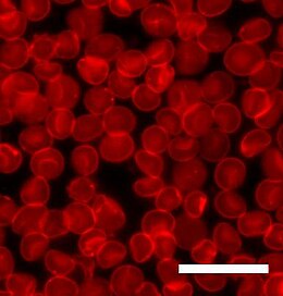
has been stained with a fluorescent dye. Scale bar is 20 μm.
302:
phosphorus, etc.) offer little contrast compared to water. If a
174:
was an important verification of the utility of model systems.
198:
when the solvent/lipid plug thins down to a single bilayer.
170:. Demonstrating that similar phenomena could be replicated
210:
which when self assembled into bilayers creates a highly
51:
has allowed the direct probing and imaging of membranes
75:
Human red blood cells viewed through a microscope. The
337:
molecules, which has led to its use in characterizing
499:, Nano Letters, vol. 8, iss. 3, pp. 941-944 (2008).
497:Interaction of Nanoparticles with Lipid Membrane
481:
479:
289:vesicles are the means of chemical release at
472:http://pubs.acs.org/doi/abs/10.1021/jp302478a
132:fluorescence interference contrast microscopy
8:
107:fluorescence recovery after photobleaching
358:
59:has been used to measure the optical
7:
14:
124:Förster resonance energy transfer
304:Transmission electron microscope
216:dual polarisation interferometry
57:dual polarisation interferometry
17:Lipid bilayer characterization
1:
314:Neutron and X-ray scattering
308:Scanning Electron Microscopy
324:X-ray scattering techniques
635:
317:
234:Illustration of a typical
43:and, more recently, with
136:destructive interference
243:Atomic force microscopy
166:to send signals via an
84:Fluorescence microscopy
67:Fluorescence Microscopy
45:fluorescence microscopy
273:
239:
190:silver/silver chloride
158:
80:
369:. 16. (1976) 1055-69.
328:Neutron reflectometry
318:Further information:
270:
233:
149:
74:
34:measurements such as
367:Biophysical Journal
278:electron microscopy
263:Electron microscopy
119:confocal microscopy
41:electron microscope
320:Neutron scattering
280:a beam of focused
274:
240:
206:Lipids are highly
159:
81:
339:phase transitions
36:x-ray diffraction
626:
619:Membrane biology
603:
600:
594:
590:
584:
581:
575:
572:
566:
562:
556:
553:
547:
544:
538:
534:
528:
525:
519:
515:
509:
506:
500:
493:
487:
483:
474:
468:
462:
459:
453:
449:
443:
440:
434:
431:
425:
421:
415:
412:
406:
403:
397:
394:
388:
385:
379:
376:
370:
363:
168:action potential
100:phase separation
634:
633:
629:
628:
627:
625:
624:
623:
609:
608:
607:
606:
601:
597:
591:
587:
582:
578:
573:
569:
563:
559:
554:
550:
545:
541:
535:
531:
526:
522:
516:
512:
507:
503:
494:
490:
484:
477:
469:
465:
460:
456:
450:
446:
441:
437:
432:
428:
422:
418:
413:
409:
404:
400:
395:
391:
386:
382:
377:
373:
364:
360:
355:
330:
316:
265:
228:
208:polar molecules
204:
144:
69:
12:
11:
5:
632:
630:
622:
621:
611:
610:
605:
604:
595:
585:
576:
567:
557:
548:
539:
529:
520:
510:
501:
488:
475:
463:
454:
444:
435:
426:
416:
407:
398:
389:
380:
371:
357:
356:
354:
351:
315:
312:
264:
261:
227:
224:
203:
200:
143:
140:
68:
65:
13:
10:
9:
6:
4:
3:
2:
631:
620:
617:
616:
614:
599:
596:
589:
586:
580:
577:
571:
568:
561:
558:
552:
549:
543:
540:
533:
530:
524:
521:
514:
511:
505:
502:
498:
492:
489:
482:
480:
476:
473:
467:
464:
458:
455:
448:
445:
439:
436:
430:
427:
420:
417:
411:
408:
402:
399:
393:
390:
384:
381:
375:
372:
368:
362:
359:
352:
350:
348:
344:
340:
336:
329:
325:
321:
313:
311:
309:
305:
300:
294:
292:
288:
283:
279:
269:
262:
260:
256:
253:
248:
244:
237:
232:
225:
223:
219:
217:
213:
209:
201:
199:
196:
191:
186:
181:
175:
173:
169:
165:
156:
152:
148:
141:
139:
137:
133:
129:
125:
120:
115:
112:
111:photobleached
108:
103:
101:
97:
93:
89:
85:
78:
77:cell membrane
73:
66:
64:
62:
61:birefringence
58:
54:
50:
46:
42:
37:
33:
28:
23:
22:lipid bilayer
18:
598:
588:
579:
570:
560:
551:
542:
532:
523:
513:
504:
491:
466:
457:
447:
438:
429:
419:
410:
401:
392:
383:
374:
361:
331:
295:
275:
257:
247:tapping mode
241:
220:
212:birefringent
205:
185:ion channels
176:
171:
160:
116:
104:
82:
52:
31:
27:non-covalent
16:
15:
155:ion channel
151:Patch clamp
424:1829-1836.
353:References
287:exocytotic
180:resistance
142:Electrical
88:absorption
347:deuterium
299:cryogenic
282:electrons
195:impedance
96:diffusion
613:Category
593:376-380.
565:275-300.
537:115-125.
518:217-226.
486:350-360.
291:synapses
252:isotopic
172:in vitro
92:emission
202:Optical
164:neurons
53:in situ
32:in situ
452:(2008)
326:, and
335:lipid
272:1950s
128:fuse
90:and
276:In
236:AFM
226:AFM
98:or
49:AFM
615::
478:^
322:,
343:z
157:.
Text is available under the Creative Commons Attribution-ShareAlike License. Additional terms may apply.