20:
54:
X-ray fluorescence with relatively small spatial discrimination (less than 50 μm) such that composition (chemistry), thickness and micro-structural measurements can be made on a wide variety of heterogeneous materials in a few seconds. It was shown that, by scanning samples with an X-Y stage, quantitative or qualitative micro-structural information could be gathered. Both these papers provided a preview into the coming integration of
987:
999:
49:
analytical figures of merit are extended to the point where trace level quantification and bulk analysis are possible. By combining the analytical information obtained from the X-ray spectra excited with electrons and with photons respectively, the main elements as well as trace elements, of low and
61:
By 1991, Pozsgai published a review article detailing the possibilities of carrying out x-ray micro-fluorescence analysis within the SEM context. The main approaches involved converting the electron optical column of an electron microscope into a transmission x-ray tube, using micro-focusing x-ray
53:
In 1986, Sandia and
Lawrence Livermore National Labs coauthored a paper (with Kevex Corporation) regarding parameters affecting X-ray micro-fluorescence. As a followup in 1988, Cross & Wherry described an X-ray micro-fluorescence analyzer which combines the nondestructive analytical method of
304:
284:
38:(SEM). Technological progress in the fields of small-spot low-power X-ray tubes and of polycapillary X-ray optics has enabled the development of compact micro-focus X-ray sources that can be attached to a SEM equipped for
196:
237:
M. Spanier, C. Herzog, D. Grötzsch, F. Kramer, I. Mantouvalou, J. Lubeck, J. Weser, C. Streeck, W. Malzer, B. Beckhoff, B. Kanngießer. Review of
Scientific Instruments, Vol 87, No 3, (035108), (2016).
216:
631:
614:
609:
758:
461:
748:
351:
292:
Performance of μ-XRF with SEM/EDS for trace analysis on the example of RoHS relevant elements–measurement, optimization and prediction of the detection limits.
97:
651:
624:
324:
559:
305:
Micro XRF Element Maps Are A New Method Of
Detecting Elements At Lower Concentrations Than The Electron Beam Produced Corollary: A Garnet Schist Example.
210:
619:
763:
254:
Determination of the real transmission of an X‐ray lens for micro‐focus XRF at the SEM by coupling measurement with calculation of scatter spectra.
1025:
62:
tubes, combining x-ray tubes with capillary techniques, as well as combining x-ray tubes with monochromators and applying synchrotron radiation.
579:
703:
569:
516:
250:
Ursula
Elisabeth Adriane Fittschen & Gerald Falkenberg. Spectrochimica Acta Part B: Atomic Spectroscopy, Vol 66, No 8, p 567-580 (2011).
958:
686:
671:
597:
39:
65:
SEM-XRF was first commercialized by IXRF Systems (Austin, TX) in March 2005. Bruker
Corporation (Billerica, MA) followed in August 2013.
113:
Hodoroaba, V., Rackwitz, V., & Reuter, D. (2010). "Micro-Focus X-Ray
Fluorescence (μ-XRF) as an Extension of the Analytical SEM".
708:
541:
394:
344:
696:
691:
589:
564:
531:
768:
753:
733:
471:
268:
Procop, Mathias, Vasile-Dan
Hodoroaba, and Vanessa Rackwitz. Microscopy and analysis / European edition. p 10-13 (May, 2011).
536:
379:
185:
1003:
713:
676:
521:
991:
551:
337:
35:
299:
Validation and traceability of XRF and SEM‐EDS elemental analysis results for solder in high‐reliability applications
738:
656:
259:
253:
963:
917:
743:
217:
Comparing the detection of iron-based pottery pigment on a carbon-coated Sherd by SEM-EDS and by Micro-XRF-SEM.
55:
953:
278:
Gaining improved chemical composition by exploitation of
Compton-to-Rayleigh intensity ratio in XRF analysis.
666:
602:
476:
435:
856:
285:
Detecting iron-based pigments on ruthenium-coated archaeological pottery by SEM-EDS and by micro-XRF-SEM.
152:
861:
681:
45:
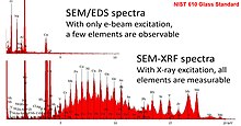
As shown in the adjacent image, when micro-focus X-ray fluorescence (microXRF) is performed with a SEM,
231:
Michael
Haschke and Stephan Boehm. Advances in Imaging and Electron Physics. Vol. 199, p 1-60, (2017).
260:
Improvements of the low-energy performance of a micro-focus x-ray source for XRF analysis with the SEM
866:
425:
831:
816:
723:
718:
661:
526:
508:
440:
430:
374:
360:
134:
Nichols, Monte C., Boehme, Dale R., Ryon, Richard W., Wherry, David, Cross, Brian, and Aden, Gary.
902:
481:
445:
291:
46:
846:
841:
225:
M. Haschke, F. Eggert and W. T. Elam. X-ray
Spectrometry. Vol. 36, No. 4, p. 254-259 (2007).
219:
Michael Pendleton, et al. The Yale Journal of Biology and Medicine. 87(1):15-20, March 2014.
796:
728:
235:
A flexible setup for angle-resolved X-ray fluorescence spectrometry with laboratory sources.
118:
298:
280:
Hodoroaba, Vasile-Dan, and Vanessa Rackwitz. Analytical Chemistry 86.14 (2014): 6858-6864.
265:
262:
Procop, Mathias; et al. X‐Ray Spectrometry: An International Journal 38.4 (2009): 308-311.
241:
851:
222:
31:
272:
X-ray fluorescence as an additional analytical method for a scanning electron microscope.
806:
420:
256:
V.‐D. Hodoroaba & M. Procop. X-Ray Spectrometry, Vol 38, No 3, p 216-221, (2009).
1019:
882:
826:
486:
933:
943:
907:
801:
791:
466:
415:
50:
high atomic number, can be analyzed – albeit with different spatial resolutions.
887:
821:
811:
968:
389:
384:
228:
213:. V.-D. Hodoroaba, et al. Microscopy and Microanalysis 17:600-601, July 2011.
122:
98:"Micro-Focus X-Ray Fluorescence (μ-XRF) as an Extension of the Analytical SEM"
19:
170:"X-ray microfluorescence analysis inside and outside the electron microscope"
135:
836:
491:
271:
247:
81:
197:"Bruker Introduces Two New Analytical Accessories for Electron Microscopes"
938:
404:
169:
948:
892:
319:
266:
A microfocus X-ray source for improved EDS and XRF analysis in the SEM.
23:
SEM/EDS spectra is compared to SEM-XRF spectra for a NIST 610 standard.
329:
30:
is an established technical term for adding a (typically micro-focus)
912:
248:
Trends in environmental science using microscopic X-ray fluorescence.
277:
274:
Procop, M., Hodoroaba, V. Microchim Acta Vol 161, p 413–419 (2008).
234:
211:
Advanced Elemental Analysis with ED-EPMA, WD-EPMA and μ-XRF at a SEM
294:
Journal of Analytical Atomic Spectrometry 28.9 (2013): 1466-1474.
18:
897:
82:"Integrated Electron and X-Ray Induced Microbeam XRF in the SEM"
333:
136:"Parameters Affecting X-Ray Microfluorescence (XRMF) Analysis"
153:
X-ray microfluorescence analyzer for multilayer metal films
287:
Microscopy and Microanalysis 20.S3 (2014): 2030-2031.
307:” Microscopy and Microanalysis 15.S2 (2009): 34-35.
926:
875:
784:
777:
644:
588:
550:
507:
500:
454:
403:
367:
242:Laboratory Micro-X-Ray Fluorescence Spectroscopy
759:Serial block-face scanning electron microscopy
462:Detectors for transmission electron microscopy
315:Two commercial vendors offer this technology:
345:
8:
301:.” X‐Ray Spectrometry 43.5 (2014): 259-268.
229:Micro-XRF in Scanning Electron Microscopes.
781:
504:
352:
338:
330:
74:
297:Sieber, John R., and Adam Mortensen. “
7:
998:
40:energy-dispersive X-ray spectroscopy
151:Brian J. Cross, David C. Wherry, "
14:
395:Timeline of microscope technology
997:
986:
985:
159:, Volume 166, 1988, pp. 263–272.
754:Precession electron diffraction
223:Micro-XRF excitation in an SEM.
1026:Electron microscopy techniques
104:. 16(S2):904–905, August 2010.
1:
115:Microscopy and Microanalysis
102:Microscopy and Microanalysis
84:. Cross BJ, Witherspoon KC.
36:Scanning Electron Microscope
1042:
739:Immune electron microscopy
657:Annular dark-field imaging
472:Everhart–Thornley detector
290:Rackwitz, Vanessa, et al.
186:"X-Beam Polycapillary XRF"
140:Advances in X-ray Analysis
100:. V.-D. Hodoroaba, et al.
42:(EDS, EDX, EDXS or XEDS).
981:
893:Hitachi High-Technologies
283:Pendleton, M. W., et al.
123:10.1017/S1431927610054115
918:Thermo Fisher Scientific
744:Geometric phase analysis
632:Aberration-Corrected TEM
88:. Jul;12(4):20–3 (2004).
56:Micro-X-ray fluorescence
667:Charge contrast imaging
477:Field electron emission
857:Thomas Eugene Everhart
142:. 1986 Vol. 30, p. 45.
24:
862:Vernon Ellis Cosslett
682:Dark-field microscopy
22:
16:X-ray sources for SEM
867:Vladimir K. Zworykin
517:Correlative light EM
426:Electron diffraction
168:Pozsgai, I. (1991),
34:(X-ray source) to a
832:Manfred von Ardenne
817:Gerasimos Danilatos
724:Electron tomography
719:Electron holography
662:Cathodoluminescence
441:Secondary electrons
431:Electron scattering
375:Electron microscopy
361:Electron microscopy
117:, 16(S2), 904–905.
954:Digital Micrograph
560:Environmental SEM
482:Field emission gun
446:X-ray fluorescence
244:, Vol. 55 (2014).
174:X-Ray Spectrometry
47:elemental analysis
25:
1013:
1012:
977:
976:
847:Nestor J. Zaluzec
842:Maximilian Haider
640:
639:
325:QUANTAX Micro-XRF
240:Michael Haschke,
1033:
1001:
1000:
989:
988:
797:Bodo von Borries
782:
542:Photoemission EM
505:
354:
347:
340:
331:
311:Commercial links
199:
194:
188:
183:
177:
166:
160:
157:Thin Solid Films
149:
143:
132:
126:
111:
105:
95:
89:
86:Microscopy Today
79:
1041:
1040:
1036:
1035:
1034:
1032:
1031:
1030:
1016:
1015:
1014:
1009:
973:
922:
871:
852:Ondrej Krivanek
773:
636:
584:
546:
532:Liquid-Phase EM
496:
455:Instrumentation
450:
408:
399:
363:
358:
313:
208:
203:
202:
195:
191:
184:
180:
167:
163:
150:
146:
133:
129:
112:
108:
96:
92:
80:
76:
71:
32:X-ray generator
17:
12:
11:
5:
1039:
1037:
1029:
1028:
1018:
1017:
1011:
1010:
1008:
1007:
995:
982:
979:
978:
975:
974:
972:
971:
966:
961:
959:Direct methods
956:
951:
946:
941:
936:
930:
928:
924:
923:
921:
920:
915:
910:
905:
900:
895:
890:
885:
879:
877:
873:
872:
870:
869:
864:
859:
854:
849:
844:
839:
834:
829:
824:
819:
814:
809:
807:Ernst G. Bauer
804:
799:
794:
788:
786:
779:
775:
774:
772:
771:
766:
761:
756:
751:
746:
741:
736:
731:
726:
721:
716:
711:
706:
701:
700:
699:
689:
684:
679:
674:
669:
664:
659:
654:
648:
646:
642:
641:
638:
637:
635:
634:
629:
628:
627:
617:
612:
607:
606:
605:
594:
592:
586:
585:
583:
582:
577:
572:
567:
562:
556:
554:
548:
547:
545:
544:
539:
534:
529:
524:
519:
513:
511:
502:
498:
497:
495:
494:
489:
484:
479:
474:
469:
464:
458:
456:
452:
451:
449:
448:
443:
438:
433:
428:
423:
421:Bremsstrahlung
418:
412:
410:
401:
400:
398:
397:
392:
387:
382:
377:
371:
369:
365:
364:
359:
357:
356:
349:
342:
334:
328:
327:
322:
312:
309:
207:
206:External links
204:
201:
200:
189:
178:
176:, 20: 215–223.
161:
144:
127:
106:
90:
73:
72:
70:
67:
15:
13:
10:
9:
6:
4:
3:
2:
1038:
1027:
1024:
1023:
1021:
1006:
1005:
996:
994:
993:
984:
983:
980:
970:
967:
965:
962:
960:
957:
955:
952:
950:
947:
945:
942:
940:
937:
935:
932:
931:
929:
925:
919:
916:
914:
911:
909:
906:
904:
901:
899:
896:
894:
891:
889:
886:
884:
883:Carl Zeiss AG
881:
880:
878:
876:Manufacturers
874:
868:
865:
863:
860:
858:
855:
853:
850:
848:
845:
843:
840:
838:
835:
833:
830:
828:
827:James Hillier
825:
823:
820:
818:
815:
813:
810:
808:
805:
803:
800:
798:
795:
793:
790:
789:
787:
783:
780:
776:
770:
767:
765:
762:
760:
757:
755:
752:
750:
747:
745:
742:
740:
737:
735:
732:
730:
727:
725:
722:
720:
717:
715:
712:
710:
707:
705:
702:
698:
695:
694:
693:
690:
688:
685:
683:
680:
678:
675:
673:
670:
668:
665:
663:
660:
658:
655:
653:
650:
649:
647:
643:
633:
630:
626:
623:
622:
621:
618:
616:
613:
611:
608:
604:
601:
600:
599:
596:
595:
593:
591:
587:
581:
580:Ultrafast SEM
578:
576:
573:
571:
568:
566:
563:
561:
558:
557:
555:
553:
549:
543:
540:
538:
537:Low-energy EM
535:
533:
530:
528:
525:
523:
520:
518:
515:
514:
512:
510:
506:
503:
499:
493:
490:
488:
487:Magnetic lens
485:
483:
480:
478:
475:
473:
470:
468:
465:
463:
460:
459:
457:
453:
447:
444:
442:
439:
437:
436:Kikuchi lines
434:
432:
429:
427:
424:
422:
419:
417:
414:
413:
411:
406:
402:
396:
393:
391:
388:
386:
383:
381:
378:
376:
373:
372:
370:
366:
362:
355:
350:
348:
343:
341:
336:
335:
332:
326:
323:
321:
318:
317:
316:
310:
308:
306:
302:
300:
295:
293:
288:
286:
281:
279:
275:
273:
269:
267:
263:
261:
257:
255:
251:
249:
245:
243:
238:
236:
232:
230:
226:
224:
220:
218:
214:
212:
205:
198:
193:
190:
187:
182:
179:
175:
171:
165:
162:
158:
154:
148:
145:
141:
137:
131:
128:
124:
120:
116:
110:
107:
103:
99:
94:
91:
87:
83:
78:
75:
68:
66:
63:
59:
57:
51:
48:
43:
41:
37:
33:
29:
21:
1002:
990:
944:EM Data Bank
908:Nion Company
802:Dennis Gabor
792:Albert Crewe
574:
570:Confocal SEM
467:Electron gun
416:Auger effect
320:Xb Micro-XRF
314:
303:
296:
289:
282:
276:
270:
264:
258:
252:
246:
239:
233:
227:
221:
215:
209:
192:
181:
173:
164:
156:
147:
139:
130:
114:
109:
101:
93:
85:
77:
64:
60:
52:
44:
27:
26:
888:FEI Company
822:Harald Rose
812:Ernst Ruska
501:Microscopes
409:with matter
407:interaction
969:Multislice
785:Developers
645:Techniques
390:Microscope
385:Micrograph
69:References
58:with SEM.
837:Max Knoll
492:Stigmator
1020:Category
992:Category
939:CrysTBox
927:Software
598:Cryo-TEM
405:Electron
1004:Commons
652:4D STEM
625:4D STEM
603:Cryo-ET
575:SEM-XRF
565:CryoSEM
522:Cryo-EM
380:History
28:SEM-XRF
949:EMsoft
934:CASINO
913:TESCAN
778:Others
677:cryoEM
368:Basics
903:Leica
749:PINEM
615:HRTEM
610:EFTEM
964:IUCr
898:JEOL
769:WBDF
764:WDXS
714:EBIC
709:EELS
704:ECCI
692:EBSD
672:CBED
620:STEM
734:FEM
729:FIB
697:TKD
687:EDS
590:TEM
552:SEM
527:EMP
155:",
119:doi
1022::
509:EM
172:.
138:.
353:e
346:t
339:v
125:.
121::
Text is available under the Creative Commons Attribution-ShareAlike License. Additional terms may apply.