289:
238:), a full field soft X-ray microscope operated by the Center for X-ray Optics and dedicated to various applications in modern nanoscience, such as nanomagnetic materials, environmental and materials sciences and biology. XM-1 uses an X-ray lens to focus X-rays on a CCD, in a manner similar to an optical microscope. XM-1 held the world record in spatial resolution with Fresnel zone plates down to 15 nm and is able to combine high spatial resolution with a sub-100ps time resolution to study e.g. ultrafast spin dynamics. In July 2012, a group at
206:
33:
367:
254:
277:
microscopes represent the only commercially available options in this domain. These microscopes enable the acquisition of soft X-ray tomograms from cryogenically vitrified as well as room temperature samples, employing flat specimen holders such as standard transmission electron microscopy (TEM) grids or glass capillaries.
281:
case of specimens imaged in glass capillaries, the full-tilt tomography may take slightly longer. Resolutions in biological specimens, determined by
Fourier Ring Correlations (1/2 Signal-to-Noise ratio), can achieve 55 nm for two-hour tomograms, with the ability to resolve Siemens star lines and spaces as small as 25 nm.
338:
allows the observation of biological specimens in their hydrated natural state, albeit embedded in water ice. Until now, resolutions of 30 nanometer are possible using the
Fresnel zone plate lens which forms the image using the soft X-rays emitted from a synchrotron. Recently, the use of soft X-rays
213:
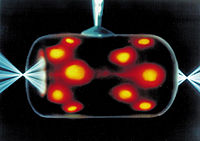
uses a "hohlraum" which is irradiated with laser beam cones from either side on its inner surface to bathe a fusion microcapsule inside with smooth high intensity X-rays. The highest energy X-rays which penetrate the hohlraum can be visualized using an X-ray microscope such as here, where X-radiation
333:
in that it can view biological samples in their natural state. Electron microscopy is widely used to obtain images with nanometer level resolution but the relatively thick living cell cannot be observed as the sample has to be chemically fixed, dehydrated, embedded in resin, then sliced ultra thin.
280:
The SXT-100 system stands out for its versatility, accommodating a variety of specimen formats. For instance, researchers can acquire tomograms on TEM grids, allowing for ±60 degrees range tilt series collected in 1-degree steps within a time frame ranging from under one hour to two hours. In the
305:
radiation sources, have fairly low brightness of the required wavelengths, so an alternative method of image formation is scanning transmission soft X-ray microscopy. Here the X-rays are focused to a point and the sample is mechanically scanned through the produced focal spot. At each point the
284:
These table-top soft X-ray microscopes enhance the accessibility of high-resolution soft X-ray tomography beyond large-scale facilities. Their integration into currently established correlative light and electron microscopy workflows bridges the resolution gap and significantly improves imaging
276:
setups offer a compact and easily integratable solution for researchers within laboratory settings. These systems typically utilize laser-driven X-ray sources, with the brightness of the source dependent on the type of target and the power of the laser. Notably, the SXT-100 table-top soft X-ray
249:
The ALS is also home to the world's first soft X-ray microscope designed for biological and biomedical research. This new instrument, XM-2 was designed and built by scientists from the
National Center for X-ray Tomography. XM-2 is capable of producing 3-Dimensional tomograms of cells.
362:
can be determined down to the placement of individual atoms within its molecules. X-ray microscopes are sometimes used for these analyses because the samples are too small to be analyzed in any other way.
156:
region (wavelength region: 2.34–4.4 nm, photon energy region: 280 – 530 eV) by the carbon atom (main element composing the living cell) and the oxygen atom (main element for water).
413:
222:
produced a shadow X-ray microscope which placed the specimen between the source and a target plate, this became the basis for the first commercial X-ray microscopes from the
374:
foil mounted in a steel case to be used as a window between a vacuum chamber and an X-ray microscope. Beryllium, due to its low Z number, is highly transparent to X-rays.
148:, X-rays do not reflect or refract easily, and they are invisible to the human eye. Therefore, the basic process of an X-ray microscope is to expose film or use a
152:(CCD) detector to detect X-rays that pass through the specimen. It is a contrast imaging technology using the difference in absorption of soft X-ray in the
867:
930:
877:
384:
549:
513:
314:. This type of Scanning Transmission X-ray Microscope (STXM) was first developed by researchers at Stony Brook University and was employed at the
789:
50:
804:
887:
882:
358:. By analyzing the internal reflections of a diffraction pattern (usually with a computer program), the three-dimensional structure of a
960:
945:
814:
315:
116:
97:
257:
Cryo-SXT of
Euglena Algae by SiriusXT's SXT-100, soft x-ray microscope, in collaboration with Roland Fleck, King's College London.
69:
862:
841:
772:
319:
76:
54:
892:
857:
955:
925:
784:
757:
83:
935:
902:
872:
836:
542:
210:
184:
173:
809:
288:
981:
821:
709:
65:
43:
134:
430:
Yamamoto Y, Shinohara K (October 2002). "Application of X-ray microscopy in analysis of living hydrated cells".
648:
335:
223:
950:
799:
535:
910:
779:
587:
355:
149:
205:
176:
826:
747:
679:
612:
311:
307:
1018:
699:
674:
653:
394:
330:
326:
180:
90:
469:"Hard X-ray microbeam experiments with a sputtered-sliced Fresnel zone plate and its applications"
242:
claimed a record spatial resolution of 10 nm, by using the hard X-ray scanning microscope at
940:
719:
684:
617:
455:
501:
339:
emitted from laser-produced plasmas rather than synchrotron radiation is becoming more popular.
988:
920:
915:
742:
714:
607:
490:
447:
219:
704:
582:
480:
439:
389:
347:
165:
1013:
976:
831:
694:
658:
577:
468:
195:
325:
The resolution of X-ray microscopy lies between that of the optical microscope and the
199:
1007:
752:
202:
produced some of the first usable X-ray images with his apparatus in the late 1940s.
17:
459:
794:
689:
602:
592:
343:
153:
643:
351:
302:
169:
32:
767:
633:
485:
191:
371:
366:
243:
494:
451:
253:
234:
The
Advanced Light Source (ALS) in Berkeley, California, is home to XM-1 (
638:
346:
in most materials, and these emissions can be analyzed to determine the
762:
359:
188:
597:
443:
572:
558:
365:
287:
252:
204:
145:
138:
235:
527:
267:
239:
531:
26:
187:. An alternative method of focusing X-rays is to use a tiny
306:
transmitted X-rays are recorded with a detector such as a
301:
Sources of soft X-rays suitable for microscopy, such as
272:
266:
In contrast to synchrotron-based soft X-ray tomography,
467:
Kamijo N, Suzuki Y, Awaji M, et al. (May 2002).
506:
969:
901:
850:
735:
728:
667:
626:
565:
57:. Unsourced material may be challenged and removed.
350:of an imaged object. Another use is to generate
502:Scientific applications of soft X-ray microscopy
141:band to produce images of very small objects.
543:
179:to focus the X-rays, which grazed X-rays off
8:
732:
550:
536:
528:
292:SiriusXT's Table-Top Soft X-ray Microscope
484:
117:Learn how and when to remove this message
385:Synchrotron X-ray tomographic microscopy
329:. It has an advantage over conventional
194:of concentric gold or nickel rings on a
405:
334:However, it should be mentioned that
7:
507:National Center for X-ray Tomography
55:adding citations to reliable sources
25:
316:National Synchrotron Light Source
31:
268:lab-based soft X-ray tomography
262:Table-top soft X-ray tomography
42:needs additional citations for
320:Brookhaven National Laboratory
236:http://www.cxro.lbl.gov/BL612/
230:Notable soft X-ray microscopes
183:curved mirrors at a very high
1:
214:is represented in orange/red.
354:patterns, a process used in
342:Additionally, X-rays cause
211:inertial confinement fusion
164:Early X-ray microscopes by
1035:
822:X-Ray Fluorescence Imaging
710:Anomalous X-ray scattering
486:10.1107/S090904950200376X
135:electromagnetic radiation
649:Synchrotron light source
336:cryo-electron microscopy
297:Characteristics and uses
224:General Electric Company
668:Interaction with matter
627:Sources and instruments
66:"Soft X-ray microscopy"
800:Diffraction tomography
375:
293:
258:
215:
911:X-ray crystallography
780:Soft x-ray microscopy
748:Panoramic radiography
588:Synchrotron radiation
414:"Desy Photon Science"
369:
356:X-ray crystallography
291:
256:
209:Indirect drive laser
208:
150:charge-coupled device
18:Soft x-ray microscopy
680:Photoelectric effect
613:Characteristic X-ray
473:J Synchrotron Radiat
312:avalanche photodiode
308:proportional counter
51:improve this article
700:Photodisintegration
675:Rayleigh scattering
654:Free-electron laser
395:Electron microscope
331:electron microscopy
327:electron microscope
941:X-ray reflectivity
720:X-ray fluorescence
685:Compton scattering
618:High-energy X-rays
514:"X-ray microscopy"
376:
294:
259:
216:
185:angle of incidence
1001:
1000:
997:
996:
989:X-ray lithography
921:Backscatter X-ray
916:X-ray diffraction
743:X-ray radiography
715:X-ray diffraction
608:Siegbahn notation
348:chemical elements
177:reflective optics
174:grazing incidence
127:
126:
119:
101:
16:(Redirected from
1026:
827:X-ray holography
733:
705:Radiation damage
552:
545:
538:
529:
524:
522:
520:
498:
488:
463:
444:10.1002/ar.10166
418:
417:
410:
390:X-ray microscope
166:Paul Kirkpatrick
131:X-ray microscope
122:
115:
111:
108:
102:
100:
59:
35:
27:
21:
1034:
1033:
1029:
1028:
1027:
1025:
1024:
1023:
1004:
1003:
1002:
993:
977:X-ray astronomy
965:
897:
846:
832:X-ray telescope
724:
695:Photoionization
663:
659:X-ray nanoprobe
622:
578:Absorption edge
566:Characteristics
561:
556:
518:
516:
511:
479:(Pt 3): 182–6.
466:
429:
426:
421:
412:
411:
407:
403:
381:
299:
264:
232:
198:substrate. Sir
196:silicon dioxide
162:
144:Unlike visible
123:
112:
106:
103:
60:
58:
48:
36:
23:
22:
15:
12:
11:
5:
1032:
1030:
1022:
1021:
1016:
1006:
1005:
999:
998:
995:
994:
992:
991:
986:
985:
984:
973:
971:
967:
966:
964:
963:
958:
953:
948:
943:
938:
933:
928:
923:
918:
913:
907:
905:
899:
898:
896:
895:
890:
885:
880:
875:
870:
865:
860:
854:
852:
848:
847:
845:
844:
839:
834:
829:
824:
819:
818:
817:
812:
807:
797:
792:
787:
782:
777:
776:
775:
770:
760:
755:
750:
745:
739:
737:
730:
726:
725:
723:
722:
717:
712:
707:
702:
697:
692:
687:
682:
677:
671:
669:
665:
664:
662:
661:
656:
651:
646:
641:
636:
630:
628:
624:
623:
621:
620:
615:
610:
605:
600:
595:
590:
585:
580:
575:
569:
567:
563:
562:
557:
555:
554:
547:
540:
532:
526:
525:
509:
504:
499:
464:
425:
424:External links
422:
420:
419:
404:
402:
399:
398:
397:
392:
387:
380:
377:
298:
295:
263:
260:
231:
228:
200:Lawrence Bragg
161:
158:
125:
124:
39:
37:
30:
24:
14:
13:
10:
9:
6:
4:
3:
2:
1031:
1020:
1017:
1015:
1012:
1011:
1009:
990:
987:
983:
980:
979:
978:
975:
974:
972:
968:
962:
959:
957:
954:
952:
949:
947:
944:
942:
939:
937:
934:
932:
929:
927:
924:
922:
919:
917:
914:
912:
909:
908:
906:
904:
900:
894:
891:
889:
886:
884:
881:
879:
876:
874:
871:
869:
866:
864:
861:
859:
856:
855:
853:
849:
843:
840:
838:
835:
833:
830:
828:
825:
823:
820:
816:
813:
811:
808:
806:
803:
802:
801:
798:
796:
793:
791:
788:
786:
783:
781:
778:
774:
771:
769:
766:
765:
764:
761:
759:
756:
754:
753:Tomosynthesis
751:
749:
746:
744:
741:
740:
738:
734:
731:
727:
721:
718:
716:
713:
711:
708:
706:
703:
701:
698:
696:
693:
691:
688:
686:
683:
681:
678:
676:
673:
672:
670:
666:
660:
657:
655:
652:
650:
647:
645:
642:
640:
637:
635:
632:
631:
629:
625:
619:
616:
614:
611:
609:
606:
604:
601:
599:
596:
594:
591:
589:
586:
584:
583:Moseley's law
581:
579:
576:
574:
571:
570:
568:
564:
560:
559:X-ray science
553:
548:
546:
541:
539:
534:
533:
530:
515:
510:
508:
505:
503:
500:
496:
492:
487:
482:
478:
474:
470:
465:
461:
457:
453:
449:
445:
441:
438:(5): 217–23.
437:
433:
428:
427:
423:
415:
409:
406:
400:
396:
393:
391:
388:
386:
383:
382:
378:
373:
368:
364:
361:
357:
353:
349:
345:
340:
337:
332:
328:
323:
321:
317:
313:
309:
304:
296:
290:
286:
282:
278:
275:
274:
270:developed by
269:
261:
255:
251:
247:
245:
241:
237:
229:
227:
225:
221:
218:In the 1950s
212:
207:
203:
201:
197:
193:
190:
186:
182:
178:
175:
171:
167:
159:
157:
155:
151:
147:
142:
140:
136:
132:
121:
118:
110:
99:
96:
92:
89:
85:
82:
78:
75:
71:
68: –
67:
63:
62:Find sources:
56:
52:
46:
45:
40:This article
38:
34:
29:
28:
19:
851:Spectroscopy
795:Ptychography
729:Applications
690:Auger effect
593:Water window
517:. Retrieved
512:Arndt Last.
476:
472:
435:
431:
408:
344:fluorescence
341:
324:
300:
285:throughput.
283:
279:
271:
265:
248:
233:
217:
163:
154:water window
143:
137:in the soft
130:
128:
113:
104:
94:
87:
80:
73:
61:
49:Please help
44:verification
41:
644:Synchrotron
352:diffraction
303:synchrotron
170:Albert Baez
160:Development
1019:Microscopy
1008:Categories
903:Scattering
768:Helical CT
634:X-ray tube
519:17 October
401:References
192:zone plate
107:April 2022
77:newspapers
432:Anat. Rec
372:beryllium
370:A square
244:PETRA III
181:parabolic
639:Betatron
495:11972376
460:43009840
452:12379938
379:See also
273:SiriusXT
220:Newberry
982:History
736:Imaging
360:crystal
189:fresnel
91:scholar
1014:X-rays
970:Others
931:GISAXS
603:L-edge
598:K-edge
493:
458:
450:
310:or an
93:
86:
79:
72:
64:
961:EDXRD
883:XANES
878:EXAFS
868:ARPES
815:3DXRD
573:X-ray
456:S2CID
172:used
146:light
139:X-ray
133:uses
98:JSTOR
84:books
946:RIXS
936:WAXS
926:SAXS
837:DFXM
805:XDCT
790:STXM
785:XPCI
773:XACT
521:2012
491:PMID
448:PMID
240:DESY
168:and
70:news
951:XRS
893:XFH
888:EDS
873:AES
863:XPS
858:XAS
842:DXA
810:DCT
758:CDI
481:doi
440:doi
436:269
318:at
129:An
53:by
1010::
956:XS
763:CT
489:.
475:.
471:.
454:.
446:.
434:.
322:.
246:.
226:.
551:e
544:t
537:v
523:.
497:.
483::
477:9
462:.
442::
416:.
120:)
114:(
109:)
105:(
95:·
88:·
81:·
74:·
47:.
20:)
Text is available under the Creative Commons Attribution-ShareAlike License. Additional terms may apply.