2206:
119:
445:
orbit of the parallel beam irradiation optical system. In the figure above, X-Y plane rotates around the point of origin in the plane in such a way “to keep mutual positional relationship between the light source (2) and screen (7) passing through the trajectory (5).” Rotation angle of this case is defined as θ. In the figure set out above, absorption coefficient at a cross-sectional coordinate (x, y) of the subject is modeled as μ(x, y).
432:: Considering a parallel beam irradiation optical system where the angle between the object and all transmission lights equals θ. Here, the numbers in the figure (see the numbers within the parentheses) respectively indicate: (1) = an object; (2) = the parallel beam light source; (3) = the screen; (4) = transmission beam; (5) = the datum circle; (6) = the origin; and (7) = a fluoroscopic image (a one-dimensional image; p
3577:
49:
2160:(CT) technology in which the source and detector travel along a helical path relative to the object. Typical implementations involve moving the patient couch through the bore of the scanner whilst the gantry rotates. Spiral CT can achieve improved image resolution for a given radiation dose, compared to individual slice acquisition. Most modern hospitals currently use spiral CT scanners.
426:
131:
28:
418:
beam irradiation optical system. Parallel beam irradiation optical system may be the easiest and most practical example of a tomographic optical system therefore, in this article, explanation of "How to obtain the
Tomographic image" will be based on "the parallel beam irradiation optical system". The resolution in tomography is typically described by the
226:. Radiocontrasts for X-ray CT are, in general, iodine-based. This is useful to highlight structures such as blood vessels that otherwise would be difficult to delineate from their surroundings. Using contrast material can also help to obtain functional information about tissues. Often, images are taken both with and without radiocontrast.
390:
artifacts, high noise and impaired image resolution. Iterative techniques provide images with improved resolution, reduced noise and fewer artifacts, as well as the ability to greatly reduce the radiation dose in certain circumstances. The disadvantage is a very high computational requirement, but advances in computer technology and
979:
2637:
In this article, the following discussion is developed based on anticlockwise motion. But, whether the direction of the rotation is anti-clockwise or a clockwise is not an essential problem. Even if the rotational direction is assumed to be in an opposite direction, no specific impact is caused on
449:
Fig. 3 is intended to illustrate the mathematical model and to illustrate the principle of tomography. In Fig.3, absorption coefficient at a cross-sectional coordinate (x, y) of the subject is modeled as μ(x, y). Consideration based on the above assumptions may clarify the following items. Therefore,
234:
In this section, the schematic configuration and motion of the parallel beam irradiation optical system configured to obtain the p(s,θ) of above-mentioned (eq. 5) will be explained. In this section, how to obtain the p(s,θ) of (eq.5) by utilizing parallel beam irradiation optical system will also be
2322:
Initial machines would rotate the X-ray source and detectors around a stationary object. Following a complete rotation, the object would be moved along its axis, and the next rotation started. Newer machines permitted continuous rotation with the object to be imaged slowly and smoothly slid through
2269:
In practical helical cone beam X-ray CT machines, the source and array of detectors are mounted on a rotating gantry while the patient is moved axially at a uniform rate. Earlier X-ray CT scanners imaged one slice at a time by rotating source and one-dimensional array of detectors while the patient
389:
techniques. These techniques are advantageous because they use an internal model of the scanner's physical properties and of the physical laws of X-ray interactions. Earlier methods, such as filtered back projection, assume a perfect scanner and highly simplified physics, which leads to a number of
1636:
444:
are also imagined in order to explain the positional relations and movements of features (0)–(7) in the figure. In addition, a virtual circle centered at the above-mentioned origin (6) is set on the datum plane (it will be called “the datum circle” henceforth). This datum circle (6) represents the
417:
Tomography is a technology that uses a tomographic optical system to obtain virtual 'slices' (a tomographic image) of specific cross section of a scanned object, allowing the user to see inside the object without cutting. There are several types of tomographic optical system including the parallel
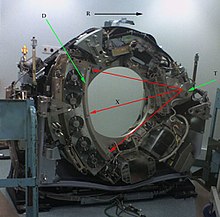
351:
Consequently, a fluoroscopic image (7) is recorded on the screen as a one-dimensional image (one image is recorded for every θ corresponding to all s values). When the angle between the object and transmission beam is θ and if the intensity of transmission beam (4) having reached each "s" point on
271:
plane are the same plane. Henceforth, this virtual plane will be called “the datum plane”. In addition, a virtual circle centered at the abovementioned origin (6) is set on the datum plane (it will be called “the datum circle” henceforth). This datum circle (5) will be represents the orbit of the
243:
Numbers (1)–(7) shown in Fig. 3 (see the numbers within the parentheses) respectively indicate: (1) = an object; (2) = the parallel beam light source; (3) = the screen; (4) = transmission beam; (5) = the datum circle (a datum feature); (6) = the origin (a datum feature); and (7) = a fluoroscopic
200:
equipment; by rotating the fluoroscope around the patient, a geometry similar to CT can be obtained, and by treating the 2D X-ray detector in a manner similar to a CT detector with a massive number of rows, it is possible to reconstruct a 3D volume from a single rotation using suitable software.
307:
X-ray source (2) to the screen (3) matches the positive direction of the t-axis while the s-axis parallels these two features. Henceforth, the angle between the x- and the s-axes will be indicated as θ. That is, parallel beam irradiation optical system where the angle between the object and the
287:
The parallel beam irradiation optical system is the key component of a CT scanner. It consists of a parallel beam X-ray source (2) and the screen (3). They are positioned so that they face each other in parallel with the origin (6) in between, both being in contact with the datum circle (6).
343:
During the above-mentioned motion (that is pivoting around the object(1)) of parallel beam irradiation optical system, the collimated X-ray source (2) emits transmission beam (4) which are effectively “parallel rays” in a geometrical optical sense. The traveling direction of each ray of the
1186:
1965:
1420:
plane rotates counter clockwise. around the point of origin in the plane in such a way “to keep mutual positional relationship between the light source (2) and screen (7) passing through the trajectory (5).” Rotation angle of this case is same as above-mentioned θ.
347:
Optical transmission can be presumed to occur ideally. That is, transmission beam penetrates without diffraction, diffusion, or reflection although it is absorbed by the object and its attenuation is assumed to occur in accordance with the Beer-Lambert law.
2225:
A helical CT beam trajectory is characterized by its pitch, which is equal to the table feed distance along the scan range over one gantry rotation divided by the section collimation. When pitch is greater than 1, the radiation dose for a given axial
785:
178:(EBT), used electromagnetic deflection of an electron beam within a very large conical X-ray tube and a stationary array of detectors to achieve very high temporal resolution, for imaging of rapidly moving structures, for example the
374:. It is also computationally undemanding, with modern scanners requiring only a few milliseconds per image. However, this is not the only technique available: the original EMI scanner solved the tomographic reconstruction problem by
1503:
2333:
machines. A subsequent development of helical CT was multi-slice (or multi-detector) CT; instead of a single row of detectors, multiple rows of detectors are used effectively capturing multiple cross-sections simultaneously.
158:, which produces a series of cross-sectional images. In terms of mathematics, the raw data acquired by the scanner consists of multiple "projections" of the object being scanned. These projections are effectively the
1004:
2270:
remained static. The helical scan method reduces the X-ray dose to the patient required for a given resolution while scanning more quickly. This is, however, at the cost of greater mathematical complexity in the
344:
transmission beam (4) is parallel to the t-axis. The transmission beam (4), emitted by the X-ray source (2), penetrates the object and reaches the screen (3) after attenuation due to absorption by the object.
2196:
In order to illuminate multiple rows of detector elements in a multi-slice scanner, the x-ray source must emit a beam which is divergent along the axial direction (i.e. a cone beam instead of a fan beam).
335:. Setting an increment that is smaller than the slice thickness results in overlap between the slices. A beneficial effect of this is a smoother transition between images when scrolling through the stack.
2185:
16 multi-slice scanner was introduced in 2001 and in 2004, 64 multislice scanners are on the market. These can produce an image in less than a second and thus can obtain images of the heart and its
1835:
1009:
790:
537:
and suppose that the transmission beam penetrates without diffraction, diffusion, or reflection. Also assume the beam is absorbed by the object and its attenuation occurs in accordance with the
315:
coordination system. Hence, object (1) will not be moved while the parallel beam irradiation optical system are rotated around the object (1). The object (1) must be smaller than datum circle.
272:
parallel beam irradiation optical system. Naturally, the origin (6), the datum circle (5), and the datum coordinate systems are virtual features which are imagined for mathematical purposes.
2118:. In the above-mentioned descriptions, “What we measured” is p(s,θ) . On the other hand, “What we want to know ” is μ(x,y). So, the next will be "How to reconstruct μ(x,y) from p(s,θ)".
2090:
454:(1)Results of measurement, i.e. a series of images obtained by transmitted light are expressed (modeled) as a function p (s,θ) obtained by performing radon transform to μ(x, y), and
378:, but this approach was limited by its high computational complexity, especially given the computer technology available at the time. More recently, manufacturers have developed
1486:
974:{\displaystyle {\begin{aligned}I=I_{0}\exp \left({-\int \mu (x,y)\,dl}\right)=I_{0}\exp \left({-{\int }_{-\infty }^{\infty }\mu (l(t))\,|{\dot {l}}(t)|dt}\right)\end{aligned}}}
1731:
1689:
1366:
2023:
1820:
594:
414:
In this section, the basic principle of tomography in the case that especially uses tomography utilizing the parallel beam irradiation optical system will be explained.
66:
1785:
535:
1442:
1386:
754:
634:
2266:
trajectory relative to the object while a two-dimensional array of detectors measures the transmitted radiation on part of a cone of rays emanating from the source.
2547:
1418:
1327:
1295:
690:
500:
719:
559:
2466:
3251:
2935:
1263:
1243:
1223:
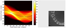
774:
654:
3065:
2509:
1631:{\displaystyle {l}_{\theta ,s}(t)=t{\begin{bmatrix}-\sin \theta \\\cos \theta \\\end{bmatrix}}+{\begin{bmatrix}s\cos \theta \\s\sin \theta \\\end{bmatrix}}}
3077:
3405:
2181:
Since its invention by
Kalender in the 1980s, helical scan CT machines have steadily increased the number of rows of detectors (slices) they deploy. The
1332:
By using a parallel beam irradiation optical system, one can experimentally obtain the series of fluoroscopic images (these are one-dimensional images
2677:
3513:
3043:
2205:
196:, due to the shape of the X-ray beam (strictly, the beam is pyramidal in shape, rather than conical). Cone-beam CT is commonly found in medical
1181:{\displaystyle {\begin{aligned}p_{l}=\ln(I/I_{0})=-\int \mu (x,y)\,dl=-{\int }_{-\infty }^{\infty }\mu (l(t))\,|{\dot {l}}(t)|dt\end{aligned}}}
600:
2230:(FOV) is decreased compared to conventional CT. At high pitches there is, however, a trade-off in terms of noise and longitudinal resolution.
3048:
2889:
118:
174:
and detector are physically rotated behind a circular shroud (see the image above right). An alternative, short lived design, known as
3601:
3441:
3320:
3060:
2969:
2899:
2493:
2437:
88:
2884:
2762:
308:
transmission beam equals θ. This datum circle (6) will be represents the orbit of the parallel beam irradiation optical system.
2166:
is credited with invention of the technique, and uses the term spiral CT. Kalender argues that the terms spiral and helical are
154:, yet it is not sufficient for interpretation. Once the scan data has been acquired, the data must be processed using a form of
2964:
2947:
3325:
3172:
3072:
3055:
2283:
1960:{\displaystyle p_{\theta }(s)=-{\int }_{-\infty }^{\infty }\mu (s\cos \theta -t\sin \theta ,s\sin \theta +t\cos \theta )\,dt}
70:
2453:
3451:
3305:
2904:
2779:
2771:
2670:
2328:
2239:
399:
192:
3562:
3496:
3428:
3418:
3396:
3167:
3135:
2766:
3226:
3194:
3177:
2638:
essential level except for some minor deformation of formula including reversing a part of positive or negative signs.
1329:
coordinate systems are chosen such that they are reflections of each other without mirror-reflective transformation.)
3300:
3035:
2775:
2271:
391:
361:
155:
59:
3541:
3209:
3199:
2038:
3580:
3531:
3310:
3189:
3162:
2923:
2708:
2663:
2612:
2134:
386:
370:
is one of the most established algorithmic techniques for this problem. It is conceptually simple, tunable and
367:
255:
are imagined in order to explain the positional relations and movements of features (0)–(7) in the figure. The
175:
162:
of the structure of the object. Reconstruction essentially involves solving the inverse Radon transformation.
3479:
3315:
3256:
3241:
3130:
2952:
2928:
371:
2525:
3536:
3350:
3219:
3150:
2957:
2289:
467:
276:
3214:
3145:
3011:
2894:
2813:
2720:
2553:
2512:. In Proceedings of the IEEE Conference on Computer Vision and Pattern Recognition 2013 (pp. 2195–2202).
1447:
295:
coordinate system while maintaining the relative positional relations between themselves and with the
291:
These two features ((2) and (3)) can rotate counterclockwise around the origin (6) together with the
3557:
3246:
3184:
3006:
2374:
2293:
2190:
1823:
159:
1700:
1658:
1335:
235:
explained. Configuration and motions of parallel beam irradiation optical system, referring Fig. 3.
3503:
3381:
3204:
2940:
2255:
2157:
352:
the screen is expressed as p(s, θ), it expresses a fluoroscopic image (7) corresponding to each θ.
1993:
1790:
564:
3491:
3340:
3335:
3236:
3119:
2974:
2725:
2364:
2313:
419:
383:
2608:
2114:“What we want to know (μ(x,y))” can be reconstructed from “What we measured ( p(s,θ))” by using
263:
coordinate systems share the origin (6) and they are positioned on the same plane. That is, the
1740:
505:
3330:
3295:
3290:
3231:
2979:
2828:
2489:
2479:
2433:
2427:
2408:
2390:
179:
1427:
1371:
730:
610:
3268:
3157:
3016:
2788:
2740:
2398:
2382:
2353:"Alignment Solution for CT Image Reconstruction using Fixed Point and Virtual Rotation Axis"
604:
538:
304:
1391:
1300:
1268:
668:
473:
299:
coordinate system (so, these two features ((2) and (3)) are always opposed each other). The
3366:
2818:
2784:
2757:
2752:
2686:
2557:
2115:
695:
544:
323:
The distance the table moves for every 360° rotation of the X-ray generator is called the
215:
108:
2650:
2378:
3508:
2543:
2521:
2403:
2352:
2163:
1248:
1228:
1208:
759:
639:
375:
112:
462:(1) The Results of measurement of p(s,θ) in a parallel beam irradiation optical system
279:
of the object (3) at each (x,y), p(s,θ) (7) is the collection of fluoroscopic images.
3595:
3140:
3109:
3097:
3092:
2850:
2745:
2585:
2297:
223:
457:(2)μ(x, y) is restored by performing inverse radon transform to measurement results.
3523:
3376:
3371:
3278:
3102:
3087:
3021:
2218:
2110:(2)μ(x, y) is restored by performing inverse radon transform to measurement results
2140:
3446:
3001:
2833:
2823:
2803:
2793:
2735:
2730:
2699:
2301:
450:
in this section, the explanation is advanced according to the order as follows:
219:
210:
197:
48:
3082:
2845:
2840:
2808:
2309:
1205:
Here, the direction from the light source toward the screen is defined as the
663:
171:
2394:
3409:
3345:
2798:
2312:
instead of photomultipliers and modern scintillation materials (for example
2182:
379:
311:
On the other hand, the object (1) will be scanned by CT scanner is fixed to
2412:
2308:
gas. These systems were in turn replaced by scintillation systems based on
2573:
425:
130:
27:
3432:
2597:
115:
are positioned on the opposite side of the circle from the X-ray source.
17:
3461:
2864:
2167:
102:
73: in this section. Unsourced material may be challenged and removed.
2454:"CT scan parameters: Translation of terms for different manufacturers"
2386:
2173:
There are a class of image artifacts specific to helical acquisition.
3456:
2316:
2528:
2369:
2319:
or rare-earth oxide ceramics) with more desirable characteristics.
182:. Systems with a very large number of detector rows, such that the
31:
CT scanner with cover removed to show internal components. Legend:
3483:
2694:
2655:
2305:
2263:
2259:
2214:
2204:
2186:
424:
129:
117:
26:
1388:
between the object and the transmitted light beam. In Fig.3, the
2461:
1368:
of a specific cross section of a scanned object) for each angle
403:
2659:
756:
is the intensity of the light beam before transmission, while
395:
42:
2550:
150:
A visual representation of the raw data obtained is called a
2426:
Webb, W. Richard; Brant, Wiliam E.; Major, Nancy M. (2014).
331:
for axial scan modes. For helical scan modes, it is called
1787:
of (eq. 3) as the same manner of (eq.2). This means that,
541:. What we want to know then is the values of the function
2300:
crystals. Cesium iodide was replaced during the 1980s by
1245:
direction and parallel with the screen is defined as the
561:. What we can measure will be the values of the function
2244:
In cone-beam computed tomography (commonly abbreviated
2486:
Nuclear
Medicine and PET/CT: Technology and Techniques
1589:
1545:
2041:
1996:
1838:
1793:
1743:
1703:
1661:
1506:
1450:
1430:
1394:
1374:
1338:
1303:
1271:
1251:
1231:
1211:
1007:
788:
762:
733:
698:
671:
642:
613:
567:
547:
508:
476:
1984:
One can define following function of two variables (
3550:
3522:
3478:
3427:
3404:
3394:
3359:
3276:
3267:
3118:
3034:
2994:
2913:
2872:
2863:
2707:
2693:
2572:Barrett and Keat (2004) RadioGraphics 24:1679-1691
2133:
2128:
2586:https://dx.doi.org/10.1148/radiology.189.3.8234684
2484:Kristen M. Waterstram-Rich; David Gilmore (2016).
2084:
2017:
1959:
1814:
1779:
1725:
1683:
1630:
1480:
1436:
1412:
1380:
1360:
1321:
1289:
1257:
1237:
1217:
1180:
973:
768:
748:
713:
684:
648:
628:
588:
553:
529:
494:
398:algorithms or use of specialized hardware such as
283:Motion of parallel beam irradiation optical system
2252:Helical (or spiral) cone beam computed tomography
2234:Helical (or spiral) cone beam computed tomography
303:plane is positioned so that the direction from a
2651:Principles of Computerized Tomographic Imaging
2467:American Association of Physicists in Medicine
2671:
8:
2596:Maldjian and Goldman (2013) AJR 200:741-747
2508:Barkan, O; Weill, J; Averbuch, A; Dekel, S.
2584:Heiken et al. (1993) Radiology 189:647-656
2085:{\displaystyle p(s,\theta )=p_{\theta }(s)}
394:techniques, such as use of highly parallel
244:image (a one-dimensional image; p (s, θ)).
3401:
3283:
3273:
2869:
2713:
2704:
2678:
2664:
2656:
776:is the beam intensity after transmission.
466:Consider the mathematical model where the
2611:at the U.S. National Library of Medicine
2432:. Elsevier Health Sciences. p. 152.
2402:
2368:
2067:
2040:
1995:
1950:
1878:
1870:
1865:
1843:
1837:
1792:
1750:
1745:
1742:
1708:
1702:
1666:
1660:
1584:
1540:
1513:
1508:
1505:
1457:
1452:
1449:
1429:
1393:
1373:
1343:
1337:
1302:
1270:
1250:
1230:
1210:
1163:
1143:
1142:
1137:
1136:
1109:
1101:
1096:
1082:
1046:
1037:
1016:
1008:
1006:
951:
931:
930:
925:
924:
897:
889:
884:
879:
863:
844:
819:
803:
789:
787:
761:
740:
735:
732:
697:
676:
670:
641:
620:
615:
612:
566:
546:
507:
475:
89:Learn how and when to remove this message
3514:Orthogonal polarization spectral imaging
2510:"Adaptive Compressed Tomography Sensing"
2488:(8 ed.). Elsevier Health Sciences.
1225:direction and that perpendicular to the
146:) as used for planning every scan slice.
2627:
2574:https://dx.doi.org/10.1148/rg.246045065
2548:"Spiral or helical CT: right or wrong?"
2351:Jun, Kyungtaek; Yoon, Seokhwan (2017).
2343:
1822:of following (eq. 5) is a resultant of
2598:https://dx.doi.org/10.2214/AJR.12.9768
2209:The field of view (FOV) multiplied by
2177:Single-slice and multi-slice Spiral CT
2125:
2258:(CT) in which the source (usually of
7:
2633:
2631:
2526:"Technical foundations of spiral CT"
2031:
1828:
1496:
997:
778:
186:-axis coverage is comparable to the
71:adding citations to reliable sources
436:(s)). Two datum coordinate systems
126:(left) and an image sample (right).
3321:Sestamibi parathyroid scintigraphy
1879:
1874:
1481:{\displaystyle {l}_{\theta ,s}(t)}
1110:
1105:
898:
893:
230:Schematic configuration and motion
218:used for X-ray CT, as well as for
25:
2323:the X-ray ring. These are called
1733:is equal to the line integral of
3576:
3575:
339:Obtaining transmission image ‘s’
190:-axis coverage are often termed
170:In conventional CT machines, an
111:that rotates around the object;
47:
3078:Cholangiopancreatography (MRCP)
2254:is a type of three-dimensional
2193:vessels) as if frozen in time.
502:is represented by the function
58:needs additional citations for
3326:Radioactive iodine uptake test
2284:History of computed tomography
2248:), the X-ray beam is conical.
2079:
2073:
2057:
2045:
2012:
2000:
1947:
1887:
1855:
1849:
1809:
1797:
1774:
1768:
1763:
1751:
1726:{\displaystyle p_{\theta }(s)}
1720:
1714:
1684:{\displaystyle p_{\theta }(s)}
1678:
1672:
1531:
1525:
1475:
1469:
1407:
1395:
1361:{\displaystyle p_{\theta }(s)}
1355:
1349:
1316:
1304:
1284:
1272:
1164:
1160:
1154:
1138:
1133:
1130:
1124:
1118:
1079:
1067:
1052:
1031:
952:
948:
942:
926:
921:
918:
912:
906:
841:
829:
708:
702:
583:
571:
524:
512:
489:
477:
1:
3306:Radionuclide ventriculography
2780:Lower gastrointestinal series
2772:Upper gastrointestinal series
2240:Cone beam computed tomography
247:Two datum coordinate systems
3497:Optical coherence tomography
3419:Myocardial perfusion imaging
3007:Dental panoramic radiography
2116:inverse radon transformation
2018:{\displaystyle p(s,\theta )}
1815:{\displaystyle p(s,\theta )}
589:{\displaystyle p(s,\theta )}
470:of the object at each point
2272:reconstruction of the image
2154:helical computed tomography
1986:
1693:
1490:
1488:, represented by equation (
1444:is the collection of lines
723:
658:
406:, now allow practical use.
3618:
3301:Ventilation/perfusion scan
2776:Small-bowel follow-through
2288:The earliest sensors were
2281:
2237:
2150:Spiral computed tomography
2129:Spiral computed tomography
692:along the light beam path
392:high-performance computing
362:Tomographic reconstruction
359:
356:Tomographic reconstruction
208:
156:tomographic reconstruction
3602:X-ray computed tomography
3571:
3542:Dynamic angiothermography
3286:
3210:Abdominal ultrasonography
2716:
2304:containing high-pressure
1780:{\displaystyle {l}_{}(t)}
1424:The beam having an angle
530:{\displaystyle \mu (x,y)}
103:X-ray computed tomography
3532:Non-contact thermography
3311:Radionuclide angiography
3163:Doppler echocardiography
2613:Medical Subject Headings
2170:and equally acceptable.
1691:is defined by equation (
387:expectation maximization
368:filtered back projection
176:electron beam tomography
3316:Radioisotope renography
2429:Fundamentals of Body CT
2296:excited by (typically)
2290:scintillation detectors
2274:from the measurements.
1437:{\displaystyle \theta }
1381:{\displaystyle \theta }
749:{\displaystyle {I}_{0}}
629:{\displaystyle {I}_{0}}
607:, the relation between
3351:Gastric emptying study
2529:Semin Ultrasound CT MR
2222:
2086:
2019:
1961:
1816:
1781:
1727:
1685:
1632:
1482:
1438:
1414:
1382:
1362:
1323:
1291:
1259:
1239:
1219:
1182:
975:
770:
750:
721:is given by equation (
715:
686:
656:is given by equation (
650:
630:
590:
555:
531:
496:
468:absorption coefficient
446:
277:absorption coefficient
147:
127:
40:
3012:X-ray motion analysis
2895:X-ray microtomography
2814:Hysterosalpingography
2721:Pneumoencephalography
2294:photomultiplier tubes
2282:Further information:
2238:Further information:
2208:
2087:
2025:is the collection of
2020:
1962:
1817:
1782:
1728:
1686:
1633:
1483:
1439:
1415:
1413:{\displaystyle (x,y)}
1383:
1363:
1324:
1322:{\displaystyle (x,y)}
1292:
1290:{\displaystyle (t,s)}
1265:direction. (Both the
1260:
1240:
1220:
1183:
976:
771:
751:
716:
687:
685:{\displaystyle p_{l}}
651:
631:
591:
556:
532:
497:
495:{\displaystyle (x,y)}
428:
382:physical model-based
360:Further information:
319:Increment/Table speed
133:
121:
30:
3537:Contact thermography
3247:Emergency ultrasound
3185:Transcranial Doppler
2936:Abdominal and pelvis
2609:Helical+Cone-Beam+CT
2213:creates a volume of
2039:
1994:
1990:). In this article,
1836:
1824:Radon transformation
1791:
1741:
1701:
1659:
1504:
1448:
1428:
1392:
1372:
1336:
1301:
1269:
1249:
1229:
1209:
1005:
786:
760:
731:
714:{\displaystyle l(t)}
696:
669:
640:
611:
565:
554:{\displaystyle \mu }
545:
506:
474:
160:Radon transformation
67:improve this article
3504:Confocal microscopy
3382:Indium-111 WBC scan
3205:Echoencephalography
2941:Virtual colonoscopy
2379:2017NatSR...741218J
2256:computed tomography
2158:computed tomography
2027:fluoroscopic images
1883:
1114:
902:
35:D: X-ray detectors
3492:Optical tomography
3341:Dacryoscintigraphy
3336:Immunoscintigraphy
2975:Whole body imaging
2726:Dental radiography
2556:2010-10-11 at the
2357:Scientific Reports
2223:
2082:
2015:
1957:
1864:
1812:
1777:
1723:
1681:
1628:
1622:
1575:
1478:
1434:
1410:
1378:
1358:
1319:
1287:
1255:
1235:
1215:
1178:
1176:
1095:
971:
969:
883:
766:
746:
711:
682:
646:
626:
586:
551:
527:
492:
447:
420:Crowther criterion
384:maximum likelihood
148:
128:
41:
39:R: Gantry rotation
3589:
3588:
3551:Target conditions
3474:
3473:
3470:
3469:
3390:
3389:
3331:Bone scintigraphy
3296:Scintimammography
3291:Cholescintigraphy
3136:contrast-enhanced
3030:
3029:
2990:
2989:
2980:Full-body CT scan
2880:General operation
2859:
2858:
2829:Angiocardiography
2387:10.1038/srep41218
2147:
2146:
2107:
2106:
1982:
1981:
1653:
1652:
1258:{\displaystyle s}
1238:{\displaystyle t}
1218:{\displaystyle t}
1203:
1202:
1151:
996:
995:
939:
769:{\displaystyle I}
649:{\displaystyle I}
366:The technique of
180:coronary arteries
99:
98:
91:
16:(Redirected from
3609:
3579:
3578:
3402:
3284:
3274:
3158:Echocardiography
3017:Hounsfield scale
2870:
2789:Cholecystography
2714:
2705:
2680:
2673:
2666:
2657:
2639:
2635:
2616:
2606:
2600:
2594:
2588:
2582:
2576:
2570:
2564:
2541:
2535:
2519:
2513:
2506:
2500:
2499:
2477:
2471:
2470:
2458:
2450:
2444:
2443:
2423:
2417:
2416:
2406:
2372:
2348:
2143:
2126:
2101:
2091:
2089:
2088:
2083:
2072:
2071:
2032:
2024:
2022:
2021:
2016:
1976:
1966:
1964:
1963:
1958:
1882:
1877:
1869:
1848:
1847:
1829:
1821:
1819:
1818:
1813:
1786:
1784:
1783:
1778:
1767:
1766:
1749:
1732:
1730:
1729:
1724:
1713:
1712:
1690:
1688:
1687:
1682:
1671:
1670:
1647:
1637:
1635:
1634:
1629:
1627:
1626:
1580:
1579:
1524:
1523:
1512:
1497:
1487:
1485:
1484:
1479:
1468:
1467:
1456:
1443:
1441:
1440:
1435:
1419:
1417:
1416:
1411:
1387:
1385:
1384:
1379:
1367:
1365:
1364:
1359:
1348:
1347:
1328:
1326:
1325:
1320:
1296:
1294:
1293:
1288:
1264:
1262:
1261:
1256:
1244:
1242:
1241:
1236:
1224:
1222:
1221:
1216:
1197:
1187:
1185:
1184:
1179:
1177:
1167:
1153:
1152:
1144:
1141:
1113:
1108:
1100:
1051:
1050:
1041:
1021:
1020:
998:
990:
980:
978:
977:
972:
970:
966:
962:
955:
941:
940:
932:
929:
901:
896:
888:
868:
867:
855:
851:
808:
807:
779:
775:
773:
772:
767:
755:
753:
752:
747:
745:
744:
739:
720:
718:
717:
712:
691:
689:
688:
683:
681:
680:
655:
653:
652:
647:
635:
633:
632:
627:
625:
624:
619:
605:Beer-Lambert law
603:conforms to the
595:
593:
592:
587:
560:
558:
557:
552:
539:Beer-Lambert law
536:
534:
533:
528:
501:
499:
498:
493:
220:plain film X-ray
216:Contrast mediums
134:Picture of a CT
94:
87:
83:
80:
74:
51:
43:
21:
3617:
3616:
3612:
3611:
3610:
3608:
3607:
3606:
3592:
3591:
3590:
3585:
3567:
3546:
3518:
3466:
3452:PET mammography
3423:
3386:
3372:Gallium-67 scan
3367:Octreotide scan
3355:
3263:
3114:
3026:
2986:
2909:
2890:High-resolution
2855:
2819:Skeletal survey
2785:Cholangiography
2698:
2689:
2687:Medical imaging
2684:
2647:
2642:
2636:
2629:
2625:
2620:
2619:
2607:
2603:
2595:
2591:
2583:
2579:
2571:
2567:
2558:Wayback Machine
2542:
2538:
2520:
2516:
2507:
2503:
2496:
2483:
2478:
2474:
2456:
2452:
2451:
2447:
2440:
2425:
2424:
2420:
2350:
2349:
2345:
2340:
2286:
2280:
2242:
2236:
2203:
2179:
2139:
2124:
2112:
2099:
2063:
2037:
2036:
1992:
1991:
1974:
1839:
1834:
1833:
1789:
1788:
1744:
1739:
1738:
1704:
1699:
1698:
1662:
1657:
1656:
1645:
1621:
1620:
1605:
1604:
1585:
1574:
1573:
1561:
1560:
1541:
1507:
1502:
1501:
1451:
1446:
1445:
1426:
1425:
1390:
1389:
1370:
1369:
1339:
1334:
1333:
1299:
1298:
1267:
1266:
1247:
1246:
1227:
1226:
1207:
1206:
1195:
1175:
1174:
1042:
1012:
1003:
1002:
988:
968:
967:
875:
859:
815:
799:
784:
783:
758:
757:
734:
729:
728:
694:
693:
672:
667:
666:
638:
637:
614:
609:
608:
563:
562:
543:
542:
504:
503:
472:
471:
464:
435:
412:
410:Basic principle
364:
358:
341:
321:
285:
241:
232:
213:
207:
168:
113:X-ray detectors
109:X-ray generator
95:
84:
78:
75:
64:
52:
38:
36:
34:
32:
23:
22:
15:
12:
11:
5:
3615:
3613:
3605:
3604:
3594:
3593:
3587:
3586:
3584:
3583:
3572:
3569:
3568:
3566:
3565:
3560:
3554:
3552:
3548:
3547:
3545:
3544:
3539:
3534:
3528:
3526:
3520:
3519:
3517:
3516:
3511:
3509:Endomicroscopy
3506:
3501:
3500:
3499:
3488:
3486:
3476:
3475:
3472:
3471:
3468:
3467:
3465:
3464:
3459:
3454:
3449:
3444:
3438:
3436:
3425:
3424:
3422:
3421:
3415:
3413:
3399:
3392:
3391:
3388:
3387:
3385:
3384:
3379:
3374:
3369:
3363:
3361:
3357:
3356:
3354:
3353:
3348:
3343:
3338:
3333:
3328:
3323:
3318:
3313:
3308:
3303:
3298:
3293:
3287:
3281:
3271:
3265:
3264:
3262:
3261:
3260:
3259:
3254:
3244:
3239:
3234:
3229:
3224:
3223:
3222:
3217:
3207:
3202:
3197:
3192:
3187:
3182:
3181:
3180:
3175:
3170:
3165:
3155:
3154:
3153:
3148:
3143:
3138:
3133:
3124:
3122:
3116:
3115:
3113:
3112:
3107:
3106:
3105:
3100:
3095:
3085:
3080:
3075:
3070:
3069:
3068:
3058:
3053:
3052:
3051:
3040:
3038:
3032:
3031:
3028:
3027:
3025:
3024:
3019:
3014:
3009:
3004:
2998:
2996:
2992:
2991:
2988:
2987:
2985:
2984:
2983:
2982:
2972:
2967:
2962:
2961:
2960:
2955:
2945:
2944:
2943:
2933:
2932:
2931:
2926:
2917:
2915:
2911:
2910:
2908:
2907:
2902:
2897:
2892:
2887:
2882:
2876:
2874:
2867:
2861:
2860:
2857:
2856:
2854:
2853:
2848:
2843:
2838:
2837:
2836:
2831:
2821:
2816:
2811:
2806:
2801:
2796:
2791:
2782:
2769:
2760:
2755:
2750:
2749:
2748:
2738:
2733:
2728:
2723:
2717:
2711:
2702:
2691:
2690:
2685:
2683:
2682:
2675:
2668:
2660:
2654:
2653:
2646:
2645:External links
2643:
2641:
2640:
2626:
2624:
2621:
2618:
2617:
2601:
2589:
2577:
2565:
2536:
2514:
2501:
2494:
2472:
2445:
2438:
2418:
2342:
2341:
2339:
2336:
2279:
2276:
2262:) describes a
2235:
2232:
2202:
2199:
2178:
2175:
2164:Willi Kalender
2145:
2144:
2137:
2131:
2130:
2123:
2120:
2111:
2108:
2105:
2104:
2095:
2093:
2081:
2078:
2075:
2070:
2066:
2062:
2059:
2056:
2053:
2050:
2047:
2044:
2014:
2011:
2008:
2005:
2002:
1999:
1980:
1979:
1970:
1968:
1956:
1953:
1949:
1946:
1943:
1940:
1937:
1934:
1931:
1928:
1925:
1922:
1919:
1916:
1913:
1910:
1907:
1904:
1901:
1898:
1895:
1892:
1889:
1886:
1881:
1876:
1873:
1868:
1863:
1860:
1857:
1854:
1851:
1846:
1842:
1811:
1808:
1805:
1802:
1799:
1796:
1776:
1773:
1770:
1765:
1762:
1759:
1756:
1753:
1748:
1722:
1719:
1716:
1711:
1707:
1680:
1677:
1674:
1669:
1665:
1651:
1650:
1641:
1639:
1625:
1619:
1616:
1613:
1610:
1607:
1606:
1603:
1600:
1597:
1594:
1591:
1590:
1588:
1583:
1578:
1572:
1569:
1566:
1563:
1562:
1559:
1556:
1553:
1550:
1547:
1546:
1544:
1539:
1536:
1533:
1530:
1527:
1522:
1519:
1516:
1511:
1477:
1474:
1471:
1466:
1463:
1460:
1455:
1433:
1409:
1406:
1403:
1400:
1397:
1377:
1357:
1354:
1351:
1346:
1342:
1318:
1315:
1312:
1309:
1306:
1286:
1283:
1280:
1277:
1274:
1254:
1234:
1214:
1201:
1200:
1191:
1189:
1173:
1170:
1166:
1162:
1159:
1156:
1150:
1147:
1140:
1135:
1132:
1129:
1126:
1123:
1120:
1117:
1112:
1107:
1104:
1099:
1094:
1091:
1088:
1085:
1081:
1078:
1075:
1072:
1069:
1066:
1063:
1060:
1057:
1054:
1049:
1045:
1040:
1036:
1033:
1030:
1027:
1024:
1019:
1015:
1011:
1010:
994:
993:
984:
982:
965:
961:
958:
954:
950:
947:
944:
938:
935:
928:
923:
920:
917:
914:
911:
908:
905:
900:
895:
892:
887:
882:
878:
874:
871:
866:
862:
858:
854:
850:
847:
843:
840:
837:
834:
831:
828:
825:
822:
818:
814:
811:
806:
802:
798:
795:
792:
791:
765:
743:
738:
710:
707:
704:
701:
679:
675:
645:
623:
618:
585:
582:
579:
576:
573:
570:
550:
526:
523:
520:
517:
514:
511:
491:
488:
485:
482:
479:
463:
460:
459:
458:
455:
433:
411:
408:
376:linear algebra
357:
354:
340:
337:
320:
317:
284:
281:
275:The μ(x,y) is
267:plane and the
240:
237:
231:
228:
224:radiocontrasts
209:Main article:
206:
205:Contrast media
203:
167:
164:
97:
96:
79:September 2016
55:
53:
46:
37:X: X-ray beam
33:T: X-ray tube
24:
14:
13:
10:
9:
6:
4:
3:
2:
3614:
3603:
3600:
3599:
3597:
3582:
3574:
3573:
3570:
3564:
3561:
3559:
3556:
3555:
3553:
3549:
3543:
3540:
3538:
3535:
3533:
3530:
3529:
3527:
3525:
3521:
3515:
3512:
3510:
3507:
3505:
3502:
3498:
3495:
3494:
3493:
3490:
3489:
3487:
3485:
3481:
3477:
3463:
3460:
3458:
3455:
3453:
3450:
3448:
3445:
3443:
3440:
3439:
3437:
3434:
3430:
3426:
3420:
3417:
3416:
3414:
3411:
3407:
3403:
3400:
3398:
3393:
3383:
3380:
3378:
3377:Ga-68-DOTATOC
3375:
3373:
3370:
3368:
3365:
3364:
3362:
3358:
3352:
3349:
3347:
3344:
3342:
3339:
3337:
3334:
3332:
3329:
3327:
3324:
3322:
3319:
3317:
3314:
3312:
3309:
3307:
3304:
3302:
3299:
3297:
3294:
3292:
3289:
3288:
3285:
3282:
3280:
3275:
3272:
3270:
3266:
3258:
3255:
3253:
3250:
3249:
3248:
3245:
3243:
3240:
3238:
3235:
3233:
3230:
3228:
3225:
3221:
3218:
3216:
3213:
3212:
3211:
3208:
3206:
3203:
3201:
3198:
3196:
3193:
3191:
3190:Intravascular
3188:
3186:
3183:
3179:
3176:
3174:
3171:
3169:
3166:
3164:
3161:
3160:
3159:
3156:
3152:
3149:
3147:
3144:
3142:
3139:
3137:
3134:
3132:
3129:
3128:
3126:
3125:
3123:
3121:
3117:
3111:
3110:Synthetic MRI
3108:
3104:
3101:
3099:
3096:
3094:
3091:
3090:
3089:
3086:
3084:
3081:
3079:
3076:
3074:
3071:
3067:
3064:
3063:
3062:
3059:
3057:
3054:
3050:
3047:
3046:
3045:
3042:
3041:
3039:
3037:
3033:
3023:
3020:
3018:
3015:
3013:
3010:
3008:
3005:
3003:
3000:
2999:
2997:
2993:
2981:
2978:
2977:
2976:
2973:
2971:
2968:
2966:
2963:
2959:
2956:
2954:
2951:
2950:
2949:
2946:
2942:
2939:
2938:
2937:
2934:
2930:
2927:
2925:
2922:
2921:
2919:
2918:
2916:
2912:
2906:
2903:
2901:
2900:Electron beam
2898:
2896:
2893:
2891:
2888:
2886:
2883:
2881:
2878:
2877:
2875:
2871:
2868:
2866:
2862:
2852:
2851:Orbital x-ray
2849:
2847:
2844:
2842:
2839:
2835:
2832:
2830:
2827:
2826:
2825:
2822:
2820:
2817:
2815:
2812:
2810:
2807:
2805:
2802:
2800:
2797:
2795:
2792:
2790:
2786:
2783:
2781:
2777:
2773:
2770:
2768:
2764:
2761:
2759:
2756:
2754:
2751:
2747:
2746:Bronchography
2744:
2743:
2742:
2739:
2737:
2734:
2732:
2729:
2727:
2724:
2722:
2719:
2718:
2715:
2712:
2710:
2706:
2703:
2701:
2696:
2692:
2688:
2681:
2676:
2674:
2669:
2667:
2662:
2661:
2658:
2652:
2649:
2648:
2644:
2634:
2632:
2628:
2622:
2614:
2610:
2605:
2602:
2599:
2593:
2590:
2587:
2581:
2578:
2575:
2569:
2566:
2562:
2559:
2555:
2552:
2549:
2545:
2540:
2537:
2533:
2530:
2527:
2523:
2518:
2515:
2511:
2505:
2502:
2497:
2495:9780323400350
2491:
2487:
2481:
2476:
2473:
2469:. 2011-08-11.
2468:
2464:
2463:
2455:
2449:
2446:
2441:
2439:9780323263580
2435:
2431:
2430:
2422:
2419:
2414:
2410:
2405:
2400:
2396:
2392:
2388:
2384:
2380:
2376:
2371:
2366:
2362:
2358:
2354:
2347:
2344:
2337:
2335:
2332:
2331:
2326:
2320:
2318:
2315:
2311:
2307:
2303:
2299:
2298:cesium iodide
2295:
2291:
2285:
2277:
2275:
2273:
2267:
2265:
2261:
2257:
2253:
2249:
2247:
2241:
2233:
2231:
2229:
2228:field of view
2220:
2216:
2212:
2207:
2200:
2198:
2194:
2192:
2188:
2184:
2176:
2174:
2171:
2169:
2165:
2161:
2159:
2155:
2151:
2142:
2138:
2136:
2132:
2127:
2121:
2119:
2117:
2109:
2103:
2096:
2094:
2092:
2076:
2068:
2064:
2060:
2054:
2051:
2048:
2042:
2034:
2033:
2030:
2028:
2009:
2006:
2003:
1997:
1989:
1988:
1978:
1971:
1969:
1967:
1954:
1951:
1944:
1941:
1938:
1935:
1932:
1929:
1926:
1923:
1920:
1917:
1914:
1911:
1908:
1905:
1902:
1899:
1896:
1893:
1890:
1884:
1871:
1866:
1861:
1858:
1852:
1844:
1840:
1831:
1830:
1827:
1825:
1806:
1803:
1800:
1794:
1771:
1760:
1757:
1754:
1746:
1736:
1717:
1709:
1705:
1696:
1695:
1675:
1667:
1663:
1655:The function
1649:
1642:
1640:
1638:
1623:
1617:
1614:
1611:
1608:
1601:
1598:
1595:
1592:
1586:
1581:
1576:
1570:
1567:
1564:
1557:
1554:
1551:
1548:
1542:
1537:
1534:
1528:
1520:
1517:
1514:
1509:
1499:
1498:
1495:
1493:
1492:
1472:
1464:
1461:
1458:
1453:
1431:
1422:
1404:
1401:
1398:
1375:
1352:
1344:
1340:
1330:
1313:
1310:
1307:
1281:
1278:
1275:
1252:
1232:
1212:
1199:
1192:
1190:
1188:
1171:
1168:
1157:
1148:
1145:
1127:
1121:
1115:
1102:
1097:
1092:
1089:
1086:
1083:
1076:
1073:
1070:
1064:
1061:
1058:
1055:
1047:
1043:
1038:
1034:
1028:
1025:
1022:
1017:
1013:
1000:
999:
992:
985:
983:
981:
963:
959:
956:
945:
936:
933:
915:
909:
903:
890:
885:
880:
876:
872:
869:
864:
860:
856:
852:
848:
845:
838:
835:
832:
826:
823:
820:
816:
812:
809:
804:
800:
796:
793:
781:
780:
777:
763:
741:
736:
726:
725:
705:
699:
677:
673:
665:
661:
660:
643:
621:
616:
606:
602:
597:
580:
577:
574:
568:
548:
540:
521:
518:
515:
509:
486:
483:
480:
469:
461:
456:
453:
452:
451:
443:
439:
431:
427:
423:
421:
415:
409:
407:
405:
401:
397:
393:
388:
385:
381:
377:
373:
372:deterministic
369:
363:
355:
353:
349:
345:
338:
336:
334:
330:
326:
318:
316:
314:
309:
306:
302:
298:
294:
289:
282:
280:
278:
273:
270:
266:
262:
258:
254:
250:
245:
238:
236:
229:
227:
225:
222:, are called
221:
217:
212:
204:
202:
199:
195:
194:
189:
185:
181:
177:
173:
165:
163:
161:
157:
153:
145:
141:
137:
132:
125:
120:
116:
114:
110:
106:
104:
93:
90:
82:
72:
68:
62:
61:
56:This section
54:
50:
45:
44:
29:
19:
3558:Acute stroke
3524:Thermography
3279:scintigraphy
3269:Radionuclide
3257:pre-hospital
3103:Tractography
3022:Radiodensity
2924:calcium scan
2885:Quantitative
2879:
2604:
2592:
2580:
2568:
2560:
2539:
2534:: (2) 81-89.
2531:
2517:
2504:
2485:
2475:
2460:
2448:
2428:
2421:
2360:
2356:
2346:
2329:
2324:
2321:
2302:ion chambers
2287:
2268:
2251:
2250:
2245:
2243:
2227:
2224:
2219:abdominal CT
2210:
2195:
2180:
2172:
2162:
2153:
2149:
2148:
2113:
2097:
2035:
2026:
1985:
1983:
1972:
1832:
1734:
1692:
1654:
1643:
1500:
1489:
1423:
1331:
1204:
1193:
1001:
986:
782:
722:
657:
598:
465:
448:
441:
437:
429:
416:
413:
365:
350:
346:
342:
332:
328:
324:
322:
312:
310:
300:
296:
292:
290:
286:
274:
268:
264:
260:
256:
252:
248:
246:
242:
233:
214:
193:cone beam CT
191:
187:
183:
169:
151:
149:
143:
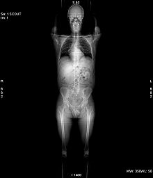
139:
135:
123:
107:by using an
101:
100:
85:
76:
65:Please help
60:verification
57:
3447:Cardiac PET
3220:renal tract
3195:Gynecologic
3127:Techniques
3098:restriction
3073:Angiography
3056:Neurography
3002:Fluoroscopy
2948:Angiography
2929:angiography
2873:Techniques:
2834:Aortography
2824:Angiography
2804:Cystography
2794:Mammography
2736:Myelography
2731:Sialography
2700:radiography
2544:Kalender WA
2522:Kalender WA
2310:photodiodes
1826:of μ(x,y).
601:attenuation
333:table speed
211:Contrast CT
198:fluoroscopy
3360:Full body:
3146:endoscopic
3120:Ultrasound
3049:functional
2846:Lymphogram
2841:Venography
2809:Arthrogram
2563:: (2) 583.
2370:1605.04833
2338:References
2314:rare-earth
2221:pictured).
2211:scan range
2168:synonymous
664:absorbance
662:) and the
329:table feed
305:collimated
239:Statements
172:X-ray tube
3563:Pregnancy
3442:Brain PET
3410:gamma ray
3346:DMSA scan
3200:Obstetric
3093:diffusion
3088:Sequences
3066:perfusion
2958:Pulmonary
2905:Cone beam
2799:Pyelogram
2551:Radiology
2395:2045-2322
2363:: 41218.
2330:spiral CT
2189:vessels (
2183:prototype
2122:Spiral CT
2069:θ
2055:θ
2010:θ
1945:θ
1942:
1930:θ
1927:
1915:θ
1912:
1903:−
1900:θ
1897:
1885:μ
1880:∞
1875:∞
1872:−
1867:∫
1862:−
1845:θ
1807:θ
1755:θ
1710:θ
1668:θ
1618:θ
1615:
1602:θ
1599:
1571:θ
1568:
1558:θ
1555:
1549:−
1515:θ
1494:) below.
1459:θ
1432:θ
1376:θ
1345:θ
1149:˙
1116:μ
1111:∞
1106:∞
1103:−
1098:∫
1093:−
1065:μ
1062:∫
1059:−
1029:
937:˙
904:μ
899:∞
894:∞
891:−
886:∫
881:−
873:
827:μ
824:∫
821:−
813:
599:When the
581:θ
549:μ
510:μ
380:iterative
325:increment
166:Structure
140:scanogram
18:Spiral CT
3596:Category
3581:Category
3433:positron
2953:Coronary
2554:Archived
2546:(1994).
2524:(1994).
2480:Page 310
2413:28120881
2191:coronary
1697:). That
727:). Here
152:sinogram
144:topogram
124:sinogram
105:operates
3480:Optical
3462:PET-MRI
3242:Carotid
3237:Scrotal
3131:doppler
3061:Cardiac
2970:Thyroid
2914:Targets
2865:CT scan
2404:5264594
2375:Bibcode
2325:helical
2292:, with
2278:History
2264:helical
2156:, is a
2141:D036542
3457:PET-CT
3232:Breast
3227:Rectal
3151:duplex
3083:Breast
2920:Heart
2615:(MeSH)
2492:
2436:
2411:
2401:
2393:
2317:garnet
2260:X-rays
2215:voxels
1737:along
1735:μ(x,y)
430:Fig. 3
3484:Laser
3406:SPECT
3215:renal
3044:Brain
2995:Other
2695:X-ray
2623:Notes
2457:(PDF)
2365:arXiv
2306:xenon
2201:Pitch
2187:blood
2152:, or
404:ASICs
400:FPGAs
136:scout
3252:FAST
2965:Head
2490:ISBN
2482:in:
2462:IAEA
2434:ISBN
2409:PMID
2391:ISSN
2246:CBCT
2135:MeSH
1297:and
636:and
440:and
259:and
251:and
3429:PET
3397:ECT
3395:3D/
3277:2D/
3178:ICE
3173:TEE
3168:TTE
3036:MRI
2767:DXR
2763:DXA
2758:KUB
2753:AXR
2741:CXR
2561:193
2399:PMC
2383:doi
2327:or
1939:cos
1924:sin
1909:sin
1894:cos
1612:sin
1596:cos
1565:cos
1552:sin
870:exp
810:exp
402:or
396:GPU
327:or
142:or
69:by
3598::
3435:):
3412:):
3141:3D
2709:2D
2630:^
2532:15
2465:.
2459:.
2407:.
2397:.
2389:.
2381:.
2373:.
2359:.
2355:.
2029:.
1026:ln
596:.
442:ts
438:xy
422:.
313:xy
301:ts
297:ts
293:ts
269:ts
265:xy
261:ts
257:xy
253:ts
249:xy
188:xy
122:A
3482:/
3431:(
3408:(
2787:/
2778:/
2774:/
2765:/
2697:/
2679:e
2672:t
2665:v
2498:.
2442:.
2415:.
2385::
2377::
2367::
2361:7
2217:(
2102:)
2100:5
2098:(
2080:)
2077:s
2074:(
2065:p
2061:=
2058:)
2052:,
2049:s
2046:(
2043:p
2013:)
2007:,
2004:s
2001:(
1998:p
1987:5
1977:)
1975:4
1973:(
1955:t
1952:d
1948:)
1936:t
1933:+
1921:s
1918:,
1906:t
1891:s
1888:(
1859:=
1856:)
1853:s
1850:(
1841:p
1810:)
1804:,
1801:s
1798:(
1795:p
1775:)
1772:t
1769:(
1764:]
1761:s
1758:,
1752:[
1747:l
1721:)
1718:s
1715:(
1706:p
1694:4
1679:)
1676:s
1673:(
1664:p
1648:)
1646:3
1644:(
1624:]
1609:s
1593:s
1587:[
1582:+
1577:]
1543:[
1538:t
1535:=
1532:)
1529:t
1526:(
1521:s
1518:,
1510:l
1491:3
1476:)
1473:t
1470:(
1465:s
1462:,
1454:l
1408:)
1405:y
1402:,
1399:x
1396:(
1356:)
1353:s
1350:(
1341:p
1317:)
1314:y
1311:,
1308:x
1305:(
1285:)
1282:s
1279:,
1276:t
1273:(
1253:s
1233:t
1213:t
1198:)
1196:2
1194:(
1172:t
1169:d
1165:|
1161:)
1158:t
1155:(
1146:l
1139:|
1134:)
1131:)
1128:t
1125:(
1122:l
1119:(
1090:=
1087:l
1084:d
1080:)
1077:y
1074:,
1071:x
1068:(
1056:=
1053:)
1048:0
1044:I
1039:/
1035:I
1032:(
1023:=
1018:l
1014:p
991:)
989:1
987:(
964:)
960:t
957:d
953:|
949:)
946:t
943:(
934:l
927:|
922:)
919:)
916:t
913:(
910:l
907:(
877:(
865:0
861:I
857:=
853:)
849:l
846:d
842:)
839:y
836:,
833:x
830:(
817:(
805:0
801:I
797:=
794:I
764:I
742:0
737:I
724:2
709:)
706:t
703:(
700:l
678:l
674:p
659:1
644:I
622:0
617:I
584:)
578:,
575:s
572:(
569:p
525:)
522:y
519:,
516:x
513:(
490:)
487:y
484:,
481:x
478:(
434:θ
184:z
138:(
92:)
86:(
81:)
77:(
63:.
20:)
Text is available under the Creative Commons Attribution-ShareAlike License. Additional terms may apply.