978:
across echocardiography labs in the United States. Accreditation is offered in adult and pediatric transthoracic and transesophageal echocardiography, as well as adult stress and fetal echo. Accreditation is a two-part process. Each facility will conduct a detailed self-evaluation, paying close attention to the IAC Standards and
Guidelines. The facility will then complete the application and submit actual case studies to the board of directors for review. Once all requirements have been met, the lab will receive certification. IAC certification is a continual process and must be maintained by the facility: it may include audits or site visits by the IAC. There are several states in which Medicare and/or private insurance carriers require accreditation (credentials) of the laboratory and/or sonographer for reimbursement of echocardiograms.
607:
treadmill or uses another exercise modality to increase the heart rate to his or her target heart rate, or 85% of the age-predicted maximum heart rate (220 − patient's age). Finally, images of the heart are taken "at stress" to assess wall motion at the peak heart rate. A stress echo assesses wall motion of the heart; it does not, however, create an image of the coronary arteries directly. Ischemia of one or more coronary arteries could cause a wall motion abnormality, which could indicate coronary artery disease. The gold standard test to directly create an image of the coronary arteries and directly assess for stenosis or occlusion is a cardiac catheterization. A stress echo is not invasive and is performed in the presence of a licensed medical professional, such as a cardiologist, and a cardiac sonographer.
851:
measurements. There are two forms, pulse and continuous. Pulsed allows velocities to be calculated in a specific place, but has a limited velocity range is can be used. Continuous wave allows the velocity to be measured from zero to the fastest blood velocities a diseased heart can generate. However, it can not tell you where in the A-scan the high velocity is coming from. Continuous wave would be used to calculate aortic stenosis because you know the high velocity is coming from the stenosis region. Pulsed would be used to find a ventricular septal defect where there should be no velocity across the septum and the pulsed tells you the location.
935:
through the cardiovascular system and return the ultrasound waves, creating a highly reflective image. There are multiple applications in which contrast-enhanced ultrasound can be useful. The most commonly used application is in the enhancement of LV endocardial borders for assessment of global and regional systolic function. Contrast may also be used to enhance visualization of wall thickening during stress echocardiography, for the assessment of LV thrombus, or for the assessment of other masses in the heart. Contrast echocardiography has also been used to assess blood perfusion throughout myocardium in the case of coronary artery disease.
379:
159:
728:
3796:
2999:
328:
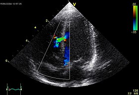
flow through the heart. Color
Doppler, as well as spectral Doppler, is used to visualize any abnormal communications between the left and right sides of the heart, any leaking of blood through the valves (valvular regurgitation), and estimate how well the valves open (or do not open in the case of valvular stenosis). The Doppler technique can also be used for tissue motion and velocity measurement, by
283:. It is one of the most widely used diagnostic imaging modalities in cardiology. It can provide a wealth of helpful information, including the size and shape of the heart (internal chamber size quantification), pumping capacity, location and extent of any tissue damage, and assessment of valves. An echocardiogram can also give physicians other estimates of heart function, such as a calculation of the
764:
53:
688:
468:
560:
1011:(ASE) is a professional organization made up of physicians, sonographers, nurses, and scientists involved in the field of echocardiography. One of the most important roles that the ASE plays is providing their recommendations through the ASE Guidelines and Standards, providing resource and educational opportunities for sonographers and physicians in the field.
662:
387:
906:
997:). Accreditation is granted through the American National Standards Institute (ANSI). Recognition of ARDMS programs in providing credentials has also earned the ARDMS accreditation with the National Commission for Certifying Agencies (NCCA). The NCCA is the accrediting arm of the National Organization for Competency Assurance (NOCA).
395:
echocardiography studies. For example, the differentiation of mild, moderate, and severe valvular disease is based upon measured criteria. Another example is the estimation of heart function by the left ventricular ejection fraction (LVEF) has vast uses including classification of heart failure and cut offs for implantation of
944:
same images. It necessitated the development of accreditation programs around the world. The aim of such programs is to standardize the practice of echocardiography and to ensure that practitioners have the proper training prior to practicing echocardiography which will eventually limit inter-observer variability.
584:
to further refine the perspective of the heart. Additionally, the ultrasound crystal is often a two-dimension crystal and the ultrasound plane being used can be rotated electronically to permit an additional dimension to optimize views of the heart structures. Often, movement in all of these dimensions is needed.
850:
A-scan or one dimensional ultrasound represents over half the standard ECHO exam. For example, it is how aortic stenosis valve area (or any obstruction). It is also how pressures are calculated in the heart such as right ventricle systolic pressure (RVSP). It is usually used in the form of
Doppler
641:
ICE is often inserted through the femoral vein and into the right atrium. From the right atrium, visualization of the interatrial septum, all four cardiac chambers, all four valves, and the pericardial space (for an effusion) can be readily visualized. It can also be advanced across the atrial septum
583:
TEE, unlike TTE, does not have discrete "windows" to view the heart. The entire esophagus and stomach can be utilized, and the probe advanced or removed along this dimension to alter the perspective on the heart. Most probes include the ability to deflect the tip of the probe in one or two dimensions
453:
TTE utilizes one- ("M mode"), two-, and three-dimensional ultrasound (time is implicit and not included) from the different windows. These can be combined with pulse wave or continuous wave
Doppler to visualize the velocity of blood flow and structure movements. Images can be enhanced with "contrast"
977:
The "Intersocietal
Accreditation Commission for Echocardiography" (IAC) sets standards for echo labs across the US. Cardiologists and sonographers who wish to have their laboratory accredited by IAC must comply with these standards. The purpose of accreditation is to maintain quality and consistency
943:
Echocardiography can at many times be subjective, meaning that the person reading the echo may have personal input that affects the interpretation of the findings, leading to so-called "inter-observer variability", where different echocardiographers might produce different reports when examining the
917:
echocardiography when the picture is moving) is possible using a matrix array ultrasound probe and an appropriate processing system. It enables detailed anatomical assessment of cardiac pathology, particularly valvular defects, and cardiomyopathies. The ability to slice the virtual heart in infinite
449:
TTE utilizes several "windows" to image the heart from different perspectives. Each window has advantages and disadvantages for viewing specific structures within the heart and, typically, numerous windows are utilized within the same study to fully assess the heart. Parasternal long and parasternal
1467:
Douglas, P. S.; Garcia, M. J.; Haines, D. E.; Lai, W. W.; Manning, W. J.; Patel, A. R.; Picard, M. H.; Polk, D. M.; Ragosta, M.; Ward, R. P.; Douglas, R. B.; Weiner, R. B.; Society for
Cardiovascular Angiography Interventions; Society of Critical Care Medicine; American Society of Echocardiography;
1000:
Under both credentialing bodies, sonographers must first document completion of prerequisite requirements, which contain both didactic and hands-on experience in the field of ultrasound. Applicants must then take a comprehensive exam demonstrating knowledge in both the physics of ultrasound and the
669:
Intravascular ultrasound (IVUS) is a specialized form of echocardiography that uses a catheter to insert the ultrasound probe inside blood vessels. This is commonly used to measure the size of blood vessels and to measure the internal diameter of the blood vessel. For example, this can be used in a
637:
ICE has the benefit over transthoracic echocardiography in that an operator who is performing a sterile procedure can also operate the ICE catheter and it is not limited to visibility problems that can arise with transthoracic or transesophageal echo. Though, there are image quality limitations due
606:
A stress echocardiogram, also known as a stress echo, uses ultrasound imaging of the heart to assess the wall motion in response to physical stress. First, images of the heart are taken "at rest" to acquire a baseline of the patient's wall motion at a resting heart rate. The patient then walks on a
426:
There are three primary types of echocardiography: transthoracic, transesophageal, and intracardic. Stress testing utilizes tranthoracic echo in combination with an exercise modality (e.g., a treadmill). Intravascular ultrasound is included below, but is as the name indicates more "ultrasound" than
327:
Not only can an echocardiogram create ultrasound images of heart structures, but it can also produce accurate assessment of the blood flowing through the heart by
Doppler echocardiography, using pulsed- or continuous-wave Doppler ultrasound. This allows assessment of both normal and abnormal blood
934:
Contrast echocardiography or contrast-enhanced ultrasound is the addition of an ultrasound contrast medium, or imaging agent, to traditional ultrasonography. The ultrasound contrast is made up of tiny microbubbles filled with a gas core and protein shell. This allows the microbubbles to circulate
402:
Health societies do not recommend routine testing when the patient has no change in clinical status or when a physician is unlikely to change care for the patient based on the results of testing. A common example of overuse of echocardiography when not indicated is the use of routine testing in
925:
Three-dimensional echocardiography technology may feature anatomical intelligence, or the use of organ-modeling technology, to automatically identify anatomy based on generic models. All generic models refer to a dataset of anatomical information that uniquely adapts to variability in patient
394:
Health societies recommend the use of echocardiography for initial diagnosis when a change in the patient's clinical status occurs and when new data from an echocardiogram would result in the physician changing the patient's care. Diagnostic criteria for numerous cardiac diseases are based on
1911:
Gilliland, Yvonne E.; Lavie, Carl J.; Ahmad, Homaa; Bernal, Jose A.; Cash, Michael E.; Dinshaw, Homeyar; Milani, Richard V.; Shah, Sangeeta; Bienvenu, Lisa (12 January 2016). "Development and
Implementation of a Quality Improvement Process for Echocardiographic Laboratory Accreditation".
926:
anatomy to perform specific tasks. Built on feature recognition and segmentation algorithms, this technology can provide patient-specific three-dimensional modeling of the heart and other aspects of the anatomy, including the brain, lungs, liver, kidneys, rib cage, and vertebral column.
579:
via the mouth, allowing image and
Doppler evaluation from a location directly behind the heart. It is most often used when transthoracic images are suboptimal and when a clearer and more precise image is needed for assessment. This test is performed in the presence of a cardiologist,
1875:
Nagueh, Sherif F.; Farrell, Mary B.; Bremer, Merri L.; Dunsiger, Shira I.; Gorman, Beverly L.; Tilkemeier, Peter L. (September 2015). "Predictors of
Delayed Accreditation of Echocardiography Laboratories: An Analysis of the Intersocietal Accreditation Commission Database".
918:
planes in an anatomically appropriate manner and to reconstruct three-dimensional images of anatomic structures make it unique for the understanding of the congenitally malformed heart. Real-time three-dimensional echocardiography can be used to guide the location of
441:
A standard echocardiogram is also known as a transthoracic echocardiogram (TTE) or cardiac ultrasound, and it is used for rapid evaluation of a patient at their bedside. In this case, the echocardiography transducer (or probe) is placed on the chest wall (or
621:
Intracardiac echocardiography (ICE) is specialized form of echocardiography that uses catheters to insert the ultrasound probe inside the heart to view structures from within the heart. ICE is often used as a part of the cardiac procedure of crossing the
354:
had developed to detect defects in metal castings. In fact, Edler in 1953 produced the first echocardiographs using an industrial Firestone-Sperry Ultrasonic Reflectoscope. In developing echocardiography, Edler worked with the physicist
952:
At the European level individual and laboratory accreditation is provided by the European Association of Echocardiography (EAE). There are three subspecialties for individual accreditation: Adult Transthoracic Echocardiography
1468:
American Society of Nuclear Cardiology; Heart Failure Society of America; Society for Cardiovascular Magnetic Resonance; Society of Cardiovascular Computed Tomography; American Heart Association; Heart Rhythm Society (2011).
450:
short axis windows are taken next to the sternum, the apical two/three/four chamber windows are taken from the apex of the heart (lower left side), and the subcostal window is taken from underneath the edge of the last rib.
1001:
clinical competency related to their specialty. Credentialed sonographers are then required to maintain competency in their field by obtaining a certain number of Continuing Medical Education credits, or CME's.
591:
surgery the TEE can be used to assess the valve function immediately before repair/replacement and immediately after. This permits revising the valve mid-surgery, if needed, to improve outcomes of the surgery.
407:. In this case, patients are often asymptomatic for years before the onset of deterioration and the results of the echocardiogram would not result in a change in care without other change in clinical status.
1754:
Bharucha, Tara; Roman, Kevin S.; Anderson, Robert H.; Vettukattil, Joseph J. (2008). "Impact of Multiplanar Review of Three-Dimensional Echocardiographic Data on Management of Congenital Heart Disease".
1558:
Kleiman, Amanda M.; Potter, Jennifer F.; Bechtel, Allison J.; Forkin, Katherine T.; Dunn, Lauren K.; Collins, Stephen R.; Lyons, Genevieve; Nemergut, Edward C.; Huffmyer, Julie L. (1 March 2019).
580:
anesthesiologist, registered nurse, and ultrasound technologist. Conscious sedation and/or localized numbing medication may be used to make the patient more comfortable during the procedure.
674:
to assess the narrowing of the coronary artery. If the catheter is retraced in a controlled manner, then an internal map can be generated to see the contour of the vessel and its branches.
969:
In the UK, accreditation is regulated by the British Society of Echocardiography. Accredited radiographers, sonographers, or other professionals are required to pass a mandatory exam.
575:
A transesophageal echocardiogram is an alternative way to perform an echocardiogram. A specialized probe containing an ultrasound transducer at its tip is passed into the patient's
324:
The most important advantages of echocardiography are that it is not invasive (does not involve breaking the skin or entering body cavities) and has no known risks or side effects.
298:
Echocardiography is an important tool in assessing wall motion abnormality in patients with suspected cardiac disease. It is a tool which helps in reaching an early diagnosis of
867:
Motion mode is infrequently used in modern echocardiography. It has specific uses and has the benefit of very high temporal fidelity (e.g., measuring LV size at end diastole).
478:
1516:
626:
with a transseptal puncture to permit catheter access from the right atrium to the left atrium; alternative access to the left heart would be retrograde through the
321:, and dilated cardiomyopathy. The use of stress echocardiography may also help determine whether any chest pain or associated symptoms are related to heart disease.
446:) of the subject, and images are taken through the chest wall. This is a non-invasive, highly accurate, and quick assessment of the overall function of the heart.
2673:
2357:
3035:
2487:
889:. Strain rate imaging measures either regional systolic deformation (strain) or the rate of regional deformation (strain rate). The methods used are either
2499:
3744:
3070:
2827:
1840:
Behera, Sarina K.; Smith, Shea N.; Tacy, Theresa A. (September 2017). "Impact of Accreditation on Quality in Echocardiograms: A Quantitative Approach".
3166:
2099:
1964:
1335:"Echocardiography, a non-invasive method for the assessment of cardiac function and morphology in preclinical drug toxicology and safety pharmacology"
2935:
1560:"Generative retrieval results in positive academic emotions and long-term retention of cardiovascular anatomy using transthoracic echocardiography"
2465:
922:
during right ventricular endomyocardial biopsies, placement of catheter-delivered valvular devices, and in many other intraoperative assessments.
171:
and the red mark the time in the cardiac cycle when the image was captured. Colors are used to represent the velocity and direction of blood flow.
842:
The various modes describe how the ultrasound crystals are used to obtain information. These modes are common to all types of echocardiography.
182:
3564:
3154:
396:
3439:
2470:
1658:"Assessing aortic valve area in aortic stenosis by continuity equation: a novel approach using real-time three-dimensional echocardiography"
1008:
1203:
Cleve, Jayne; McCulloch, Marti L. (2018), Nihoyannopoulos, Petros; Kisslo, Joseph (eds.), "Conducting a Cardiac Ultrasound Examination",
2311:
350:, was the first of his profession to apply ultrasonic pulse echo imaging in diagnosing cardiac disease, which the acoustical physicist
3689:
3365:
3270:
2863:
2742:
2482:
2391:
1400:
1220:
3570:
2321:
894:
829:
811:
785:
715:
701:
136:
70:
1792:"Comparison of Fluoroscopic versus Real Time Three-Dimensional Transthoracic Echocardiographic Guidance of Endomyocardial Biopsies"
1615:
Spencer, Kirk T.; Kimura, Bruce J.; Korcarz, Claudia E.; Pellikka, Patricia A.; Rahko, Peter S.; Siegel, Robert J. (1 June 2013).
3823:
3028:
2306:
2301:
2184:
1534:
3577:
3384:
2386:
2369:
643:
539:
329:
117:
587:
TEE can be used as stand-alone procedures, or incorporated into catheter- or surgical-based procedures. For example, during a
3677:
3374:
2747:
2594:
2494:
2477:
1530:
1512:
789:
570:
511:
89:
74:
35:
3699:
2873:
2727:
2326:
2201:
2193:
2092:
1173:
335:
Echocardiography was also the first ultrasound subspecialty to use intravenous contrast. Echocardiography is performed by
3200:
881:
Strain rate imaging is an ultrasound method for imaging regional differences in contraction (dyssynergy) in for instance
3684:
3672:
3553:
2984:
2918:
2850:
2840:
2818:
2589:
2557:
2188:
518:
436:
96:
486:
279:
Echocardiography is routinely used in the diagnosis, management, and follow-up of patients with any suspected or known
3799:
3021:
2648:
2616:
2599:
616:
336:
318:
774:
378:
2722:
2457:
2197:
1041:
3324:
793:
778:
525:
497:
482:
103:
63:
3631:
3521:
3275:
3065:
2963:
2631:
2621:
1065:
164:
3818:
3710:
3694:
3161:
3144:
3002:
2953:
2732:
2611:
2584:
2345:
2130:
2085:
261:
189:
302:, showing regional wall motion abnormality. Also, it is important in treatment and follow-up in patients with
507:
85:
3706:
3648:
3548:
3513:
3359:
2901:
2737:
2678:
2663:
2552:
2374:
2350:
1015:
650:
414:, diagnosing patients with valvular heart disease and other congenital abnormalities. An emerging branch is
1962:
3752:
2958:
2772:
2641:
2572:
2379:
1183:
882:
415:
404:
367:
3783:
3587:
3508:
2636:
2567:
2433:
2316:
2235:
2142:
1126:
1095:
707:
299:
3757:
3314:
3127:
3122:
2979:
2668:
2606:
2428:
390:
Echocardiogram in the parasternal long-axis view, showing a measurement of the heart's left ventricle
3833:
3636:
3614:
3300:
3139:
3112:
2925:
2803:
2626:
2362:
1470:"ACCF/ASE/AHA/ASNC/HFSA/HRS/SCAI/SCCM/SCCT/SCMR 2011 Appropriate Use Criteria for Echocardiography"
1147:
914:
886:
876:
601:
356:
31:
1286:"Echo and heart failure: when do people need an echo, and when do they need natriuretic peptides?"
3828:
3619:
3532:
3528:
3474:
3469:
3410:
3331:
3239:
3188:
3102:
2913:
2762:
2757:
2658:
2541:
2396:
2147:
1945:
1597:
1362:
1141:
1004:
In 2009, New Mexico and Oregon became the first two states to require licensure of sonographers.
671:
623:
249:
3662:
3608:
3538:
3446:
3434:
3295:
3285:
3280:
3183:
3134:
3045:
2752:
2717:
2712:
2653:
2401:
2250:
2031:
2013:
1937:
1929:
1893:
1857:
1813:
1772:
1736:
1687:
1638:
1589:
1581:
1491:
1446:
1396:
1388:
1354:
1315:
1266:
1258:
1216:
1178:
1120:
1056:
1032:
588:
532:
307:
288:
177:
110:
17:
993:
accreditation for certifying bodies from the International Organization for Standardization (
859:
Brightness mode is often synonymous with "2D" and is very commonly used in echocardiography.
3762:
3405:
3335:
3318:
3250:
3223:
3218:
2690:
2438:
2210:
2162:
2021:
2005:
1992:
Alsharqi, M; Woodward, W J; Mumith, J A; Markham, D C; Upton, R; Leeson, P (December 2018).
1921:
1885:
1849:
1803:
1764:
1726:
1718:
1677:
1669:
1628:
1571:
1481:
1436:
1346:
1305:
1297:
1248:
1208:
314:
3013:
1617:"Focused Cardiac Ultrasound: Recommendations from the American Society of Echocardiography"
454:
that are typically some sort of micro bubble suspension that reflect the ultrasound waves.
158:
3734:
3656:
3503:
3493:
3479:
3370:
3340:
3246:
3075:
3060:
2788:
2240:
2206:
2179:
2174:
2108:
1968:
1525:
1102:
443:
351:
347:
257:
3641:
3626:
3422:
3399:
3230:
3214:
3149:
2930:
2026:
1993:
1731:
1706:
1682:
1657:
1441:
1424:
1310:
1285:
1047:
890:
360:
284:
1722:
727:
665:
Coronary artery IVUS with lumen inside yellow line and atherosclerotic plaque in green
3812:
3778:
3498:
3117:
2562:
2531:
2519:
2514:
2272:
2167:
1768:
303:
280:
168:
1949:
1830:
Rodriguez, Gall. "Innovations Revolutionaize Medical Imaging". NEMA electroindustry.
1601:
1366:
3543:
2945:
2798:
2793:
2700:
2524:
2509:
2443:
631:
363:
2052:
221:
2053:
Echocardiography (Views, normal values, measurements, free software...) – TECHmED
427:"echocardiography" as it is imaging the walls of a vessel rather than the heart.
195:
3720:
3463:
3427:
3107:
3092:
2868:
2423:
2255:
2245:
2225:
2215:
2157:
2152:
2121:
1808:
1791:
1212:
763:
215:
52:
1889:
1853:
1673:
1633:
1616:
1486:
1469:
1253:
1236:
3715:
3582:
3487:
3255:
2504:
2267:
2262:
2230:
981:
There are two credentialing bodies in the United States for sonographers, the
649:
Utilization of ICE imagery can be incorporated into the 3-D models built with
411:
343:
2017:
1933:
1585:
1576:
1559:
1350:
1262:
1018:
in Echo but they are at a very early stage and still needs full development.
989:(ARDMS), established in 1975. Both CCI and ARDMS have earned the prestigious
208:
2831:
2767:
2220:
1334:
1168:
1155:
576:
2035:
1941:
1897:
1861:
1817:
1776:
1740:
1691:
1642:
1593:
1495:
1450:
1358:
1319:
1270:
1284:
Modin, Daniel; Andersen, Ditte Madsen; Biering-Sørensen, Tor (June 2018).
2854:
919:
339:, cardiac physiologists (UK), or physicians trained in echocardiography.
292:
2009:
1333:
Hanton, G.; Eder, V.; Rochefort, G.; Bonnet, P.; Hyvelin, J. M. (2008).
1301:
1026:
The most commonly used terminology in echocardiography diagnostics are:
661:
2883:
2286:
1925:
496:
if you can. Unsourced or poorly sourced material may be challenged and
386:
203:
342:
Recognized as the "Father of Echocardiography", the Swedish physician
231:
3379:
2878:
905:
642:
into the left atrium to visualize the left atrial appendage during
3728:
3558:
3389:
3234:
3049:
2905:
2116:
2077:
1980:
1790:
Platts D, Brown M, Javorsky G, West C, Kelly N, Burstow D (2010).
904:
660:
627:
385:
377:
253:
2057:
2072:
909:
Three-dimensional echocardiogram of a heart viewed from the apex
39:
3017:
2081:
757:
722:
681:
638:
to size constraints of the probe being limited to a catheter.
461:
46:
1014:
There have been various institutes who are working on use of
2067:
264:. The visual image formed using this technique is called an
1707:"A case of arrhythmogenic right ventricular cardiomyopathy"
1705:
Goland S, Czer LS, Luthringer D, Siegel RJ (January 2008).
2062:
1425:"The origin of echocardiography: A Tribute to Inge Edler"
1656:
Poh KK, Levine RA, Solis J, et al. (October 2008).
961:) and Congenital Heart Disease Echocardiography (CHD).
739:
493:
418:, which involves echocardiography of an unborn fetus.
163:
An abnormal echocardiogram: Image shows a midmuscular
2058:
Virtual TEE – online self-study and teaching resource
1517:"Five Things Physicians and Patients Should Question"
1237:"Echocardiography in heart failure: Beyond diagnosis"
1207:, Springer International Publishing, pp. 33–42,
2063:
echocardia – online self-study and teaching resource
3771:
3743:
3600:
3455:
3351:
3091:
3084:
2972:
2944:
2900:
2849:
2826:
2816:
2781:
2698:
2689:
2540:
2456:
2416:
2335:
2294:
2285:
2129:
2115:
2073:
CT2TEE – transesophageal echocardiography simulator
1878:
Journal of the American Society of Echocardiography
1842:
Journal of the American Society of Echocardiography
1621:
Journal of the American Society of Echocardiography
987:
American Registry for Diagnostic Medical Sonography
214:
202:
188:
176:
151:
77:. Unsourced material may be challenged and removed.
2068:Echobasics – free online echocardiography tutorial
1339:Expert Opinion on Drug Metabolism & Toxicology
1076:TAPSE – tricuspid annular plane systolic excursion
913:Three-dimensional echocardiography (also known as
871:Strain rate imaging (deformation echocardiography)
1462:
1460:
1389:"The Development of Ultrasound for Clinical Use"
1994:"Artificial intelligence and echocardiography"
492:Please review the contents of the section and
382:Sonographer doing an echocardiogram of a child
3029:
2093:
1474:Journal of the American College of Cardiology
8:
1088:IVSd – interventricular septal end diastole
957:), Adult Transesophageal Echocardiography (
792:. Unsourced material may be challenged and
716:Learn how and when to remove these messages
3088:
3071:Cardiology diagnostic tests and procedures
3036:
3022:
3014:
2823:
2705:
2695:
2291:
2135:
2126:
2100:
2086:
2078:
1507:
1505:
1393:Pioneers of Medicine Without a Nobel Prize
1152:RVSP – right ventricular systolic pressure
983:Cardiovascular Credentialing International
157:
3167:Transcatheter pulmonary valve replacement
2025:
1807:
1730:
1681:
1632:
1575:
1485:
1440:
1309:
1252:
830:Learn how and when to remove this message
812:Learn how and when to remove this message
137:Learn how and when to remove this message
3210:shunt from heart chamber to blood vessel
2936:Orthogonal polarization spectral imaging
403:response to a patient diagnosis of mild
167:. The trace in the lower left shows the
3310:shunt from blood vessel to blood vessel
1387:Batohi, Bhavna; Sidhu, Paul S. (2014).
1195:
397:implantable cardioverter-defibrillators
295:function (how well the heart relaxes).
1524:Choosing Wisely: an initiative of the
1423:Singh, Siddharth; Goyal, Abha (2007).
1395:. World Scientific. pp. 141–159.
1134:RVOT – right ventricular outflow tract
1070:RVOT – right ventricular outflow tract
148:
27:Medical imaging technique of the heart
3440:Valve-sparing aortic root replacement
3179:enlargement of existing septal defect
7:
3174:production of septal defect in heart
1796:European Journal of Echocardiography
1241:European Journal of Echocardiography
1009:American Society of Echocardiography
985:(CCI), established in 1968, and the
790:adding citations to reliable sources
410:Echocardiography has a vast role in
75:adding citations to reliable sources
227:
3271:transposition of the great vessels
3196:creation of septal defect in heart
2743:Sestamibi parathyroid scintigraphy
901:Three-dimensional echocardiography
25:
3571:Cardiac resynchronization therapy
895:Speckle tracking echocardiography
697:This section has multiple issues.
313:Echocardiography can help detect
3795:
3794:
2998:
2997:
1769:10.1016/j.athoracsur.2008.04.106
1564:Advances in Physiology Education
1131:RAVI – right atrial volume index
762:
726:
686:
558:
466:
51:
3578:Left atrial appendage occlusion
2500:Cholangiopancreatography (MRCP)
1110:LAVI – left atrial volume index
705:or discuss these issues on the
644:left atrial appendage occlusion
330:tissue Doppler echocardiography
260:, using standard ultrasound or
62:needs additional citations for
2748:Radioactive iodine uptake test
1531:American College of Cardiology
1513:American College of Cardiology
1391:. In Thompson, Gilbert (ed.).
1091:PWd – posterior wall thickness
571:Transesophageal echocardiogram
494:add the appropriate references
458:Transesophageal echocardiogram
18:Transthoracic echocardiography
1:
3700:Radionuclide ventriculography
2728:Radionuclide ventriculography
2202:Lower gastrointestinal series
2194:Upper gastrointestinal series
1723:10.1016/s0828-282x(08)70551-8
1174:Aortic valve area calculation
1107:ST Jxn – sinotubular junction
611:Intracardiac echocardiography
3685:Myocardial perfusion imaging
2919:Optical coherence tomography
2841:Myocardial perfusion imaging
2429:Dental panoramic radiography
1235:Oh, J. K. (1 January 2007).
1082:EDD – end diastolic diameter
437:Transthoracic echocardiogram
431:Transthoracic echocardiogram
36:other types of electrography
1213:10.1007/978-3-319-71617-6_2
1085:ESD – end systolic diameter
617:Intracardiac echocardiogram
479:reliable medical references
346:(1911–2001), a graduate of
319:hypertrophic cardiomyopathy
3850:
3292:for univentricular defect
2723:Ventilation/perfusion scan
2198:Small-bowel follow-through
1998:Echo Research and Practice
1890:10.1016/j.echo.2015.05.003
1854:10.1016/j.echo.2017.06.008
1634:10.1016/j.echo.2013.04.001
1487:10.1016/j.jacc.2010.11.002
1290:Echo Research and Practice
1254:10.1016/j.euje.2006.09.002
1148:global longitudinal strain
1113:EDV – end-diastolic volume
1042:isovolumic relaxation time
874:
614:
599:
568:
434:
29:
3792:
3632:Implantable loop recorder
3276:Arterial switch operation
3066:Interventional cardiology
3056:
2993:
2964:Dynamic angiothermography
2708:
2632:Abdominal ultrasonography
2138:
1809:10.1093/ejechocard/jeq036
1116:ESV – end-systolic volume
1066:ventricular outflow tract
930:Contrast echocardiography
634:into the left ventricle.
485:or relies too heavily on
228:
165:ventricular septal defect
156:
3711:Coronary catheterization
3201:Blalock–Hanlon procedure
3162:Mitral valve replacement
3145:Aortic valve replacement
2954:Non-contact thermography
2733:Radionuclide angiography
2585:Doppler echocardiography
1674:10.1093/eurheartj/ehn022
1577:10.1152/advan.00047.2018
1351:10.1517/17425255.4.6.681
1073:PHT – pressure half time
657:Intravascular ultrasound
30:Not to be confused with
3824:Medical ultrasonography
3707:Cardiac catheterization
3649:Electrophysiology study
3549:Radiofrequency ablation
3514:Alcohol septal ablation
2738:Radioisotope renography
1137:RVD – basal RV diameter
1016:Artificial intelligence
885:or dyssynchrony due to
651:electroanatomic mapping
596:Stress echocardiography
3753:Impedance cardiography
3375:Coronary artery bypass
2773:Gastric emptying study
1184:Fetal echocardiography
1037:DT – deceleration time
910:
883:ischemic heart disease
666:
416:fetal echocardiography
405:valvular heart disease
391:
383:
368:Heinrich Rudolph Hertz
3784:Transcutaneous pacing
3588:Heart transplantation
3509:Ventricular reduction
3325:Blalock–Taussig shunt
2434:X-ray motion analysis
2317:X-ray microtomography
2236:Hysterosalpingography
2143:Pneumoencephalography
1127:fractional shortening
1096:left ventricular mass
1061:RV – right ventricle
908:
664:
389:
381:
300:myocardial infarction
3758:Ballistocardiography
3315:systemic circulation
2959:Contact thermography
2669:Emergency ultrasound
2607:Transcranial Doppler
2358:Abdominal and pelvis
786:improve this section
337:cardiac sonographers
71:improve this article
3637:Cardiac stress test
3615:Electrocardiography
3554:Pacemaker insertion
3301:Kawashima procedure
3265:compound procedures
3140:Aortic valve repair
3113:Mitral valve repair
2926:Confocal microscopy
2804:Indium-111 WBC scan
2627:Echoencephalography
2363:Virtual colonoscopy
2010:10.1530/ERP-18-0056
1884:(9): 1062–1069.e7.
1302:10.1530/erp-18-0004
1079:VC – vena contracta
887:Bundle branch block
877:Strain rate imaging
646:device deployment.
602:Cardiac stress test
366:and grandnephew of
357:Carl Hellmuth Hertz
32:electrocardiography
3690:Cardiovascular MRI
3620:Vectorcardiography
3475:Pericardial window
3470:Pericardiocentesis
3411:Drug-eluting stent
3240:Rastelli procedure
3189:Balloon septostomy
2914:Optical tomography
2763:Dacryoscintigraphy
2758:Immunoscintigraphy
2397:Whole body imaging
2148:Dental radiography
1967:2008-11-19 at the
1926:10.1111/echo.13129
1429:Tex. Heart Inst. J
1142:inferior vena cava
911:
738:. You can help by
672:coronary angiogram
667:
624:interatrial septum
508:"Echocardiography"
392:
384:
262:Doppler ultrasound
256:. It is a type of
246:cardiac ultrasound
86:"Echocardiography"
3806:
3805:
3663:Angiocardiography
3609:Electrophysiology
3596:
3595:
3539:Catheter ablation
3522:Conduction system
3447:LeCompte maneuver
3435:Bentall procedure
3296:Norwood procedure
3286:Senning procedure
3281:Mustard procedure
3184:Atrial septostomy
3135:Valve replacement
3011:
3010:
2973:Target conditions
2896:
2895:
2892:
2891:
2812:
2811:
2753:Bone scintigraphy
2718:Scintimammography
2713:Cholescintigraphy
2558:contrast-enhanced
2452:
2451:
2412:
2411:
2402:Full-body CT scan
2302:General operation
2281:
2280:
2251:Angiocardiography
1757:Ann. Thorac. Surg
1668:(20): 2526–2535.
1179:Electrocardiogram
1121:ejection fraction
1052:RA – right atrium
1033:body surface area
840:
839:
832:
822:
821:
814:
756:
755:
720:
589:valve replacement
567:
566:
543:
359:, the son of the
308:ejection fraction
289:ejection fraction
239:
238:
204:OPS-301 code
147:
146:
139:
121:
16:(Redirected from
3841:
3798:
3797:
3763:Cardiotocography
3731:
3695:Ventriculography
3668:Echocardiography
3659:
3611:
3527:Maze procedure (
3524:
3490:
3466:
3419:
3418:Obstacle removal
3406:Bare-metal stent
3402:
3362:
3319:pulmonary artery
3311:
3266:
3251:pulmonary artery
3224:Fontan procedure
3219:pulmonary artery
3211:
3175:
3089:
3038:
3031:
3024:
3015:
3001:
3000:
2824:
2706:
2696:
2580:Echocardiography
2439:Hounsfield scale
2292:
2211:Cholecystography
2136:
2127:
2102:
2095:
2088:
2079:
2040:
2039:
2029:
2004:(4): R115–R125.
1989:
1983:
1978:
1972:
1960:
1954:
1953:
1914:Echocardiography
1908:
1902:
1901:
1872:
1866:
1865:
1837:
1831:
1828:
1822:
1821:
1811:
1787:
1781:
1780:
1751:
1745:
1744:
1734:
1702:
1696:
1695:
1685:
1653:
1647:
1646:
1636:
1612:
1606:
1605:
1579:
1555:
1549:
1548:
1547:
1545:
1539:
1533:, archived from
1521:
1509:
1500:
1499:
1489:
1480:(9): 1126–1166.
1464:
1455:
1454:
1444:
1420:
1414:
1413:
1411:
1409:
1384:
1378:
1377:
1375:
1373:
1330:
1324:
1323:
1313:
1281:
1275:
1274:
1256:
1232:
1226:
1225:
1205:Echocardiography
1200:
915:four-dimensional
835:
828:
817:
810:
806:
803:
797:
766:
758:
751:
748:
730:
723:
712:
690:
689:
682:
562:
561:
553:
550:
544:
542:
501:
470:
469:
462:
315:cardiomyopathies
248:, is the use of
244:, also known as
242:Echocardiography
232:edit on Wikidata
224:
198:
161:
152:Echocardiography
149:
142:
135:
131:
128:
122:
120:
79:
55:
47:
21:
3849:
3848:
3844:
3843:
3842:
3840:
3839:
3838:
3819:Cardiac imaging
3809:
3808:
3807:
3802:
3788:
3767:
3739:
3735:Phonocardiogram
3727:
3657:Cardiac imaging
3655:
3607:
3592:
3520:
3504:Septal myectomy
3494:Cardiomyoplasty
3486:
3480:Pericardiectomy
3462:
3451:
3417:
3398:
3358:
3352:Cardiac vessels
3347:
3341:Glenn procedure
3309:
3264:
3247:right ventricle
3209:
3173:
3095:
3080:
3076:Cardiac imaging
3061:Cardiac surgery
3052:
3042:
3012:
3007:
2989:
2968:
2940:
2888:
2874:PET mammography
2845:
2808:
2794:Gallium-67 scan
2789:Octreotide scan
2777:
2685:
2536:
2448:
2408:
2331:
2312:High-resolution
2277:
2241:Skeletal survey
2207:Cholangiography
2120:
2111:
2109:Medical imaging
2106:
2049:
2044:
2043:
1991:
1990:
1986:
1979:
1975:
1969:Wayback Machine
1961:
1957:
1910:
1909:
1905:
1874:
1873:
1869:
1839:
1838:
1834:
1829:
1825:
1789:
1788:
1784:
1753:
1752:
1748:
1704:
1703:
1699:
1655:
1654:
1650:
1614:
1613:
1609:
1557:
1556:
1552:
1543:
1541:
1540:on 24 June 2012
1537:
1526:ABIM Foundation
1519:
1511:
1510:
1503:
1466:
1465:
1458:
1422:
1421:
1417:
1407:
1405:
1403:
1386:
1385:
1381:
1371:
1369:
1332:
1331:
1327:
1283:
1282:
1278:
1234:
1233:
1229:
1223:
1202:
1201:
1197:
1192:
1165:
1160:
1103:ascending aorta
1024:
975:
967:
950:
941:
932:
903:
879:
873:
865:
857:
848:
836:
825:
824:
823:
818:
807:
801:
798:
783:
767:
752:
746:
743:
736:needs expansion
691:
687:
680:
659:
630:and across the
619:
613:
604:
598:
573:
563:
559:
554:
548:
545:
502:
491:
487:primary sources
471:
467:
460:
439:
433:
424:
376:
352:Floyd Firestone
348:Lund University
306:, by assessing
272:, or simply an
258:medical imaging
252:to examine the
235:
220:
194:
172:
143:
132:
126:
123:
80:
78:
68:
56:
43:
28:
23:
22:
15:
12:
11:
5:
3847:
3845:
3837:
3836:
3831:
3826:
3821:
3811:
3810:
3804:
3803:
3793:
3790:
3789:
3787:
3786:
3781:
3775:
3773:
3769:
3768:
3766:
3765:
3760:
3755:
3749:
3747:
3745:Function tests
3741:
3740:
3738:
3737:
3732:
3724:
3723:
3718:
3713:
3704:
3703:
3702:
3692:
3687:
3682:
3681:
3680:
3675:
3665:
3660:
3652:
3651:
3646:
3645:
3644:
3642:Bruce protocol
3634:
3629:
3627:Holter monitor
3624:
3623:
3622:
3612:
3604:
3602:
3598:
3597:
3594:
3593:
3591:
3590:
3585:
3580:
3574:
3573:
3568:
3562:
3556:
3551:
3546:
3541:
3536:
3525:
3517:
3516:
3511:
3506:
3501:
3496:
3491:
3483:
3482:
3477:
3472:
3467:
3459:
3457:
3453:
3452:
3450:
3449:
3443:
3442:
3437:
3431:
3430:
3425:
3423:Endarterectomy
3420:
3414:
3413:
3408:
3403:
3400:Coronary stent
3395:
3394:
3393:
3392:
3387:
3382:
3368:
3363:
3355:
3353:
3349:
3348:
3346:
3345:
3344:
3343:
3329:
3328:
3327:
3312:
3306:
3305:
3304:
3303:
3298:
3290:
3289:
3288:
3283:
3278:
3267:
3261:
3260:
3259:
3258:
3244:
3243:
3242:
3231:left ventricle
3228:
3227:
3226:
3212:
3206:
3205:
3204:
3203:
3193:
3192:
3191:
3186:
3176:
3170:
3169:
3164:
3159:
3158:
3157:
3152:
3150:Ross procedure
3142:
3137:
3132:
3131:
3130:
3125:
3115:
3110:
3105:
3099:
3097:
3086:
3082:
3081:
3079:
3078:
3073:
3068:
3063:
3057:
3054:
3053:
3048:involving the
3043:
3041:
3040:
3033:
3026:
3018:
3009:
3008:
3006:
3005:
2994:
2991:
2990:
2988:
2987:
2982:
2976:
2974:
2970:
2969:
2967:
2966:
2961:
2956:
2950:
2948:
2942:
2941:
2939:
2938:
2933:
2931:Endomicroscopy
2928:
2923:
2922:
2921:
2910:
2908:
2898:
2897:
2894:
2893:
2890:
2889:
2887:
2886:
2881:
2876:
2871:
2866:
2860:
2858:
2847:
2846:
2844:
2843:
2837:
2835:
2821:
2814:
2813:
2810:
2809:
2807:
2806:
2801:
2796:
2791:
2785:
2783:
2779:
2778:
2776:
2775:
2770:
2765:
2760:
2755:
2750:
2745:
2740:
2735:
2730:
2725:
2720:
2715:
2709:
2703:
2693:
2687:
2686:
2684:
2683:
2682:
2681:
2676:
2666:
2661:
2656:
2651:
2646:
2645:
2644:
2639:
2629:
2624:
2619:
2614:
2609:
2604:
2603:
2602:
2597:
2592:
2587:
2577:
2576:
2575:
2570:
2565:
2560:
2555:
2546:
2544:
2538:
2537:
2535:
2534:
2529:
2528:
2527:
2522:
2517:
2507:
2502:
2497:
2492:
2491:
2490:
2480:
2475:
2474:
2473:
2462:
2460:
2454:
2453:
2450:
2449:
2447:
2446:
2441:
2436:
2431:
2426:
2420:
2418:
2414:
2413:
2410:
2409:
2407:
2406:
2405:
2404:
2394:
2389:
2384:
2383:
2382:
2377:
2367:
2366:
2365:
2355:
2354:
2353:
2348:
2339:
2337:
2333:
2332:
2330:
2329:
2324:
2319:
2314:
2309:
2304:
2298:
2296:
2289:
2283:
2282:
2279:
2278:
2276:
2275:
2270:
2265:
2260:
2259:
2258:
2253:
2243:
2238:
2233:
2228:
2223:
2218:
2213:
2204:
2191:
2182:
2177:
2172:
2171:
2170:
2160:
2155:
2150:
2145:
2139:
2133:
2124:
2113:
2112:
2107:
2105:
2104:
2097:
2090:
2082:
2076:
2075:
2070:
2065:
2060:
2055:
2048:
2047:External links
2045:
2042:
2041:
1984:
1973:
1955:
1920:(3): 459–471.
1903:
1867:
1848:(9): 913–922.
1832:
1823:
1802:(7): 637–643.
1782:
1763:(3): 875–881.
1746:
1697:
1648:
1627:(6): 567–581.
1607:
1550:
1501:
1456:
1435:(4): 431–438.
1415:
1402:978-1783263868
1401:
1379:
1345:(6): 681–696.
1325:
1296:(2): R65–R79.
1276:
1227:
1222:978-3319716176
1221:
1194:
1193:
1191:
1188:
1187:
1186:
1181:
1176:
1171:
1164:
1161:
1159:
1158:
1153:
1150:
1144:
1138:
1135:
1132:
1129:
1123:
1117:
1114:
1111:
1108:
1105:
1099:
1092:
1089:
1086:
1083:
1080:
1077:
1074:
1071:
1068:
1062:
1059:
1053:
1050:
1044:
1038:
1035:
1028:
1023:
1020:
991:ANSI-ISO 17024
974:
971:
966:
963:
949:
946:
940:
937:
931:
928:
902:
899:
891:tissue Doppler
875:Main article:
872:
869:
864:
861:
856:
853:
847:
844:
838:
837:
820:
819:
770:
768:
761:
754:
753:
733:
731:
721:
695:
694:
692:
685:
679:
676:
658:
655:
615:Main article:
612:
609:
600:Main article:
597:
594:
569:Main article:
565:
564:
557:
555:
474:
472:
465:
459:
456:
435:Main article:
432:
429:
423:
420:
375:
372:
361:Nobel laureate
285:cardiac output
281:heart diseases
266:echocardiogram
237:
236:
229:
226:
225:
218:
212:
211:
206:
200:
199:
192:
186:
185:
180:
174:
173:
162:
154:
153:
145:
144:
127:September 2017
59:
57:
50:
26:
24:
14:
13:
10:
9:
6:
4:
3:
2:
3846:
3835:
3832:
3830:
3827:
3825:
3822:
3820:
3817:
3816:
3814:
3801:
3791:
3785:
3782:
3780:
3779:Cardioversion
3777:
3776:
3774:
3770:
3764:
3761:
3759:
3756:
3754:
3751:
3750:
3748:
3746:
3742:
3736:
3733:
3730:
3726:
3725:
3722:
3719:
3717:
3714:
3712:
3708:
3705:
3701:
3698:
3697:
3696:
3693:
3691:
3688:
3686:
3683:
3679:
3676:
3674:
3671:
3670:
3669:
3666:
3664:
3661:
3658:
3654:
3653:
3650:
3647:
3643:
3640:
3639:
3638:
3635:
3633:
3630:
3628:
3625:
3621:
3618:
3617:
3616:
3613:
3610:
3606:
3605:
3603:
3599:
3589:
3586:
3584:
3581:
3579:
3576:
3575:
3572:
3569:
3566:
3563:
3560:
3557:
3555:
3552:
3550:
3547:
3545:
3542:
3540:
3537:
3534:
3530:
3526:
3523:
3519:
3518:
3515:
3512:
3510:
3507:
3505:
3502:
3500:
3499:Dor procedure
3497:
3495:
3492:
3489:
3485:
3484:
3481:
3478:
3476:
3473:
3471:
3468:
3465:
3461:
3460:
3458:
3454:
3448:
3445:
3444:
3441:
3438:
3436:
3433:
3432:
3429:
3426:
3424:
3421:
3416:
3415:
3412:
3409:
3407:
3404:
3401:
3397:
3396:
3391:
3388:
3386:
3383:
3381:
3378:
3377:
3376:
3372:
3369:
3367:
3364:
3361:
3357:
3356:
3354:
3350:
3342:
3339:
3338:
3337:
3334:to the right
3333:
3330:
3326:
3323:
3322:
3320:
3316:
3313:
3308:
3307:
3302:
3299:
3297:
3294:
3293:
3291:
3287:
3284:
3282:
3279:
3277:
3274:
3273:
3272:
3268:
3263:
3262:
3257:
3254:
3253:
3252:
3248:
3245:
3241:
3238:
3237:
3236:
3232:
3229:
3225:
3222:
3221:
3220:
3216:
3213:
3208:
3207:
3202:
3199:
3198:
3197:
3194:
3190:
3187:
3185:
3182:
3181:
3180:
3177:
3172:
3171:
3168:
3165:
3163:
3160:
3156:
3155:Transcatheter
3153:
3151:
3148:
3147:
3146:
3143:
3141:
3138:
3136:
3133:
3129:
3126:
3124:
3121:
3120:
3119:
3118:Valvuloplasty
3116:
3114:
3111:
3109:
3106:
3104:
3101:
3100:
3098:
3094:
3090:
3087:
3083:
3077:
3074:
3072:
3069:
3067:
3064:
3062:
3059:
3058:
3055:
3051:
3047:
3039:
3034:
3032:
3027:
3025:
3020:
3019:
3016:
3004:
2996:
2995:
2992:
2986:
2983:
2981:
2978:
2977:
2975:
2971:
2965:
2962:
2960:
2957:
2955:
2952:
2951:
2949:
2947:
2943:
2937:
2934:
2932:
2929:
2927:
2924:
2920:
2917:
2916:
2915:
2912:
2911:
2909:
2907:
2903:
2899:
2885:
2882:
2880:
2877:
2875:
2872:
2870:
2867:
2865:
2862:
2861:
2859:
2856:
2852:
2848:
2842:
2839:
2838:
2836:
2833:
2829:
2825:
2822:
2820:
2815:
2805:
2802:
2800:
2799:Ga-68-DOTATOC
2797:
2795:
2792:
2790:
2787:
2786:
2784:
2780:
2774:
2771:
2769:
2766:
2764:
2761:
2759:
2756:
2754:
2751:
2749:
2746:
2744:
2741:
2739:
2736:
2734:
2731:
2729:
2726:
2724:
2721:
2719:
2716:
2714:
2711:
2710:
2707:
2704:
2702:
2697:
2694:
2692:
2688:
2680:
2677:
2675:
2672:
2671:
2670:
2667:
2665:
2662:
2660:
2657:
2655:
2652:
2650:
2647:
2643:
2640:
2638:
2635:
2634:
2633:
2630:
2628:
2625:
2623:
2620:
2618:
2615:
2613:
2612:Intravascular
2610:
2608:
2605:
2601:
2598:
2596:
2593:
2591:
2588:
2586:
2583:
2582:
2581:
2578:
2574:
2571:
2569:
2566:
2564:
2561:
2559:
2556:
2554:
2551:
2550:
2548:
2547:
2545:
2543:
2539:
2533:
2532:Synthetic MRI
2530:
2526:
2523:
2521:
2518:
2516:
2513:
2512:
2511:
2508:
2506:
2503:
2501:
2498:
2496:
2493:
2489:
2486:
2485:
2484:
2481:
2479:
2476:
2472:
2469:
2468:
2467:
2464:
2463:
2461:
2459:
2455:
2445:
2442:
2440:
2437:
2435:
2432:
2430:
2427:
2425:
2422:
2421:
2419:
2415:
2403:
2400:
2399:
2398:
2395:
2393:
2390:
2388:
2385:
2381:
2378:
2376:
2373:
2372:
2371:
2368:
2364:
2361:
2360:
2359:
2356:
2352:
2349:
2347:
2344:
2343:
2341:
2340:
2338:
2334:
2328:
2325:
2323:
2322:Electron beam
2320:
2318:
2315:
2313:
2310:
2308:
2305:
2303:
2300:
2299:
2297:
2293:
2290:
2288:
2284:
2274:
2273:Orbital x-ray
2271:
2269:
2266:
2264:
2261:
2257:
2254:
2252:
2249:
2248:
2247:
2244:
2242:
2239:
2237:
2234:
2232:
2229:
2227:
2224:
2222:
2219:
2217:
2214:
2212:
2208:
2205:
2203:
2199:
2195:
2192:
2190:
2186:
2183:
2181:
2178:
2176:
2173:
2169:
2168:Bronchography
2166:
2165:
2164:
2161:
2159:
2156:
2154:
2151:
2149:
2146:
2144:
2141:
2140:
2137:
2134:
2132:
2128:
2125:
2123:
2118:
2114:
2110:
2103:
2098:
2096:
2091:
2089:
2084:
2083:
2080:
2074:
2071:
2069:
2066:
2064:
2061:
2059:
2056:
2054:
2051:
2050:
2046:
2037:
2033:
2028:
2023:
2019:
2015:
2011:
2007:
2003:
1999:
1995:
1988:
1985:
1982:BSEcho – Exam
1981:
1977:
1974:
1970:
1966:
1963:
1959:
1956:
1951:
1947:
1943:
1939:
1935:
1931:
1927:
1923:
1919:
1915:
1907:
1904:
1899:
1895:
1891:
1887:
1883:
1879:
1871:
1868:
1863:
1859:
1855:
1851:
1847:
1843:
1836:
1833:
1827:
1824:
1819:
1815:
1810:
1805:
1801:
1797:
1793:
1786:
1783:
1778:
1774:
1770:
1766:
1762:
1758:
1750:
1747:
1742:
1738:
1733:
1728:
1724:
1720:
1716:
1712:
1711:Can J Cardiol
1708:
1701:
1698:
1693:
1689:
1684:
1679:
1675:
1671:
1667:
1663:
1659:
1652:
1649:
1644:
1640:
1635:
1630:
1626:
1622:
1618:
1611:
1608:
1603:
1599:
1595:
1591:
1587:
1583:
1578:
1573:
1569:
1565:
1561:
1554:
1551:
1536:
1532:
1528:
1527:
1518:
1514:
1508:
1506:
1502:
1497:
1493:
1488:
1483:
1479:
1475:
1471:
1463:
1461:
1457:
1452:
1448:
1443:
1438:
1434:
1430:
1426:
1419:
1416:
1404:
1398:
1394:
1390:
1383:
1380:
1368:
1364:
1360:
1356:
1352:
1348:
1344:
1340:
1336:
1329:
1326:
1321:
1317:
1312:
1307:
1303:
1299:
1295:
1291:
1287:
1280:
1277:
1272:
1268:
1264:
1260:
1255:
1250:
1246:
1242:
1238:
1231:
1228:
1224:
1218:
1214:
1210:
1206:
1199:
1196:
1189:
1185:
1182:
1180:
1177:
1175:
1172:
1170:
1167:
1166:
1162:
1157:
1154:
1151:
1149:
1145:
1143:
1139:
1136:
1133:
1130:
1128:
1124:
1122:
1118:
1115:
1112:
1109:
1106:
1104:
1100:
1097:
1093:
1090:
1087:
1084:
1081:
1078:
1075:
1072:
1069:
1067:
1063:
1060:
1058:
1054:
1051:
1049:
1045:
1043:
1039:
1036:
1034:
1030:
1029:
1027:
1021:
1019:
1017:
1012:
1010:
1005:
1002:
998:
996:
992:
988:
984:
979:
973:United States
972:
970:
964:
962:
960:
956:
947:
945:
939:Accreditation
938:
936:
929:
927:
923:
921:
916:
907:
900:
898:
896:
892:
888:
884:
878:
870:
868:
862:
860:
854:
852:
845:
843:
834:
831:
816:
813:
805:
802:February 2023
795:
791:
787:
781:
780:
776:
771:This section
769:
765:
760:
759:
750:
747:February 2023
741:
737:
734:This section
732:
729:
725:
724:
719:
717:
710:
709:
704:
703:
698:
693:
684:
683:
677:
675:
673:
663:
656:
654:
652:
647:
645:
639:
635:
633:
629:
625:
618:
610:
608:
603:
595:
593:
590:
585:
581:
578:
572:
556:
552:
541:
538:
534:
531:
527:
524:
520:
517:
513:
510: –
509:
505:
504:Find sources:
499:
495:
489:
488:
484:
480:
475:This section
473:
464:
463:
457:
455:
451:
447:
445:
438:
430:
428:
421:
419:
417:
413:
408:
406:
400:
398:
388:
380:
373:
371:
369:
365:
362:
358:
353:
349:
345:
340:
338:
333:
331:
325:
322:
320:
316:
311:
309:
305:
304:heart failure
301:
296:
294:
290:
286:
282:
277:
275:
271:
267:
263:
259:
255:
251:
247:
243:
233:
223:
219:
217:
213:
210:
207:
205:
201:
197:
193:
191:
187:
184:
181:
179:
175:
170:
169:cardiac cycle
166:
160:
155:
150:
141:
138:
130:
119:
116:
112:
109:
105:
102:
98:
95:
91:
88: –
87:
83:
82:Find sources:
76:
72:
66:
65:
60:This article
58:
54:
49:
48:
45:
41:
37:
33:
19:
3667:
3567:implantation
3561:implantation
3544:Cryoablation
3385:Off-pump CAB
3195:
3178:
3103:Valve repair
3093:Heart valves
2980:Acute stroke
2946:Thermography
2701:scintigraphy
2691:Radionuclide
2679:pre-hospital
2579:
2525:Tractography
2444:Radiodensity
2346:calcium scan
2307:Quantitative
2001:
1997:
1987:
1976:
1958:
1917:
1913:
1906:
1881:
1877:
1870:
1845:
1841:
1835:
1826:
1799:
1795:
1785:
1760:
1756:
1749:
1717:(1): 61–62.
1714:
1710:
1700:
1665:
1662:Eur. Heart J
1661:
1651:
1624:
1620:
1610:
1570:(1): 47–54.
1567:
1563:
1553:
1542:, retrieved
1535:the original
1523:
1477:
1473:
1432:
1428:
1418:
1408:23 September
1406:. Retrieved
1392:
1382:
1370:. Retrieved
1342:
1338:
1328:
1293:
1289:
1279:
1244:
1240:
1230:
1204:
1198:
1064:LVOT – left
1025:
1013:
1006:
1003:
999:
994:
990:
986:
982:
980:
976:
968:
958:
954:
951:
942:
933:
924:
912:
880:
866:
858:
849:
841:
826:
808:
799:
784:Please help
772:
744:
740:adding to it
735:
713:
706:
700:
699:Please help
696:
668:
648:
640:
636:
632:aortic valve
620:
605:
586:
582:
574:
546:
536:
529:
522:
515:
503:
483:verification
476:
452:
448:
440:
425:
409:
401:
393:
374:Medical uses
364:Gustav Hertz
341:
334:
326:
323:
312:
297:
278:
273:
270:cardiac echo
269:
265:
245:
241:
240:
133:
124:
114:
107:
100:
93:
81:
69:Please help
64:verification
61:
44:
3721:Cardiac PET
3464:Pericardium
3428:Atherectomy
3366:Angioplasty
3108:Valvulotomy
2869:Cardiac PET
2642:renal tract
2617:Gynecologic
2549:Techniques
2520:restriction
2495:Angiography
2478:Neurography
2424:Fluoroscopy
2370:Angiography
2351:angiography
2295:Techniques:
2256:Aortography
2246:Angiography
2226:Cystography
2216:Mammography
2158:Myelography
2153:Sialography
2122:radiography
1247:(1): 4–14.
1055:LV – left
1022:Terminology
855:B-mode / 2D
477:needs more
216:MedlinePlus
3834:Cardiology
3813:Categories
3716:Cardiac CT
3583:Cardiotomy
3488:Myocardium
3256:Sano shunt
3046:procedures
3044:Tests and
2782:Full body:
2568:endoscopic
2542:Ultrasound
2471:functional
2268:Lymphogram
2263:Venography
2231:Arthrogram
1190:References
1046:LA – left
702:improve it
549:March 2023
519:newspapers
412:pediatrics
344:Inge Edler
317:, such as
250:ultrasound
97:newspapers
3829:Radiology
3096:and septa
2985:Pregnancy
2864:Brain PET
2832:gamma ray
2768:DMSA scan
2622:Obstetric
2515:diffusion
2510:Sequences
2488:perfusion
2380:Pulmonary
2327:Cone beam
2221:Pyelogram
2018:2055-0464
1934:0742-2822
1586:1043-4046
1544:17 August
1263:1525-2167
1169:Angiogram
1156:E/A ratio
1101:Ao asc –
1057:ventricle
920:bioptomes
773:does not
708:talk page
653:systems.
577:esophagus
293:diastolic
3800:Category
3533:minimaze
3529:Cox maze
3003:Category
2855:positron
2375:Coronary
2036:30400053
1971:ESCardio
1965:Archived
1950:10482668
1942:26757247
1898:26087758
1862:28865558
1818:20335406
1777:18721576
1741:18209772
1692:18263866
1643:23711341
1602:58659588
1594:30615478
1496:21349406
1451:18172524
1367:72290828
1359:18611111
1320:29691224
1271:17240313
1163:See also
178:ICD-9-CM
3085:Surgery
2902:Optical
2884:PET-MRI
2664:Carotid
2659:Scrotal
2553:doppler
2483:Cardiac
2392:Thyroid
2336:Targets
2287:CT scan
2027:6280250
1732:2631252
1683:2721715
1442:2170493
1372:30 June
1311:5958420
1094:LVMI –
1040:IVRT –
794:removed
779:sources
533:scholar
498:removed
196:D004452
111:scholar
3772:Pacing
3380:MIDCAB
3371:Bypass
3321:shunt
3215:atrium
3128:mitral
3123:aortic
2879:PET-CT
2654:Breast
2649:Rectal
2573:duplex
2505:Breast
2342:Heart
2034:
2024:
2016:
1948:
1940:
1932:
1896:
1860:
1816:
1775:
1739:
1729:
1690:
1680:
1641:
1600:
1592:
1584:
1494:
1449:
1439:
1399:
1365:
1357:
1318:
1308:
1269:
1261:
1219:
1146:GLS –
1140:IVC –
1048:atrium
1031:BSA –
948:Europe
863:M-mode
846:A-mode
535:
528:
521:
514:
506:
444:thorax
291:, and
222:003869
113:
106:
99:
92:
84:
3729:sound
3601:Tests
3559:S-ICD
3456:Other
3390:TECAB
3235:aorta
3050:heart
2906:Laser
2828:SPECT
2637:renal
2466:Brain
2417:Other
2117:X-ray
1946:S2CID
1598:S2CID
1538:(PDF)
1520:(PDF)
1363:S2CID
1125:FS –
1119:EF –
1098:index
678:Modes
628:aorta
540:JSTOR
526:books
422:Types
254:heart
230:[
209:3-052
183:88.72
118:JSTOR
104:books
3531:and
3269:for
2674:FAST
2387:Head
2032:PMID
2014:ISSN
1938:PMID
1930:ISSN
1894:PMID
1858:PMID
1814:PMID
1773:PMID
1737:PMID
1688:PMID
1639:PMID
1590:PMID
1582:ISSN
1546:2012
1492:PMID
1447:PMID
1410:2016
1397:ISBN
1374:2021
1355:PMID
1316:PMID
1267:PMID
1259:ISSN
1217:ISBN
1007:The
777:any
775:cite
512:news
481:for
274:echo
268:, a
190:MeSH
90:news
40:ECMO
3678:TEE
3673:TTE
3565:ICD
3360:CHD
3332:SVC
3317:to
3249:to
3233:to
3217:to
2851:PET
2819:ECT
2817:3D/
2699:2D/
2600:ICE
2595:TEE
2590:TTE
2458:MRI
2189:DXR
2185:DXA
2180:KUB
2175:AXR
2163:CXR
2022:PMC
2006:doi
1922:doi
1886:doi
1850:doi
1804:doi
1765:doi
1727:PMC
1719:doi
1678:PMC
1670:doi
1629:doi
1572:doi
1482:doi
1437:PMC
1347:doi
1306:PMC
1298:doi
1249:doi
1209:doi
995:ISO
959:TEE
955:TTE
893:or
788:by
742:.
73:by
38:or
34:or
3815::
3336:PA
2857:):
2834:):
2563:3D
2131:2D
2030:.
2020:.
2012:.
2000:.
1996:.
1944:.
1936:.
1928:.
1918:33
1916:.
1892:.
1882:28
1880:.
1856:.
1846:30
1844:.
1812:.
1800:11
1798:.
1794:.
1771:.
1761:86
1759:.
1735:.
1725:.
1715:24
1713:.
1709:.
1686:.
1676:.
1666:29
1664:.
1660:.
1637:.
1625:26
1623:.
1619:.
1596:.
1588:.
1580:.
1568:43
1566:.
1562:.
1529:,
1522:,
1515:,
1504:^
1490:.
1478:57
1476:.
1472:.
1459:^
1445:.
1433:34
1431:.
1427:.
1361:.
1353:.
1341:.
1337:.
1314:.
1304:.
1292:.
1288:.
1265:.
1257:.
1243:.
1239:.
1215:,
965:UK
897:.
711:.
500:.
399:.
370:.
332:.
310:.
287:,
276:.
3709:/
3535:)
3373:/
3037:e
3030:t
3023:v
2904:/
2853:(
2830:(
2209:/
2200:/
2196:/
2187:/
2119:/
2101:e
2094:t
2087:v
2038:.
2008::
2002:5
1952:.
1924::
1900:.
1888::
1864:.
1852::
1820:.
1806::
1779:.
1767::
1743:.
1721::
1694:.
1672::
1645:.
1631::
1604:.
1574::
1498:.
1484::
1453:.
1412:.
1376:.
1349::
1343:4
1322:.
1300::
1294:5
1273:.
1251::
1245:8
1211::
953:(
833:)
827:(
815:)
809:(
804:)
800:(
796:.
782:.
749:)
745:(
718:)
714:(
551:)
547:(
537:·
530:·
523:·
516:·
490:.
234:]
140:)
134:(
129:)
125:(
115:·
108:·
101:·
94:·
67:.
42:.
20:)
Text is available under the Creative Commons Attribution-ShareAlike License. Additional terms may apply.