80:
65:
37:
680:
Elias, Antoine; Mallard, Luc; Elias, Marie; Alquier, Catherine; Guidolin, François; Gauthier, Bruno; Viard, Alain; Mahouin, Pierre; et al. (2011). "A single complete ultrasound investigation of the venous network for the diagnostic management of patients with a clinically suspected first episode
244:
for detecting DVT in symptomatic patients. Results are not reliable when the patient is symptomless and must be checked carefully. For example, in high risk post-operative patients, mainly after orthopedic surgery where there is already lower limb pain and edema following surgery, thrombi can be
190:, in the same way that a rubber pipe cannot be compressed if the water inside is frozen. However, if the probe is parallel to the vein axis, when the examiner compresses it, the probe can slide to the right or to the left giving a
231:
The inability to compress the vein is one of the more reliable indications of venous thrombosis. There is a simplified technique called "compression ultrasonography" which can be used for quick DVT diagnosis, mainly for the
724:
Cogo, Alberto; Lensing, AW; Prandoni, P; Hirsh, J (1993). "Distribution of
Thrombosis in Patients with Symptomatic Deep Vein Thrombosis: Implications for Simplifying the Diagnostic Process with Compression Ultrasound".
194:
for thrombosis as the probe has moved away and the vein will not then be evident. Nevertheless, when the examiner needs to show the head thrombus in a printout, the probe will be presented parallel to the vein axis.
894:
The
Management of Venous Thromboembolic Diseases and the Role of Thrombophilia Testing, NICE Clinical Guidelines, No. 144. National Clinical Guideline Centre (UK). London: Royal College of Physicians (UK);
207:, and will be seen as a black area in the gray-scale image and will be hardly visible. When the examiner uses color, the imaging is not much improved. A thrombus may not be evident in the scan. Also a vein
848:
Jongbloets, L.M.M.; Koopman, M.M.W.; Büller, H.R.; Ten Cate, J.W.; Lensing, A.W.A. (1994). "Limitations of compression ultrasound for the detection of symptomless postoperative deep vein thrombosis".
186:. The most reliable sign of thrombosis (even when a good image and color is present) is the absence of compressibility - A vein cannot be compressed when the blood is in a solid state, as with a
265:
378:
Members of 2007 and 2011 AAOS Guideline
Development Work Groups on PE/VTED Prophylaxis; Mont, M; Jacobs, J; Lieberman, J; Parvizi, J; Lachiewicz, P; Johanson, N; Watters, W (Apr 18, 2012).
586:
427:
219:
where the thrombus is floating, an emergency situation will be indicated. If the thrombus is near to the sapheno-femoral junction there will be a high risk of a
137:
is negative but a high clinical suspicion of pulmonary embolism remains. It may identify a deep vein thrombosis in up to 50% of people with pulmonary embolism.
79:
240:. It is very useful in an emergency situation and is performed just by vein compression using transducer pressure. Compression ultrasonography has both high
64:
211:
may show echoes without the presence of a thrombus. The location of the thrombus and its detail will inform of the seriousness of the condition. In a
348:
150:
Serial follow-up the ultrasound exam is not necessary after an initially complete, normal study in individuals with DVT symptoms who have suspected
650:
614:
497:"Duplex Ultrasound Investigation of the Veins in Chronic Venous Disease of the Lower Limbs—UIP Consensus Document. Part I. Basic Principles"
915:
920:
245:
localized in the calf veins and often these are not completely occlusive. In this situation a complete examination is mandatory.
352:
178:
to the vein axis), displaying cross-sections of the veins. All collateral veins are better detected this way, including
241:
216:
537:
Raghavendra BN, Horii SC, Hilton S, Subramanyam BR, Rosen RJ, Lam S; Horii; Hilton; Subramanyam; Rosen; Lam (1986).
155:
17:
256:
of venous blood flow that correlates with respiration can be diagnostic of the absence of deep vein thrombosis.
445:"Serial duplex ultrasound examinations for deep vein thrombosis in patients with suspected pulmonary embolism"
762:"Deep venous thrombosis in postoperative vascular surgical patients: A frequent finding without prophylaxis"
253:
92:
68:
380:"Preventing venous thromboembolic disease in patients undergoing elective total hip and knee arthroplasty"
171:
134:
46:
580:
421:
632:"Training in Diagnostic Ultrasound: Essentials, Principles and Standards: Report of a Who Study Group"
212:
123:
98:
233:
42:
495:
Coleridge-Smith, P.; Labropoulos, N.; Partsch, H.; Myers, K.; Nicolaides, A.; Cavezzi, A. (2006).
873:
830:
706:
568:
220:
151:
130:
111:
865:
822:
803:"The Diagnostic Approach to Deep Venous Thrombosis: Diagnostic Tests for Deep Vein Thrombosis"
783:
742:
698:
656:
646:
610:
560:
518:
474:
466:
409:
330:
312:
72:
857:
814:
773:
734:
690:
550:
508:
456:
399:
391:
320:
302:
140:
760:
Hollyoak, Maureen; Woodruff, Peter; Muller, Michael; Daunt, Nicholas; Weir, Paula (2001).
361:
357:
208:
179:
144:
738:
892:
404:
379:
325:
290:
237:
191:
107:
861:
461:
444:
909:
175:
877:
834:
710:
572:
443:
Bendick, Phillip J.; Glover, John L.; Brown, O.William; Ranval, Timothy J. (1996).
204:
103:
84:
110:, because thrombi in these veins are associated with the greatest risk of harmful
71:
showing absence of flow and hyperechogenic content in deep vein thrombosis of the
802:
631:
513:
496:
307:
183:
470:
316:
83:
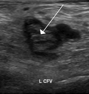
Coronal plane, seen from medial side of lower leg, showing thrombosis of the
778:
761:
555:
538:
826:
787:
702:
694:
522:
413:
334:
170:
ultrasonography, venous ultrasonography is carried out with the probe in a
129:
Lower limbs venous ultrasonography is also indicated in cases of suspected
869:
746:
660:
564:
478:
395:
818:
187:
167:
203:
A very recently formed thrombus is not very solid, it will have a low
78:
63:
291:"Point-of-care ultrasound in the diagnosis of pulmonary embolism"
289:
Squizzato, Alessandro; Galli, Luca; Gerdes, Victor E. A. (2015).
539:"Deep venous thrombosis detection by probe compression of veins"
36:
147:
are, by themselves, not indications to perform the procedure.
603:
Ultrasonographie vasculaire diagnostique: Théorie et pratique
266:
Ultrasonography of chronic venous insufficiency of the legs
87:, with hyperechoic content and only marginal blood flow.
384:
The
Journal of Bone and Joint Surgery. American Volume
501:
European
Journal of Vascular and Endovascular Surgery
353:"Five Things Physicians and Patients Should Question"
122:
The risk of deep vein thrombosis can be estimated by
284:
282:
182:, but of most importance is the detection of venous
609:] (in French). Paris: Vigot. pp. 386–437.
607:
Vascular diagnostic ultrasound: Theory and practice
54:
29:
807:Seminars in Respiratory and Critical Care Medicine
18:Ultrasonography for suspected deep vein thrombosis
639:World Health Organization Technical Report Series
490:
488:
58:focuses primarily on femoral and popliteal vein,
681:of deep venous thrombosis of the lower limbs".
8:
585:: CS1 maint: multiple names: authors list (
426:: CS1 maint: numeric names: authors list (
365:, American Academy of Orthopaedic Surgeons
35:
898:. Royal College of Physicians (UK). 2012.
777:
554:
512:
460:
403:
324:
306:
349:American Academy of Orthopaedic Surgeons
278:
30:Ultrasonography of deep vein thrombosis
578:
419:
26:
891:"Diagnosis of deep vein thrombosis".
268:, mainly targeting superficial veins.
7:
739:10.1001/archinte.1993.00410240085010
25:
1:
862:10.1016/S0140-6736(94)90240-2
727:Archives of Internal Medicine
462:10.1016/S0741-5214(96)70005-8
801:Elliott, C. Gregory (2000).
41:Deep vein thrombosis of the
766:Journal of Vascular Surgery
449:Journal of Vascular Surgery
295:Critical Ultrasound Journal
242:sensitivity and specificity
217:superficial vein thrombosis
156:ventilation/perfusion scans
45:, seen with the probe in a
937:
916:Diagnostic medical imaging
683:Thrombosis and Haemostasis
645:: i–46, back cover. 1998.
514:10.1016/j.ejvs.2005.07.019
308:10.1186/s13089-015-0025-5
102:focuses primarily on the
34:
921:Medical ultrasonography
779:10.1067/mva.2001.116803
601:Dauzat, Michel (1991).
556:10.7863/jum.1986.5.2.89
360:: an initiative of the
254:Doppler ultrasonography
69:Doppler ultrasonography
695:10.1055/s-0037-1613435
162:Technique and findings
135:CT pulmonary angiogram
88:
76:
396:10.2106/JBJS.9408edit
82:
67:
819:10.1055/s-2000-13187
213:deep vein thrombosis
99:deep vein thrombosis
535:Page pp. 89–95 in:
234:common femoral vein
43:common femoral vein
221:pulmonary embolism
154:and nondiagnostic
152:pulmonary embolism
131:pulmonary embolism
112:pulmonary embolism
89:
77:
652:978-92-4-120875-8
616:978-2-7114-1104-7
543:J. Ultrasound Med
351:(February 2013),
73:subsartorial vein
62:
61:
16:(Redirected from
928:
900:
899:
888:
882:
881:
856:(8906): 1142–4.
845:
839:
838:
798:
792:
791:
781:
757:
751:
750:
721:
715:
714:
677:
671:
670:
668:
667:
636:
627:
621:
620:
597:
591:
590:
584:
576:
558:
533:
527:
526:
516:
492:
483:
482:
464:
440:
434:
431:
425:
417:
407:
373:
372:
370:
345:
339:
338:
328:
310:
286:
180:perforator veins
39:
27:
21:
936:
935:
931:
930:
929:
927:
926:
925:
906:
905:
904:
903:
890:
889:
885:
847:
846:
842:
800:
799:
795:
759:
758:
754:
733:(24): 2777–80.
723:
722:
718:
679:
678:
674:
665:
663:
653:
634:
630:
628:
624:
617:
600:
598:
594:
577:
536:
534:
530:
494:
493:
486:
442:
441:
437:
418:
377:
368:
366:
362:ABIM Foundation
358:Choosing Wisely
347:
346:
342:
288:
287:
280:
275:
262:
251:
229:
215:(DVT), or in a
201:
164:
145:hip replacement
120:
93:Ultrasonography
50:
23:
22:
15:
12:
11:
5:
934:
932:
924:
923:
918:
908:
907:
902:
901:
883:
840:
813:(6): 495–504.
793:
752:
716:
672:
651:
622:
615:
592:
528:
484:
455:(5): 732–737.
435:
433:
432:
374:, which cites
340:
277:
276:
274:
271:
270:
269:
261:
258:
250:
247:
238:popliteal vein
228:
225:
200:
197:
192:false negative
163:
160:
119:
116:
108:popliteal vein
60:
59:
56:
52:
51:
40:
32:
31:
24:
14:
13:
10:
9:
6:
4:
3:
2:
933:
922:
919:
917:
914:
913:
911:
897:
896:
887:
884:
879:
875:
871:
867:
863:
859:
855:
851:
844:
841:
836:
832:
828:
824:
820:
816:
812:
808:
804:
797:
794:
789:
785:
780:
775:
772:(4): 656–60.
771:
767:
763:
756:
753:
748:
744:
740:
736:
732:
728:
720:
717:
712:
708:
704:
700:
696:
692:
688:
684:
676:
673:
662:
658:
654:
648:
644:
640:
633:
629:Page 1-2 in:
626:
623:
618:
612:
608:
604:
599:Page 422 in:
596:
593:
588:
582:
574:
570:
566:
562:
557:
552:
548:
544:
540:
532:
529:
524:
520:
515:
510:
506:
502:
498:
491:
489:
485:
480:
476:
472:
468:
463:
458:
454:
450:
446:
439:
436:
429:
423:
415:
411:
406:
401:
397:
393:
389:
385:
381:
376:
375:
364:
363:
359:
354:
350:
344:
341:
336:
332:
327:
322:
318:
314:
309:
304:
300:
296:
292:
285:
283:
279:
272:
267:
264:
263:
259:
257:
255:
248:
246:
243:
239:
235:
226:
224:
222:
218:
214:
210:
206:
198:
196:
193:
189:
185:
181:
177:
176:perpendicular
173:
169:
161:
159:
157:
153:
148:
146:
142:
138:
136:
132:
127:
125:
117:
115:
113:
109:
105:
101:
100:
96:in suspected
95:
94:
86:
85:fibular veins
81:
74:
70:
66:
57:
53:
48:
44:
38:
33:
28:
19:
893:
886:
853:
849:
843:
810:
806:
796:
769:
765:
755:
730:
726:
719:
689:(2): 221–7.
686:
682:
675:
664:. Retrieved
642:
638:
625:
606:
602:
595:
581:cite journal
549:(2): 89–95.
546:
542:
531:
507:(1): 83–92.
504:
500:
452:
448:
438:
422:cite journal
390:(8): 673–4.
387:
383:
367:, retrieved
356:
343:
298:
294:
252:
230:
205:echogenicity
202:
199:Echogenicity
165:
149:
139:
128:
121:
118:Medical uses
104:femoral vein
97:
91:
90:
227:Compression
223:occurring.
174:position, (
172:transversal
124:Wells score
47:transversal
910:Categories
850:The Lancet
666:2013-02-05
273:References
184:thrombosis
471:0741-5214
317:2036-3176
49:position.
878:23576444
835:12384128
827:16088760
788:11668320
711:39149938
703:12574799
573:25293123
523:16226898
414:22517384
335:26034556
301:(1): 7.
260:See also
236:and the
188:thrombus
168:arterial
133:where a
106:and the
870:7910237
747:8257253
661:9659004
565:3514943
479:8918316
405:3326687
326:4447771
249:Doppler
166:Unlike
55:Purpose
876:
868:
833:
825:
786:
745:
709:
701:
659:
649:
613:
571:
563:
521:
477:
469:
412:
402:
369:19 May
333:
323:
315:
874:S2CID
831:S2CID
707:S2CID
635:(PDF)
605:[
569:S2CID
209:lumen
895:June
866:PMID
823:PMID
784:PMID
743:PMID
699:PMID
657:PMID
647:ISBN
611:ISBN
587:link
561:PMID
519:PMID
475:PMID
467:ISSN
428:link
410:PMID
371:2013
331:PMID
313:ISSN
141:Knee
858:doi
854:343
815:doi
774:doi
735:doi
731:153
691:doi
643:875
551:doi
509:doi
457:doi
400:PMC
392:doi
321:PMC
303:doi
143:or
912::
872:.
864:.
852:.
829:.
821:.
811:21
809:.
805:.
782:.
770:34
768:.
764:.
741:.
729:.
705:.
697:.
687:89
685:.
655:.
641:.
637:.
583:}}
579:{{
567:.
559:.
545:.
541:.
517:.
505:31
503:.
499:.
487:^
473:.
465:.
453:24
451:.
447:.
424:}}
420:{{
408:.
398:.
388:94
386:.
382:.
355:,
329:.
319:.
311:.
297:.
293:.
281:^
158:.
126:.
114:.
880:.
860::
837:.
817::
790:.
776::
749:.
737::
713:.
693::
669:.
619:.
589:)
575:.
553::
547:5
525:.
511::
481:.
459::
430:)
416:.
394::
337:.
305::
299:7
75:.
20:)
Text is available under the Creative Commons Attribution-ShareAlike License. Additional terms may apply.