498:
220:
497:
1340:
196:
172:
184:
208:
342:
1450:
156:
533:
1:posterior segment 2:ora serrata 3:ciliary muscle 4:ciliary zonules 5:Schlemm's canal 6:pupil 7:anterior chamber 8:cornea 9:iris 10:lens cortex 11:lens nucleus 12:ciliary process 13:conjunctiva 14:inferior oblique muscule 15:inferior rectus muscule 16:medial rectus muscle 17:retinal arteries and
151:
It was discovered that in monkeys, outer segments of central foveolar cones are twice as long as those from parafoveal cones and do not run completely parallel to the incident light. Unique Müller cells are present in the central foveolae (area of 200 μm in diameter) of humans and monkeys.
219:
137:
The center of the foveola is sometimes referred to as the umbo, a small (150-200μm) center of the floor of the foveola; features elongated cones. The umbo is the observed point corresponding to the normal light reflex but not solely responsible for this light reflex.
134:. In this region the cone receptors are found to be longer, slimmer, and more densely packed than anywhere else in the retina, thus allowing that region to have the potential to have the highest visual acuity in the eye.
534:
veins 18:optic disc 19:dura mater 20:central retinal artery 21:central retinal vein 22:optic nerve 23:vorticose vein 24:bulbar sheath 25:macula 26:fovea 27:sclera 28:choroid 29:superior rectus muscle 30:retina
171:
777:
229:
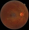
showing the macula as a spot to the left. The optic disc is the area on the right where blood vessels converge. The grey, more diffuse spot in the centre is a shadow
183:
517:
86:
1086:
514:
515:
520:
195:
529:
516:
521:
501:
377:
62:
507:
1491:
159:
Photograph of the retina of the human eye, with overlay diagrams showing the positions and sizes of the macula, fovea, and optic disc
1314:
512:
148:) and humans. Serial sections and FIB analysis were then used to construct 3D models of central Müller and photoreceptor cells.
1079:
505:
524:
523:
504:
144:
Serial semithin and ultrathin sections, and focused ion beam (FIB) tomography were prepared from 32 foveolae from monkeys (
503:
81:
1023:
968:
936:
773:
511:
921:
890:
882:
562:
522:
513:
510:
1510:
1072:
717:
707:
207:
519:
518:
1269:
1099:
502:
370:
1383:
1375:
525:
399:
177:
Schematic diagram of the macula lutea of the retina, showing perifovea, parafovea, fovea, and clinical macula
1239:
1154:
712:
668:
526:
1515:
1484:
1339:
1189:
852:
745:
476:
93:
69:
57:
298:
Tschulakow, Alexander V; Oltrup, Theo; Bende, Thomas; Schmelzle, Sebastian; Schraermeyer, Ulrich (2018).
1028:
833:
757:
695:
683:
638:
1149:
528:
509:
1409:
1274:
1249:
1214:
1018:
363:
527:
1329:
1319:
1179:
1174:
1139:
1043:
749:
700:
688:
678:
633:
567:
530:
481:
428:
508:
1520:
1209:
1184:
1164:
1013:
793:
789:
785:
733:
456:
1360:
572:
423:
1477:
1414:
1234:
1169:
1053:
769:
673:
590:
506:
461:
387:
331:
275:
226:
1424:
1294:
1264:
1254:
1219:
1095:
1038:
976:
813:
466:
418:
321:
311:
265:
1429:
1309:
1304:
1289:
1204:
1124:
951:
910:
906:
828:
230:
123:
355:
1461:
1324:
1299:
1279:
1259:
1159:
1134:
956:
946:
931:
926:
623:
615:
595:
471:
446:
326:
299:
131:
1504:
1199:
1144:
981:
916:
753:
1224:
1194:
941:
765:
600:
582:
122:. Approximately 0.35 mm in diameter, the foveola lies in the center of the
1244:
1033:
902:
859:
797:
761:
189:
Time-Domain OCT of the macular area of a retina at 800 nm, axial resolution 3 μm
346:
270:
253:
1367:
1284:
1229:
1114:
847:
605:
74:
1391:
1129:
823:
818:
737:
391:
254:"Müller Cell Cone, an Overlooked Part of the Anatomy of the Fovea Centralis"
127:
1064:
335:
279:
99:
17:
1419:
1119:
741:
993:
894:
554:
341:
316:
1354:
988:
898:
652:
438:
410:
119:
115:
1449:
628:
155:
154:
45:
345:
Material was copied from this source, which is available under a
542:
1068:
359:
1457:
118:, a yellowish, cone photoreceptor filled portion of the human
496:
141:
The anatomy of the foveola was recently reinvestigated.
1465:
347:
Creative
Commons Attribution 4.0 International License
1402:
1347:
1107:
1006:
967:
881:
872:
806:
726:
661:
651:
614:
581:
553:
541:
437:
409:
398:
80:
68:
56:
44:
39:
34:
29:
500:
293:
291:
289:
201:Spectral-Domain OCT macula cross-section scan.
1485:
1080:
371:
8:
300:"The anatomy of the foveola reinvestigated"
1492:
1478:
1087:
1073:
1065:
878:
658:
550:
406:
378:
364:
356:
325:
315:
269:
244:
167:
114:is located within a region called the
97:
26:
7:
1446:
1444:
25:
1448:
1338:
340:
218:
206:
194:
182:
170:
1:
1464:. You can help Knowledge by
1024:Optical coherence tomography
778:Photosensitive ganglion cell
774:Giant retina ganglion cells
563:Capillary lamina of choroid
1537:
1443:
718:Retinal pigment epithelium
708:External limiting membrane
271:10.1001/archopht.117.6.821
252:Gass, J. Donald M (1999).
130:and a cone-shaped zone of
1336:
494:
258:Archives of Ophthalmology
92:
1376:Ascending and Descending
1456:This article about the
713:Layer of rods and cones
669:Inner limiting membrane
535:
213:macula histology (OCT)
160:
94:Anatomical terminology
1029:Eye care professional
834:Foveal avascular zone
696:Outer plexiform layer
684:Inner plexiform layer
639:Iris sphincter muscle
532:
158:
1410:Accidental viewpoint
1049:Physiological Optics
1019:Ocular immune system
758:Retina ganglion cell
1315:Vertical–horizontal
873:Anatomical regions
734:Photoreceptor cells
701:Outer nuclear layer
689:Inner nuclear layer
679:Ganglion cell layer
634:Iris dilator muscle
429:Trabecular meshwork
146:Macaca fascicularis
1415:Auditory illusions
1210:Impossible trident
536:
317:10.7717/peerj.4482
161:
126:and contains only
1511:Human eye anatomy
1473:
1472:
1438:
1437:
1430:Temporal illusion
1425:Tactile illusions
1395:(2015 photograph)
1096:Optical illusions
1062:
1061:
1054:Visual perception
1002:
1001:
969:Posterior segment
937:Posterior chamber
868:
867:
770:Bistratified cell
674:Nerve fiber layer
647:
646:
591:Ciliary processes
492:
491:
227:fundus photograph
164:Additional images
108:
107:
103:
16:(Redirected from
1528:
1494:
1487:
1480:
1452:
1445:
1342:
1295:Schroeder stairs
1270:Peripheral drift
1265:Penrose triangle
1089:
1082:
1075:
1066:
1039:Refractive error
977:Vitreous chamber
922:Anterior chamber
883:Anterior segment
879:
659:
568:Bruch's membrane
551:
543:Uvea / vascular
499:
419:Episcleral layer
407:
380:
373:
366:
357:
350:
344:
339:
329:
319:
295:
284:
283:
273:
249:
222:
210:
198:
186:
174:
100:edit on Wikidata
27:
21:
1536:
1535:
1531:
1530:
1529:
1527:
1526:
1525:
1501:
1500:
1499:
1498:
1441:
1439:
1434:
1398:
1348:Popular culture
1343:
1334:
1305:Spinning dancer
1125:Ambiguous image
1103:
1093:
1063:
1058:
998:
963:
952:Capsule of lens
907:Lacrimal system
874:
864:
824:Parafoveal area
819:Perifoveal area
802:
746:Horizontal cell
722:
643:
610:
577:
573:Sattler's layer
544:
537:
531:
488:
433:
424:Schlemm's canal
402:
394:
386:Anatomy of the
384:
354:
353:
297:
296:
287:
251:
250:
246:
241:
234:
223:
214:
211:
202:
199:
190:
187:
178:
175:
166:
104:
23:
22:
15:
12:
11:
5:
1534:
1532:
1524:
1523:
1518:
1513:
1503:
1502:
1497:
1496:
1489:
1482:
1474:
1471:
1470:
1453:
1436:
1435:
1433:
1432:
1427:
1422:
1417:
1412:
1406:
1404:
1400:
1399:
1397:
1396:
1388:
1387:(1961 drawing)
1380:
1379:(1960 drawing)
1372:
1364:
1357:
1351:
1349:
1345:
1344:
1337:
1335:
1333:
1332:
1327:
1322:
1317:
1312:
1307:
1302:
1300:Shepard tables
1297:
1292:
1287:
1282:
1277:
1272:
1267:
1262:
1260:Penrose stairs
1257:
1252:
1247:
1242:
1237:
1232:
1227:
1222:
1217:
1212:
1207:
1202:
1197:
1192:
1187:
1182:
1177:
1172:
1167:
1162:
1157:
1155:Checker shadow
1152:
1147:
1142:
1137:
1135:Autostereogram
1132:
1127:
1122:
1117:
1111:
1109:
1105:
1104:
1094:
1092:
1091:
1084:
1077:
1069:
1060:
1059:
1057:
1056:
1051:
1046:
1041:
1036:
1031:
1026:
1021:
1016:
1010:
1008:
1004:
1003:
1000:
999:
997:
996:
991:
986:
985:
984:
973:
971:
965:
964:
962:
961:
960:
959:
957:Zonule of Zinn
954:
944:
939:
934:
929:
927:Aqueous humour
924:
919:
914:
887:
885:
876:
870:
869:
866:
865:
863:
862:
857:
856:
855:
845:
844:
843:
842:
841:
836:
826:
821:
810:
808:
804:
803:
801:
800:
730:
728:
724:
723:
721:
720:
715:
710:
704:
703:
698:
692:
691:
686:
681:
676:
671:
665:
663:
656:
649:
648:
645:
644:
642:
641:
636:
631:
626:
620:
618:
612:
611:
609:
608:
603:
598:
596:Ciliary muscle
593:
587:
585:
579:
578:
576:
575:
570:
565:
559:
557:
548:
539:
538:
495:
493:
490:
489:
487:
486:
485:
484:
479:
474:
469:
464:
459:
449:
443:
441:
435:
434:
432:
431:
426:
421:
415:
413:
404:
396:
395:
385:
383:
382:
375:
368:
360:
352:
351:
285:
243:
242:
240:
237:
236:
235:
224:
217:
215:
212:
205:
203:
200:
193:
191:
188:
181:
179:
176:
169:
165:
162:
106:
105:
96:
90:
89:
84:
78:
77:
72:
66:
65:
60:
54:
53:
48:
42:
41:
37:
36:
32:
31:
24:
14:
13:
10:
9:
6:
4:
3:
2:
1533:
1522:
1519:
1517:
1516:Visual system
1514:
1512:
1509:
1508:
1506:
1495:
1490:
1488:
1483:
1481:
1476:
1475:
1469:
1467:
1463:
1459:
1454:
1451:
1447:
1442:
1431:
1428:
1426:
1423:
1421:
1418:
1416:
1413:
1411:
1408:
1407:
1405:
1401:
1394:
1393:
1389:
1386:
1385:
1381:
1378:
1377:
1373:
1370:
1369:
1365:
1363:
1362:
1358:
1356:
1353:
1352:
1350:
1346:
1341:
1331:
1328:
1326:
1323:
1321:
1318:
1316:
1313:
1311:
1308:
1306:
1303:
1301:
1298:
1296:
1293:
1291:
1288:
1286:
1283:
1281:
1278:
1276:
1273:
1271:
1268:
1266:
1263:
1261:
1258:
1256:
1253:
1251:
1248:
1246:
1243:
1241:
1238:
1236:
1233:
1231:
1228:
1226:
1223:
1221:
1218:
1216:
1213:
1211:
1208:
1206:
1203:
1201:
1198:
1196:
1193:
1191:
1190:Fraser spiral
1188:
1186:
1183:
1181:
1178:
1176:
1173:
1171:
1168:
1166:
1163:
1161:
1158:
1156:
1153:
1151:
1148:
1146:
1143:
1141:
1138:
1136:
1133:
1131:
1128:
1126:
1123:
1121:
1118:
1116:
1113:
1112:
1110:
1106:
1101:
1097:
1090:
1085:
1083:
1078:
1076:
1071:
1070:
1067:
1055:
1052:
1050:
1047:
1045:
1044:Accommodation
1042:
1040:
1037:
1035:
1032:
1030:
1027:
1025:
1022:
1020:
1017:
1015:
1012:
1011:
1009:
1005:
995:
992:
990:
987:
983:
982:Vitreous body
980:
979:
978:
975:
974:
972:
970:
966:
958:
955:
953:
950:
949:
948:
945:
943:
940:
938:
935:
933:
930:
928:
925:
923:
920:
918:
917:Fibrous tunic
915:
912:
908:
904:
900:
896:
892:
889:
888:
886:
884:
880:
877:
871:
861:
858:
854:
851:
850:
849:
846:
840:
837:
835:
832:
831:
830:
827:
825:
822:
820:
817:
816:
815:
812:
811:
809:
805:
799:
795:
791:
787:
783:
779:
775:
771:
767:
763:
759:
755:
754:Amacrine cell
751:
747:
743:
739:
735:
732:
731:
729:
725:
719:
716:
714:
711:
709:
706:
705:
702:
699:
697:
694:
693:
690:
687:
685:
682:
680:
677:
675:
672:
670:
667:
666:
664:
660:
657:
654:
650:
640:
637:
635:
632:
630:
627:
625:
622:
621:
619:
617:
613:
607:
604:
602:
599:
597:
594:
592:
589:
588:
586:
584:
580:
574:
571:
569:
566:
564:
561:
560:
558:
556:
552:
549:
546:
540:
483:
480:
478:
475:
473:
470:
468:
465:
463:
460:
458:
455:
454:
453:
450:
448:
445:
444:
442:
440:
436:
430:
427:
425:
422:
420:
417:
416:
414:
412:
408:
405:
401:
400:Fibrous tunic
397:
393:
389:
381:
376:
374:
369:
367:
362:
361:
358:
348:
343:
337:
333:
328:
323:
318:
313:
309:
305:
301:
294:
292:
290:
286:
281:
277:
272:
267:
263:
259:
255:
248:
245:
238:
232:
228:
221:
216:
209:
204:
197:
192:
185:
180:
173:
168:
163:
157:
153:
149:
147:
142:
139:
135:
133:
129:
125:
121:
117:
113:
101:
95:
91:
88:
85:
83:
79:
76:
73:
71:
67:
64:
61:
59:
55:
52:
49:
47:
43:
38:
33:
28:
19:
1466:expanding it
1455:
1440:
1390:
1382:
1374:
1366:
1361:Trompe-l'œil
1359:
1225:Lilac chaser
1195:Gravity hill
1048:
942:Ciliary body
838:
782:Diencephalon
781:
766:Parasol cell
750:Bipolar cell
601:Pars plicata
583:Ciliary body
451:
307:
303:
264:(6): 821–3.
261:
257:
247:
150:
145:
143:
140:
136:
132:Müller cells
111:
109:
63:A15.2.04.023
50:
1371:(1864 book)
1275:Poggendorff
1250:Oppel-Kundt
1245:Necker cube
1240:Müller-Lyer
1215:Irradiation
1034:Eye disease
1014:Keratocytes
903:Conjunctiva
860:Ora serrata
798:Muller glia
762:Midget cell
482:Endothelium
472:Dua's layer
40:Identifiers
1505:Categories
1368:Spectropia
1285:Rubin vase
1235:McCollough
1230:Mach bands
1180:Ehrenstein
1175:Ebbinghaus
1140:Barberpole
1115:Afterimage
875:of the eye
848:Optic disc
606:Pars plana
477:Descemet's
457:Epithelium
128:cone cells
18:Umbo (eye)
1521:Eye stubs
1420:Illusions
1392:The dress
1384:Waterfall
1185:Flash lag
1165:Cornsweet
1150:Café wall
1130:Ames room
1108:Illusions
853:Optic cup
738:Cone cell
392:human eye
310:: e4482.
1170:Delboeuf
1120:Ambigram
742:Rod cell
547:(middle)
462:Bowman's
336:29576957
280:10369597
231:artifact
1403:Related
1330:Zöllner
1320:White's
1255:Orbison
1220:Jastrow
994:Choroid
895:Eyebrow
839:Foveola
655:(inner)
555:Choroid
403:(outer)
390:of the
327:5853608
112:foveola
51:foveola
35:Details
30:Foveola
1355:Op art
1310:Ternus
1290:Sander
1205:Hering
1145:Bezold
989:Retina
899:Eyelid
891:Adnexa
814:Macula
794:K cell
790:M cell
786:P cell
662:Layers
653:Retina
624:Stroma
467:Stroma
452:layers
447:Limbus
439:Cornea
411:Sclera
334:
324:
278:
120:retina
116:macula
1460:is a
1325:Wundt
1280:Ponzo
1160:Chubb
1007:Other
911:Orbit
829:Fovea
807:Other
744:) → (
727:Cells
629:Pupil
545:tunic
388:globe
304:PeerJ
239:Notes
124:fovea
98:[
87:77666
46:Latin
1462:stub
1200:Grid
1100:list
947:Lens
932:Iris
780:) →
756:) →
748:) →
616:Iris
332:PMID
276:PMID
110:The
75:6786
58:TA98
1458:eye
752:→ (
322:PMC
312:doi
266:doi
262:117
82:FMA
70:TA2
1507::
909:,
905:,
901:,
897:,
796:,
792:,
788:,
784::
776:,
772:,
768:,
764:,
740:,
330:.
320:.
306:.
302:.
288:^
274:.
260:.
256:.
225:A
1493:e
1486:t
1479:v
1468:.
1102:)
1098:(
1088:e
1081:t
1074:v
913:)
893:(
760:(
736:(
379:e
372:t
365:v
349:.
338:.
314::
308:6
282:.
268::
233:.
102:]
20:)
Text is available under the Creative Commons Attribution-ShareAlike License. Additional terms may apply.