20:
327:
233:
248:
molecules showed that membrane permeability mechanisms differ depending on the size of dextran molecules. Microinjection of dextran molecules from 3 to 70 kDa was reported to have crossed the cellular membrane via transient pores. In contrast, dextran molecules of 155 and 500 kDa were predominantly
210:
produced by ultrasound stimulation may push and pull on the membrane to produce a membrane opening. These rapid oscillations are also responsible for adjacent fluid flow called microstreaming which increases pressure on surrounding cells producing further sonoporation to whole cell populations. The
317:
Following sonoporation-mediated membrane permeabilization, cells can automatically repair the membrane openings through a phenomenon called "reparable sonoporation." The membrane resealing process has been shown to be calcium-dependent. This property may suggest that the membrane repair process
23:
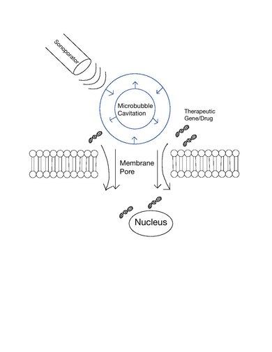
Schematic of
Sonoporation Mechanism. This figure depicts the general understanding of sonoporation where a dedicated sonoporator applies ultrasound to induce microbubble cavitation and eventually pore formation. The therapeutic gene or drug of interest thus may translocate within the
93:, in a medical treatment scenario whereby a patient is given modified DNA, and an ultrasonic transducer might target this modified DNA into specific regions of the patient's body. The bioactivity of this technique is similar to, and in some cases found superior to,
219:
The mechanism by which molecules cross cellular membrane barriers during sonoporation remains unclear. Different theories exist that may potentially explain barrier permeabilization and molecular delivery. The dominant hypotheses include pore formation,
367:
In vivo ultrasound mediated drug delivery was first reported in 1991 and many other preclinical studies involving sonoporation have followed. This method is being used to deliver therapeutic drugs or genes to treat a variety of diseases including:
154:
The microbubbles used today are composed of a gas core and a surrounding shell. The makeup of these elements may vary depending on the preferred physical and chemical properties. Microbubble shells have been formed with
197:
relative to their liquid environment, making them highly responsive to acoustic application. As a result of ultrasound stimulation, microbubbles undergo expansion and contraction, a phenomenon called stable
240:
Pore formation following ultrasound application was first reported in 1999 in a study that observed cell membrane craters following ultrasound application at 255 kHz. Later, sonoporation mediated
289:
seen in traditional endocytosis pathways. Other work reported sonoporation induced the formation of hydrogen peroxide, a cellular reaction that is also known to be involved with endocytosis.
75:
to enhance delivery of these large molecules. The exact mechanism of sonoporation-mediated membrane translocation remains unclear, with a few different hypotheses currently being explored.
1067:
Alter J, Sennoga CA, Lopes DM, Eckersley RJ, Wells DJ (2009). "Microbubble stability is a major determinant of the efficiency of ultrasound and microbubble mediated in vivo gene transfer".
146:
applications to enhance the acoustic impact of ultrasound. For sonoporation specifically, microbubbles are used to significantly enhance membrane translocation of molecular therapeutics.
236:
Schematic representation of molecular translocation via endocytosis. The second representation from the left illustrates the endocytotic mechanism involving clathrin-coated pits.
253:. This variability in membrane behavior has led to other studies investigating membrane rupture and resealing characteristics depending on ultrasound amplitude and duration.
388:
is coupled with ultrasound-mediated microbubble vascular disruption. This increase in delivery efficiency could allow for the appropriate reduction in therapeutic dosing.
384:... The preclinical utility of sonoporation is well illustrated through past tumor radiation treatments which have reported a more than 10-fold cellular destruction when
211:
physical mechanisms supposedly involved with microbubble-enhanced sonoporation have been referred to as push, pull, microstreaming, translation, and jetting.
128:, which quantifies the likelihood that exposure to diagnostic ultrasound will produce an adverse biological effect by a non-thermal action based on pressure.
309:. Multiple studies examining membrane wounds note observing resealing behavior, a process dependent on recruitment of ATP and intracellular vesicles.
261:
Various cellular reactions to ultrasound indicate the mechanism of molecular uptake via endocytosis. These observed reactionary phenomena include
285:
opening in response to microbubble oscillations. These findings act as support for ultrasound application inducing calcium-mediated uncoating of
277:
for the role of endocytosis in sonoporation. Ultrasound application to cells and adjacent microbubbles was shown to produce marked cell membrane
762:
526:. 2nd IEEE International Symposium on Biomedical Imaging: Macro to Nano (IEEE Cat No. 04EX821). Vol. 2. New York: IEEE. pp. 29–32.
473:
835:
Hauser J, Ellisman M, Steinau HU, Stefan E, Dudda M, Hauser M (2009). "Ultrasound enhanced endocytotic activity of human fibroblasts".
301:. The nature of these wounds may vary based on the degree of acoustic cavitation leading to a spectrum of cell behavior, from membrane
326:
19:
539:
563:
Klibanov AL (2006). "Microbubble contrast agents: targeted ultrasound imaging and ultrasound-assisted drug-delivery applications".
788:"Ultrasound and microbubble-targeted delivery of macromolecules is regulated by induction of endocytosis and pore formation"
355:
ultimately led to further in vitro studies that hinted at the potential for sonoporation transfection of plasmid DNA and
143:
278:
745:
Bouakaz A, Zeghimi A, Doinikov AA (2016). "Sonoporation: Concept and
Mechanisms". In Escoffre JM, Bouakaz A (eds.).
1113:
64:
339:
The first study reporting molecular delivery using ultrasound was a 1987 in vitro study attempting to transfer
113:
Sonoporation is performed with a dedicated sonoporator. Sonoporation may also be performed with custom-built
297:
Mechanically created wounds in the plasma membrane have been observed as a result of sonoporation-produced
1108:
381:
377:
699:
749:. Advances in Experimental Medicine and Biology. Vol. 880. Heidelberg: Springer. pp. 175–189.
356:
352:
114:
928:
871:
302:
101:) ultrasound has been demonstrated to result in complete cellular death (rupturing), thus cellular
606:
Lindner JR (2004). "Microbubbles in medical imaging: current applications and future directions".
817:
722:
631:
588:
545:
385:
274:
281:
along with progressive intracellular calcium increase, which is believed to be a consequence of
1084:
1049:
995:
946:
894:
852:
809:
786:
Meijering BD, Juffermans LJ, van Wamel A, Henning RH, Zuhorn IS, Emmer M, et al. (2009).
768:
758:
680:
623:
580:
535:
469:
436:
266:
44:
1076:
1039:
1029:
985:
977:
936:
886:
844:
799:
750:
714:
670:
662:
615:
572:
527:
502:
461:
426:
418:
125:
282:
194:
176:
94:
56:
872:"Effect of ultrasound-activated microbubbles on the cell electrophysiological properties"
318:
involves a cell's active repair mechanism in response to the cellular influx of calcium.
932:
848:
1080:
1044:
1017:
990:
965:
890:
675:
650:
576:
431:
406:
241:
139:
1102:
726:
203:
40:
821:
635:
592:
549:
348:
262:
87:
72:
60:
48:
981:
804:
787:
754:
330:
A study showing verified preclinical efficacy of acoustic targeted drug delivery.
1016:
Tomizawa M, Shinozaki F, Motoyoshi Y, Sugiyama T, Yamamoto S, Sueishi M (2013).
298:
270:
250:
221:
207:
187:
136:
651:"Mechanisms of microbubble-facilitated sonoporation for drug and gene delivery"
531:
344:
199:
118:
102:
68:
36:
966:"Effects of extracellular calcium on cell membrane resealing in sonoporation"
950:
405:
Song Y, Hahn T, Thompson IP, Mason TJ, Preston GM, Li G, et al. (2007).
117:
connected to bench-top function generators and acoustic amplifiers. Standard
160:
1088:
1053:
999:
898:
856:
813:
772:
718:
684:
627:
584:
440:
941:
916:
1034:
422:
286:
172:
168:
232:
124:
Measurement of the acoustics used in sonoporation is listed in terms of
340:
245:
164:
90:
666:
507:
490:
373:
369:
249:
found in vesicle-like structures, likely indicating the mechanism of
83:
619:
78:
Sonoporation is under active study for the introduction of foreign
306:
269:, and cell intracellular calcium concentration. Studies have used
156:
465:
870:
Tran TA, Roger S, Le
Guennec JY, Tranquart F, Bouakaz A (2007).
79:
455:
86:
cells. Sonoporation is also being studied for use in targeted
39:
in the ultrasonic range for increasing the permeability of the
190:
98:
52:
105:
must also be accounted for when employing this technique.
171:. The gas core can be made up of air or heavy gases like
698:
Postema M, Kotopoulis S, Delalande A, Gilja OH (2012).
121:
medical devices may also be used in some applications.
347:
cells using sonoporation. This successful plasmid DNA
51:
in order to allow uptake of large molecules such as
491:"Frequency, pulse length, and the mechanical index"
524:Microbubbles for ultrasound diagnosis and therapy
1011:
1009:
407:"Ultrasound-mediated DNA transfer for bacteria"
1018:"Sonoporation: Gene transfer using ultrasound"
740:
738:
736:
700:"Sonoporation: why microbubbles create pores"
8:
522:Fowlkes JB, Kripfgans OD, Carson PL (2004).
910:
908:
97:. Extended exposure to low-frequency (<
1043:
1033:
989:
940:
917:"Acoustic Streaming and Its Applications"
803:
674:
506:
430:
325:
231:
18:
397:
964:Zhou Y, Shi J, Cui J, Deng CX (2008).
202:. If a microbubble is attached to the
67:. Sonoporation employs the acoustic
7:
1069:Ultrasound in Medicine & Biology
879:Ultrasound in Medicine & Biology
837:Ultrasound in Medicine & Biology
82:in tissue culture cells, especially
43:. This technique is usually used in
849:10.1016/j.ultrasmedbio.2009.06.1090
1081:10.1016/j.ultrasmedbio.2008.12.015
891:10.1016/j.ultrasmedbio.2006.07.029
577:10.1097/01.rli.0000199292.88189.0f
14:
649:Fan Z, Kumon RE, Deng CX (2014).
495:Acoustics Research Letters Online
215:Membrane translocation mechanism
460:. Singapore: World Scientific.
457:Emerging Therapeutic Ultrasound
275:membrane potential ion exchange
16:Technique in molecular biology
1:
982:10.1016/j.jconrel.2007.11.007
970:Journal of Controlled Release
805:10.1161/CIRCRESAHA.108.183806
608:Nature Reviews Drug Discovery
1022:World Journal of Methodology
755:10.1007/978-3-319-22536-4_10
144:contrast-enhanced ultrasound
132:Microbubble contrast agents
1130:
707:Ultraschall in der Medizin
532:10.1109/isbi.2004.1398466
115:piezoelectric transducers
454:Wu J, Nyborg WL (2006).
565:Investigative Radiology
224:, and membrane wounds.
747:Therapeutic Ultrasound
719:10.1055/s-0031-1274749
411:Nucleic Acids Research
343:DNA to cultured mouse
331:
273:techniques to monitor
237:
142:are generally used in
25:
942:10.3390/fluids3040108
353:antibiotic resistance
329:
235:
22:
1035:10.5662/wjm.v3.i4.39
792:Circulation Research
655:Therapeutic Delivery
287:clathrin-coated pits
55:into the cell, in a
41:cell plasma membrane
933:2018Fluid...3..108W
322:Preclinical studies
183:Mechanism of action
33:cellular sonication
489:Church CC (2005).
423:10.1093/nar/gkm710
386:ionizing radiation
332:
313:Membrane resealing
238:
206:, the microbubble
26:
1114:Molecular biology
843:(12): 2084–2092.
764:978-3-319-22536-4
667:10.4155/tde.14.10
508:10.1121/1.1901757
475:978-981-256-685-0
279:hyperpolarization
267:hydrogen peroxide
45:molecular biology
1121:
1093:
1092:
1064:
1058:
1057:
1047:
1037:
1013:
1004:
1003:
993:
961:
955:
954:
944:
912:
903:
902:
876:
867:
861:
860:
832:
826:
825:
807:
783:
777:
776:
742:
731:
730:
704:
695:
689:
688:
678:
646:
640:
639:
603:
597:
596:
560:
554:
553:
519:
513:
512:
510:
486:
480:
479:
451:
445:
444:
434:
402:
351:conferring G418
283:calcium channels
193:cores have high
150:General features
126:mechanical index
1129:
1128:
1124:
1123:
1122:
1120:
1119:
1118:
1099:
1098:
1097:
1096:
1066:
1065:
1061:
1015:
1014:
1007:
963:
962:
958:
914:
913:
906:
874:
869:
868:
864:
834:
833:
829:
785:
784:
780:
765:
744:
743:
734:
702:
697:
696:
692:
648:
647:
643:
620:10.1038/nrd1417
605:
604:
600:
562:
561:
557:
542:
521:
520:
516:
488:
487:
483:
476:
453:
452:
448:
404:
403:
399:
394:
365:
337:
324:
315:
295:
293:Membrane wounds
259:
230:
217:
195:compressibility
185:
177:perfluorocarbon
152:
140:contrast agents
134:
111:
95:electroporation
59:process called
57:cell disruption
17:
12:
11:
5:
1127:
1125:
1117:
1116:
1111:
1101:
1100:
1095:
1094:
1075:(6): 976–984.
1059:
1005:
956:
904:
885:(1): 158–163.
862:
827:
798:(5): 679–687.
778:
763:
732:
690:
661:(4): 467–486.
641:
614:(6): 527–532.
598:
571:(3): 354–362.
555:
540:
514:
501:(3): 162–168.
481:
474:
446:
396:
395:
393:
390:
364:
361:
336:
333:
323:
320:
314:
311:
294:
291:
271:patch clamping
258:
255:
242:microinjection
229:
228:Pore formation
226:
216:
213:
184:
181:
151:
148:
133:
130:
110:
107:
65:transformation
47:and non-viral
15:
13:
10:
9:
6:
4:
3:
2:
1126:
1115:
1112:
1110:
1109:Biotechnology
1107:
1106:
1104:
1090:
1086:
1082:
1078:
1074:
1070:
1063:
1060:
1055:
1051:
1046:
1041:
1036:
1031:
1027:
1023:
1019:
1012:
1010:
1006:
1001:
997:
992:
987:
983:
979:
975:
971:
967:
960:
957:
952:
948:
943:
938:
934:
930:
926:
922:
918:
915:Wu J (2018).
911:
909:
905:
900:
896:
892:
888:
884:
880:
873:
866:
863:
858:
854:
850:
846:
842:
838:
831:
828:
823:
819:
815:
811:
806:
801:
797:
793:
789:
782:
779:
774:
770:
766:
760:
756:
752:
748:
741:
739:
737:
733:
728:
724:
720:
716:
712:
708:
701:
694:
691:
686:
682:
677:
672:
668:
664:
660:
656:
652:
645:
642:
637:
633:
629:
625:
621:
617:
613:
609:
602:
599:
594:
590:
586:
582:
578:
574:
570:
566:
559:
556:
551:
547:
543:
541:0-7803-8388-5
537:
533:
529:
525:
518:
515:
509:
504:
500:
496:
492:
485:
482:
477:
471:
467:
463:
459:
458:
450:
447:
442:
438:
433:
428:
424:
420:
416:
412:
408:
401:
398:
391:
389:
387:
383:
379:
375:
371:
362:
360:
358:
354:
350:
346:
342:
334:
328:
321:
319:
312:
310:
308:
304:
300:
292:
290:
288:
284:
280:
276:
272:
268:
264:
256:
254:
252:
247:
243:
234:
227:
225:
223:
214:
212:
209:
205:
204:cell membrane
201:
196:
192:
189:
182:
180:
178:
174:
170:
166:
162:
158:
149:
147:
145:
141:
138:
131:
129:
127:
122:
120:
116:
108:
106:
104:
100:
96:
92:
89:
85:
81:
76:
74:
70:
66:
62:
58:
54:
50:
46:
42:
38:
34:
30:
21:
1072:
1068:
1062:
1028:(4): 39–44.
1025:
1021:
976:(1): 34–43.
973:
969:
959:
924:
920:
882:
878:
865:
840:
836:
830:
795:
791:
781:
746:
713:(1): 97–98.
710:
706:
693:
658:
654:
644:
611:
607:
601:
568:
564:
558:
523:
517:
498:
494:
484:
466:10.1142/6047
456:
449:
417:(19): e129.
414:
410:
400:
366:
349:transfection
338:
316:
299:shear forces
296:
263:ion exchange
260:
239:
218:
208:oscillations
186:
153:
135:
123:
112:
88:Gene therapy
77:
73:microbubbles
61:transfection
49:gene therapy
37:use of sound
32:
29:Sonoporation
28:
27:
382:Alzheimer's
378:Parkinson's
305:to instant
257:Endocytosis
251:endocytosis
222:endocytosis
188:Microbubble
137:Microbubble
1103:Categories
927:(4): 108.
392:References
345:fibroblast
307:cell lysis
200:cavitation
119:ultrasound
69:cavitation
951:2311-5521
727:260344222
359:in vivo.
161:galactose
109:Equipment
103:viability
84:mammalian
35:, is the
1089:19285783
1054:25237622
1000:18158198
899:17189059
857:19828232
822:23063345
814:19168443
773:26486338
685:24856171
636:29807146
628:15173842
593:27546582
585:16481920
550:29683103
441:17890732
335:In vitro
303:blebbing
173:nitrogen
169:polymers
1045:4145571
991:2270413
929:Bibcode
676:4116608
432:2095817
363:In vivo
341:plasmid
246:dextran
165:albumin
91:in vivo
1087:
1052:
1042:
998:
988:
949:
921:Fluids
897:
855:
820:
812:
771:
761:
725:
683:
673:
634:
626:
591:
583:
548:
538:
472:
439:
429:
374:Cancer
370:Stroke
157:lipids
875:(PDF)
818:S2CID
723:S2CID
703:(PDF)
632:S2CID
589:S2CID
546:S2CID
357:siRNA
167:, or
80:genes
31:, or
24:cell.
1085:PMID
1050:PMID
996:PMID
947:ISSN
895:PMID
853:PMID
810:PMID
769:PMID
759:ISBN
681:PMID
624:PMID
581:PMID
536:ISBN
470:ISBN
437:PMID
1077:doi
1040:PMC
1030:doi
986:PMC
978:doi
974:126
937:doi
887:doi
845:doi
800:doi
796:104
751:doi
715:doi
671:PMC
663:doi
616:doi
573:doi
528:doi
503:doi
462:doi
427:PMC
419:doi
244:of
191:gas
175:or
99:MHz
71:of
63:or
53:DNA
1105::
1083:.
1073:35
1071:.
1048:.
1038:.
1024:.
1020:.
1008:^
994:.
984:.
972:.
968:.
945:.
935:.
923:.
919:.
907:^
893:.
883:33
881:.
877:.
851:.
841:35
839:.
816:.
808:.
794:.
790:.
767:.
757:.
735:^
721:.
711:33
709:.
705:.
679:.
669:.
657:.
653:.
630:.
622:.
610:.
587:.
579:.
569:41
567:.
544:.
534:.
497:.
493:.
468:.
435:.
425:.
415:35
413:.
409:.
380:,
376:,
372:,
265:,
179:.
163:,
159:,
1091:.
1079::
1056:.
1032::
1026:3
1002:.
980::
953:.
939::
931::
925:3
901:.
889::
859:.
847::
824:.
802::
775:.
753::
729:.
717::
687:.
665::
659:5
638:.
618::
612:3
595:.
575::
552:.
530::
511:.
505::
499:6
478:.
464::
443:.
421::
Text is available under the Creative Commons Attribution-ShareAlike License. Additional terms may apply.