195:
469:, and the mesocephalon, and which may be stated somewhat in this way- that, speaking approximately, equally important nerve-fibres are developed simultaneously, but those of dissimilar importance are developed one after another in a succession defined by an imperative law (Fundamental Law of Myelogenesis). The formation of medullary substance is almost completed in certain convolutions at a time when in some it is not even begun and in others has made only slight progress.
146:
294:
been myelinated – however, myelinogenesis continued to rapidly increase. During the fourth week postnatal, nearly 85% of the axons in the rat optic had been myelinated. During the fifth week and onward toward week sixteen, the myelination decelerated and the remaining unmyelinated axons were ensheathed in myelin. Through the rat optic nerve, early research made significant contributions to knowledge in the field of myelinogenesis.
242:
290:. In the optic nerve, the oligodendrocyte cells divided for the final time at five days, with the onset of myelin formation occurring on or around day 6 or 7. However, the exact process by which the oligodendrocytes were stimulated to produce myelin was not yet fully understood, but early myelination in the optic nerve has been linked to a rise in the production of various lipids – cholesterol, cerebroside, and sulfatide.
20:
333:, sulfated glycoproteins, and sulfated mucopolysaccharides appear to be associated with neurons rather than myelin. When graphing the amount of sulfatide made from and the activity of sulfotransferase, we get to distinguished peaks. The peaks occur on the 15th post-natal day. These peaks corresponded with the maximal myelination period of the optic nerve that has been seen throughout the experiment.
184:
306:(which forms sulfatide) appeared on the 9th post-natal day and reached a peak on the 15th post-natal day. This expression was similar to a period where the optic nerve showed a maximal myelination period of the axon. As the activity of axon myelination decreased, and one could conclude that the activity of the enzyme is paralleled with the incorporation of
218:
demonstrated that
Schwann cells and oligodendrocytes have a shared mechanism to stimulate myelination. A similar study working to provide evidence for neuronal regulation of myelinogenesis suggested that myelin formation was due to Schwann cells that were controlled by an undefined property of an associated axon.
460:
the investment with medullary substance (myelinisation) has already begun in some places three months before the maturity of the foetus, whilst in other places numerous fibres are devoid of medullary substance even three months after birth. The order of succession in the convolutions is governed by a
336:
In conclusion, the early phase of myelination was correlated with the increases synthesis of lipids, cholesterol, cerebroside, and sulfatide. It is likely that these compounds are synthesized and packaged in the Golgi
Apparatus of oligodendroglia. Even though the transport of these lipids is unknown,
293:
As researchers began to do postnatal research, they found that myelinogenesis in the rat optic nerve initially commences with axons the largest diameters before proceeding to the remaining smaller axons. In the second week postnatal, oligodendrocyte formation slowed – at this point, 15% of axons have
138:
Myelinogenesis thus encompasses the process of transition between phases 3 and 4. Upon initiation of myelinogenesis, each pioneer process forms lamellar extensions which extend and elaborate circumferentially around the target axon. This forms the first turn of the myelin sheath. The sheath continues
257:
re-synthesize proteins associated with myelin-specific proteins when axonal presence is re-established. Synthesis of myelin-specific proteins only occurs in
Schwann cells associated with axons. Furthermore, membrane-membrane interactions between axons may be required to promote the synthesis of P1,
455:
In the cerebral convolutions, as in all other parts of the central nervous system, the nerve-fibres do not develop everywhere simultaneously, but step by step in a definite succession, this order of events being particularly maintained in regard to the appearance of the medullary substance. In the
119:
Sometimes referred to as premyelinating oligodendrocytes, these cells extend "pioneer processes" which contact axons and anchor premyelinating oligodendrocytes to neurons such that they are poised to commence myelinogenesis in response to axonal signals. These pioneer processes grow longitudinally
217:
Axon-derived signals regulate the onset of myelinogenesis. Researchers studied regenerating PNS axons for 28 weeks in order to investigate whether or not peripheral axons stimulate oligodendrocytes to begin myelination. Experimental induction of myelination by regenerating peripheral axons
253:, researchers revealed distinct bands with band sizes of 27,000 daltons (P1), 19,000 daltons (P2), and 14,000 daltons (P0). Studies have also shown that P1 and P2 are active before P0 since this protein comes from the peripheral nervous system. In the process of regeneration,
213:
plays an integral role in the process of oligodendrocyte myelinogenesis by regulating expression of myelin-related genes. OLIG1 is necessary in order to initiate myelination by oligodendrocytes in the brain, but is somewhat dispensable in the spinal cord.
394:
313:
The studies on a rat optic nerve revealed that 15 days post-natal is when an increase in myelination is observed. Before this time period, most of the axons, roughly about 70%, are not myelinated. At this time, Sulfate was incorporated into
841:
Weinberg, E., & Spencer, P. (1979). Studies on the control of myelinogenesis. 3. Signaling of oligodendrocyte myelination by regenerating peripheral axons. Brain
Research, 162(2), 273-279. doi:10.1016/0006-8993(79)90289-0
139:
to expand along the length of the target axon while new membrane is synthesized at the leading edge of the inner tongue of the developing myelin sheath, which begins to take on a spiral cross-sectional structure.
278:
nerve that consists entirely of unmyelinated axons. Furthermore, the use of the rat optic nerve helped provide insight for early myelinogenesis researchers into improper and atypical courses of myelinogenesis.
486:
Eilam, R.; Bar-Lev, D.D.; Levin-Zaidman, S.; Tsoory, M.; LoPresti, P.; Sela, M.; Arnon, R.; Aharoni, R. (2014). "Oligodendrogenesis and myelinogenesis during postnatal development effect of glatiramer acetate".
829:
Xin, M. (2005). Myelinogenesis and Axonal
Recognition by Oligodendrocytes in Brain Are Uncoupled in Olig1-Null Mice. Journal of Neuroscience, 25(6), 1354-1365. doi:10.1523/jneurosci.3034-04.2005
890:
Tennekoon, GI., Cohen, SR., Price, DL., McKhann, GM. (1977). Myelinogenesis in optic nerve. A morphological, autoradiographic, and biochemical analysis. Journal of Cell
Biology, 72(3), 604-616.
690:
Watkins, T., Mulinyawe, S., Emery, B., Barres, B. (2008). Distinct Stages of
Myelination Regulated by Y-Secretase and Astrocytes in a Rapidly Myelinating CNS Coculture System. 555-569
1077:
167:
To drive proper assembly of membrane layers, PLP is inserted into the membrane to stabilize interactions between external leaflets of the myelin membranes; MBP is locally
850:
Marziali, L.N., Garcia, C.I., Pasquini, J.M. (2015). Transferrin and thyroid hormone converge in the control of myelinogenesis. Experimental
Neurology. Vol 265. 129–141.
899:
Dangata, Y., Kaufman, M. (1997). Myelinogenesis in the Optic Nerve of (C57BL x CBA) F1 Hybrid Mice: A Morphometric
Analysis.European Journal of Morphology, 35(1), 3-18.
816:
677:
145:
420:. He identified 45 separate cortical areas and, in fact, mapped the cerebral cortex by the myelination pattern. The first cortical region to myelinate is in the
864:
Politis, MJ, N. Sternberger, Kathy Ederle, and Peter S. Spencer. "Studies on the
Control of Myelinogenesis." The Journal of Neuroscience 2.9 (1982): 1252-266.
909:
1070:
702:
Kinney, H. C., & Volpe, J. J. (2018). Myelination Events. Volpe’s Neurology of the Newborn, 176–188. doi:10.1016/b978-0-323-42876-7.00008-9
65:, which is essential for timely signal conduction between spatially separate brain regions, as well as provides metabolic support to neurons.
152:
107:
OPCs exit their proliferative, self-renewing state and begin to express genes and proteins associated with oligodendrocyte fate commitment.
1326:
171:
and inserted into the cytoplasmic membrane leaflets to strengthen myelin membranes internally. In concert with the formation of axonal
1063:
45:
and continuing throughout postnatal development. Myelinogenesis continues throughout the lifespan to support learning and memory via
1240:
1235:
42:
1118:
187:
94:
1189:
206:
716:
Friedrich, VL., Hardy, RJ., (1996). Progressive Remodeling of the Oligodendrocyte Process Arbor during Myelinogenesis. 243-54.
326:
reached a peak in enzyme activity. This time frame also showed a period of maximal myelination based on the biochemical data.
230:
100:
The oligodendrocyte lineage can be further classified into four stages based on their relation to the onset of myelination:
630:
Serrano-Regal MP, Luengas-Escuza I, Bayón-Cordero L, Ibarra-Aizpurua N, Alberdi E, Pérez-Samartín A; et al. (2020).
397:
113:
These cells express the O4 antigen and develop multiple processes which extend radially with no particular organization.
194:
126:
After myelinogenesis, mature oligodendrocytes surround axons in organized, multilamellar myelin sheaths that contain
131:
274:. The implementation of this method of study has long allowed for experimental observation of myelinogenesis in a
86:
917:
90:
97:(OPCs) or Schwann cell progenitors into their mature counterparts, followed by myelin formation around axons.
1194:
448:
229:
act both separately and synergistically to promote myelinogenesis, as apotransferrin promotes expression of
1278:
1230:
1174:
1149:
374:
350:
156:
78:
1113:
810:
671:
404:(CA: anterior centrale) of a 7-month-old human fetus. Nissl-stained parasagittal section (Flechsig 1921)
362:
168:
1283:
1258:
769:"Myelin membrane wrapping of CNS axons by PI(3,4,5)P3-dependent polarized growth at the inner tongue"
433:
401:
127:
62:
1273:
1179:
1220:
659:
512:
440:
425:
370:
303:
1263:
1210:
997:
962:
941:"Strategies for protecting oligodendrocytes and enhancing remyelination in multiple sclerosis"
798:
749:
651:
632:"Oligodendrocyte Differentiation and Myelination Is Potentiated via GABAB Receptor Activation"
612:
563:
504:
466:
444:
249:
Peripheral myelinogenesis is controlled by the synthesis of proteins P1, P2, and P0. By using
19:
1225:
1184:
1166:
1030:
989:
952:
788:
780:
739:
643:
602:
594:
553:
543:
496:
429:
323:
283:
266:
The process and mechanistic function of myelinogenesis has traditionally been studied using
74:
58:
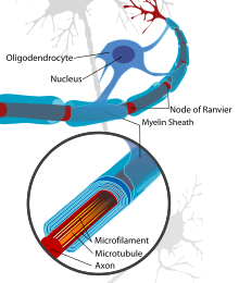
1269:
413:
226:
199:
172:
160:
46:
377:(CNS). Although research is being conducted on protecting oligodendrocytes and promoting
767:
Snaidero N, Möbius W, Czopka T, Hekking LH, Mathisen C, Verkleij D; et al. (2014).
1087:
1017:"Developmental (myelogenetic) localisation of the cerebral cortex in the human subject"
957:
940:
793:
768:
647:
607:
582:
558:
531:
366:
275:
267:
222:
38:
34:
1034:
1320:
663:
409:
382:
378:
254:
50:
516:
1055:
421:
412:
spent most of his career studying and publishing the details of the process in the
271:
198:
Neuron with oligodendrocyte and myelin sheath showing cytoskeletal structures at a
82:
24:
744:
727:
245:
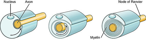
1. Axon 2. Nucleus of Schwann cell 3. Schwann cell 4. Myelin sheath 5. Neurilemma
1106:
993:
462:
393:
319:
241:
183:
784:
728:"On the biogenesis of myelin membranes: sorting, trafficking and cell polarity"
1021:
980:
Cohen JA (July 2009). "Emerging therapies for relapsing multiple sclerosis".
548:
282:
One early study showed that in the developing rat optic nerves, formation of
1215:
631:
330:
315:
287:
1001:
966:
802:
753:
655:
616:
567:
508:
457:
250:
1101:
1050:
500:
416:
of humans. This takes place mostly between two months before and after
307:
89:. Therefore, the first stage of myelinogenesis is often defined as the
1144:
1128:
598:
358:
346:
1016:
1123:
939:
Rodgers, Jane M.; Robinson, Andrew P.; Miller, Stephen D. (2013).
417:
392:
240:
210:
193:
182:
461:
law identical with the law which I have shown holds good for the
354:
337:
it appears that myelination is delayed without their synthesis.
54:
1059:
532:"Myelin Dynamics Throughout Life: An Ever-Changing Landscape?"
381:
in MS, current therapies mainly address the role of the
914:
National Institute of Neurological Disorders and Stroke
886:
884:
882:
880:
878:
876:
874:
872:
870:
302:
Studies on the developing optic nerve revealed that
1294:
1251:
1203:
1165:
1158:
1137:
1094:
837:
835:
583:"Oligodendroglia: metabolic supporters of neurons"
175:, the myelin sheath's edges form paranodal loops.
16:Formation of myelin sheaths in the nervous system
453:
1071:
712:
710:
708:
8:
815:: CS1 maint: multiple names: authors list (
676:: CS1 maint: multiple names: authors list (
53:following injury. Successful myelination of
910:"NINDS Multiple Sclerosis Information Page"
221:Recent research in rats has suggested that
1162:
1078:
1064:
1056:
860:
858:
856:
698:
696:
956:
792:
743:
606:
557:
547:
373:(MS), where demyelination occurs in the
18:
478:
41:, typically initiated in late prenatal
23:Myelination of a peripheral nerve by a
808:
669:
7:
439:The last areas to myelinate are the
33:is the formation and development of
916:. 19 November 2015. Archived from
648:10.1016/j.neuroscience.2019.07.014
365:can affect the functioning of the
286:and subsequent myelination occurs
270:and biochemical techniques in rat
14:
262:Myelinogenesis in the optic nerve
1119:Lateralization of brain function
581:Philips T, Rothstein JD (2017).
530:Williamson JM, Lyons DA (2018).
188:Transmission electron micrograph
144:
95:oligodendrocyte progenitor cells
1190:Somatosensory evoked potentials
428:'s area 4), the second is the
231:thyroid hormone receptor alpha
1:
1035:10.1016/s0140-6736(01)01429-5
1015:Flechsig, Paul (1901-10-19).
745:10.1016/j.febslet.2009.10.085
432:and the third is part of the
400:(CP: posterior centrale) and
726:Baron W, Hoekstra D (2010).
398:Primary somatosensory cortex
994:10.1001/archneurol.2009.104
310:() into sulfatide in vivo.
1343:
1327:Developmental neuroscience
785:10.1016/j.cell.2013.11.044
132:myelin proteolipid protein
1185:Auditory evoked potential
353:layer that surrounds the
237:Peripheral myelinogenesis
120:along their target axons.
117:Immature oligodendrocyte:
87:peripheral nervous system
47:neural circuit plasticity
549:10.3389/fncel.2018.00424
1195:Visual evoked potential
449:dorsolateral prefrontal
351:electrically insulating
124:Mature oligodendrocyte:
1279:Long-term potentiation
1231:Postsynaptic potential
1175:Bereitschaftspotential
471:
447:cortex (F#44) and the
405:
375:central nervous system
369:. One such disease is
246:
207:basic helix–loop–helix
202:
191:
157:central nervous system
79:central nervous system
27:
1114:Intracranial pressure
396:
363:demyelinating disease
341:Clinical significance
244:
209:transcription factor
197:
186:
22:
1284:Long-term depression
1259:Axoplasmic transport
456:convolutions of the
434:somatosensory cortex
408:Another researcher,
402:primary motor cortex
318:and the activity of
190:of a myelinated axon
128:myelin basic protein
73:Myelin is formed by
63:saltatory conduction
1274:Synaptic plasticity
1266:/Nerve regeneration
920:on 13 February 2016
536:Front Cell Neurosci
443:cortex (F#43), the
155:Myelination in the
111:Preoligodendrocyte:
1221:Membrane potential
1086:Physiology of the
945:Discovery Medicine
501:10.1002/glia.22632
441:anterior cingulate
406:
385:in demyelination.
371:multiple sclerosis
304:galactocerebroside
298:Role of sulfatides
247:
203:
192:
61:speed by enabling
28:
1314:
1313:
1310:
1309:
1264:Neuroregeneration
1211:Neurotransmission
1051:Myelination Atlas
467:medulla oblongata
445:inferior temporal
1334:
1226:Action potential
1204:Other short term
1167:Evoked potential
1163:
1080:
1073:
1066:
1057:
1039:
1038:
1012:
1006:
1005:
977:
971:
970:
960:
936:
930:
929:
927:
925:
906:
900:
897:
891:
888:
865:
862:
851:
848:
842:
839:
830:
827:
821:
820:
814:
806:
796:
764:
758:
757:
747:
723:
717:
714:
703:
700:
691:
688:
682:
681:
675:
667:
627:
621:
620:
610:
599:10.1172/JCI90610
593:(9): 3271–3280.
578:
572:
571:
561:
551:
527:
521:
520:
483:
430:olfactory cortex
389:Research History
324:sulfotransferase
284:oligodendrocytes
173:nodes of Ranvier
148:
105:Differentiation:
75:oligodendrocytes
59:action potential
43:neurodevelopment
1342:
1341:
1337:
1336:
1335:
1333:
1332:
1331:
1317:
1316:
1315:
1306:
1290:
1270:Neuroplasticity
1247:
1199:
1154:
1133:
1090:
1084:
1047:
1042:
1014:
1013:
1009:
979:
978:
974:
938:
937:
933:
923:
921:
908:
907:
903:
898:
894:
889:
868:
863:
854:
849:
845:
840:
833:
828:
824:
807:
779:(1–2): 277–90.
766:
765:
761:
725:
724:
720:
715:
706:
701:
694:
689:
685:
668:
629:
628:
624:
580:
579:
575:
529:
528:
524:
485:
484:
480:
476:
451:cortex (F#45).
414:cerebral cortex
391:
343:
300:
264:
239:
227:thyroid hormone
200:node of Ranvier
181:
165:
164:
163:
161:oligodendrocyte
154:
149:
91:differentiation
71:
17:
12:
11:
5:
1340:
1338:
1330:
1329:
1319:
1318:
1312:
1311:
1308:
1307:
1305:
1304:
1302:Myelinogenesis
1298:
1296:
1292:
1291:
1289:
1288:
1287:
1286:
1281:
1267:
1261:
1255:
1253:
1249:
1248:
1246:
1245:
1244:
1243:
1238:
1228:
1223:
1218:
1213:
1207:
1205:
1201:
1200:
1198:
1197:
1192:
1187:
1182:
1177:
1171:
1169:
1160:
1156:
1155:
1153:
1152:
1147:
1141:
1139:
1135:
1134:
1132:
1131:
1126:
1121:
1116:
1111:
1110:
1109:
1098:
1096:
1092:
1091:
1088:nervous system
1085:
1083:
1082:
1075:
1068:
1060:
1054:
1053:
1046:
1045:External links
1043:
1041:
1040:
1029:(4077): 1028.
1007:
972:
931:
901:
892:
866:
852:
843:
831:
822:
759:
738:(9): 1760–70.
718:
704:
692:
683:
622:
573:
522:
495:(4): 649–665.
477:
475:
472:
390:
387:
367:nervous system
342:
339:
299:
296:
276:model organism
268:ultrastructure
263:
260:
238:
235:
223:apotransferrin
180:
177:
151:
150:
143:
142:
141:
136:
135:
121:
114:
108:
70:
67:
39:nervous system
35:myelin sheaths
31:Myelinogenesis
15:
13:
10:
9:
6:
4:
3:
2:
1339:
1328:
1325:
1324:
1322:
1303:
1300:
1299:
1297:
1293:
1285:
1282:
1280:
1277:
1276:
1275:
1271:
1268:
1265:
1262:
1260:
1257:
1256:
1254:
1250:
1242:
1239:
1237:
1234:
1233:
1232:
1229:
1227:
1224:
1222:
1219:
1217:
1214:
1212:
1209:
1208:
1206:
1202:
1196:
1193:
1191:
1188:
1186:
1183:
1181:
1178:
1176:
1173:
1172:
1170:
1168:
1164:
1161:
1157:
1151:
1148:
1146:
1143:
1142:
1140:
1138:Primarily PNS
1136:
1130:
1127:
1125:
1122:
1120:
1117:
1115:
1112:
1108:
1105:
1104:
1103:
1100:
1099:
1097:
1095:Primarily CNS
1093:
1089:
1081:
1076:
1074:
1069:
1067:
1062:
1061:
1058:
1052:
1049:
1048:
1044:
1036:
1032:
1028:
1024:
1023:
1018:
1011:
1008:
1003:
999:
995:
991:
987:
983:
976:
973:
968:
964:
959:
954:
951:(86): 53–63.
950:
946:
942:
935:
932:
919:
915:
911:
905:
902:
896:
893:
887:
885:
883:
881:
879:
877:
875:
873:
871:
867:
861:
859:
857:
853:
847:
844:
838:
836:
832:
826:
823:
818:
812:
804:
800:
795:
790:
786:
782:
778:
774:
770:
763:
760:
755:
751:
746:
741:
737:
733:
729:
722:
719:
713:
711:
709:
705:
699:
697:
693:
687:
684:
679:
673:
665:
661:
657:
653:
649:
645:
641:
637:
633:
626:
623:
618:
614:
609:
604:
600:
596:
592:
588:
587:J Clin Invest
584:
577:
574:
569:
565:
560:
555:
550:
545:
541:
537:
533:
526:
523:
518:
514:
510:
506:
502:
498:
494:
490:
482:
479:
473:
470:
468:
464:
459:
452:
450:
446:
442:
437:
435:
431:
427:
423:
419:
415:
411:
410:Paul Flechsig
403:
399:
395:
388:
386:
384:
383:immune system
380:
379:remyelination
376:
372:
368:
364:
360:
356:
352:
348:
340:
338:
334:
332:
327:
325:
321:
317:
311:
309:
305:
297:
295:
291:
289:
285:
280:
277:
273:
269:
261:
259:
256:
255:Schwann cells
252:
243:
236:
234:
232:
228:
224:
219:
215:
212:
208:
201:
196:
189:
185:
178:
176:
174:
170:
162:
158:
153:
147:
140:
133:
129:
125:
122:
118:
115:
112:
109:
106:
103:
102:
101:
98:
96:
92:
88:
84:
83:Schwann cells
80:
76:
68:
66:
64:
60:
56:
52:
51:remyelination
48:
44:
40:
36:
32:
26:
21:
1301:
1026:
1020:
1010:
988:(7): 821–8.
985:
982:Arch. Neurol
981:
975:
948:
944:
934:
922:. Retrieved
918:the original
913:
904:
895:
846:
825:
811:cite journal
776:
772:
762:
735:
731:
721:
686:
672:cite journal
639:
636:Neuroscience
635:
625:
590:
586:
576:
539:
535:
525:
492:
488:
481:
454:
438:
436:(BA 3,1,2).
422:motor cortex
407:
344:
335:
329:In the CNS,
328:
312:
301:
292:
281:
272:optic nerves
265:
258:P2, and P0.
248:
220:
216:
204:
166:
137:
123:
116:
110:
104:
99:
72:
30:
29:
25:Schwann cell
1107:Wakefulness
642:: 163–180.
463:spinal cord
359:nerve cells
320:cerebroside
49:as well as
1241:Inhibitory
1236:Excitatory
1022:The Lancet
474:References
169:translated
130:(MBP) and
57:increases
1252:Long term
1216:Chronaxie
1150:Sensation
732:FEBS Lett
664:198934117
424:(part of
349:forms an
331:sulfatide
316:sulfatide
288:postnatal
179:Mechanism
1321:Category
1002:19597083
967:23911232
803:24439382
754:19896485
656:31349008
617:28862639
568:30510502
517:25559134
509:24481644
458:cerebrum
426:Brodmann
357:of some
345:Because
251:SDS-PAGE
1102:Arousal
958:3970909
924:6 March
794:4862569
608:5669561
559:6252314
542:: 424.
308:sulfate
85:in the
77:in the
37:in the
1145:Reflex
1129:Memory
1000:
965:
955:
801:
791:
752:
662:
654:
615:
605:
566:
556:
515:
507:
465:, the
361:, any
347:myelin
159:by an
134:(PLP).
69:Stages
1295:Other
1124:Sleep
660:S2CID
513:S2CID
418:birth
211:OLIG1
55:axons
1180:P300
1159:Both
998:PMID
963:PMID
926:2016
817:link
799:PMID
773:Cell
750:PMID
678:link
652:PMID
613:PMID
564:PMID
505:PMID
489:Glia
355:axon
225:and
205:The
81:and
1031:doi
1027:158
990:doi
953:PMC
789:PMC
781:doi
777:156
740:doi
736:584
644:doi
640:439
603:PMC
595:doi
591:127
554:PMC
544:doi
497:doi
93:of
1323::
1025:.
1019:.
996:.
986:66
984:.
961:.
949:86
947:.
943:.
912:.
869:^
855:^
834:^
813:}}
809:{{
797:.
787:.
775:.
771:.
748:.
734:.
730:.
707:^
695:^
674:}}
670:{{
658:.
650:.
638:.
634:.
611:.
601:.
589:.
585:.
562:.
552:.
540:12
538:.
534:.
511:.
503:.
493:62
491:.
322:,
233:.
1272:/
1079:e
1072:t
1065:v
1037:.
1033::
1004:.
992::
969:.
928:.
819:)
805:.
783::
756:.
742::
680:)
666:.
646::
619:.
597::
570:.
546::
519:.
499::
Text is available under the Creative Commons Attribution-ShareAlike License. Additional terms may apply.