68:
170:
29:
193:
tubular structures of mature and bland appearance with scanty interposed stroma. Cells are small with dark staining nuclei and inconspicuous nucleoli. Blastema is absent whereas calcospherites may be present. Glomeruloid figures are a striking finding, reminiscent of early fetal metenephric tissue. The lumen of the acini may contain otherwise epithelial infoldings or fibrillary material but it is quite often empty. Mitoses are conspicuously absent. In the series reported by Jones
147:
201:
found that intense and diffuse immunoreactivity for alpha-methylacyl-CoA racemase (AMACR) is useful in differentiating renal cell carcinoma from MA but a panel including AMACR, CK7 and CD57 is better in this differential diagnosis. Differential diagnosis may be quite difficult indeed as exemplified
192:
Metanephric adenoma is diagnosed histologically. The tumours can be located at upper pole, lower pole and mid-hilar region of the kidney; they are well circumscribed but unencapsulated, tan pink, with possible cystic and hemorrhagic foci. They show a uniform architecture of closely packed acinar or
231:
MA has been described in the past under other names such as néphrome néphronogène, metanephroider
Nierentumor and nephroblastomartiges Nierenadenom (5) but the term metanephric adenoma was suggested by Brisigotti, Cozzutto et al. in 1992 and then widely accepted. Prior to this report, Nagashima et
214:
stated that genetic analysis of chromosome 7, 17, and Y may facilitate discrimination of MA from papillary renal cell carcinoma in difficult cases. Their study showed that MA lacks the frequent gain of chromosomes 7 and 17 and losses of the Y chromosome that are typical of papillary renal cell
132:
and a palpable mass. Mean age at onset is around 40 years with a range of 5 to 83 years and the mean size of the tumour is 5.5 cm with a range 0.3 to 15 cm (1). Polycythemia is more frequent in MA than in any other type of renal tumour. Of further relevance is that this tumour is more
748:
733:
406:
Jones EC, Pins M, Dickersin GR, Young RH (June 1995). "Metanephric adenoma of the kidney. A clinicopathological, immunohistochemical, flow cytometric, cytogenetic, and electron microscopic study of seven cases".
197:
tumour cells were reactive for Leu7 in 3 cases of 5, to vimentine in 4 of 6, to cytocheratin in 2 of 6, to epithelial membrane antigen in 1 of 6 cases and muscle specific antigen in 1 of 6. Olgac
232:
al. in 1991 had not offered a nosological innovation for their two cases whereas the denomination of néphrome néphronogène proposed by Pages and
Granier in 1980 had gone largely undetected.
533:
Pins MR, Jones EC, Martul EV, Kamat BR, Umlas J, Renshaw AA (May 1999). "Metanephric adenoma-like tumors of the kidney: report of 3 malignancies with emphasis on discriminating features".
133:
commonly calcified than any other kidney neoplasm. Surgery is curative and no other treatment is recommended. There is so far no evidence of metastases or local recurrence.
652:
Nagashima Y, Arai N, Tanaka Y, Yoshida S, Sumino K, Ohaki Y, Matsushita K, Morita T, Misugi K (1991). "Two cases of a renal epithelial tumour resembling immature nephron".
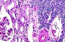
180:, right of image, showing the characteristic features (round nuclear membrane, no nucleoli, and fine chromatin). Normal kidney is seen on the left of the image. Kidney
249:
Bastos Netto JM, Esteves TC, Mattos R, Tibiriçá SH, Costa SM, Vieira LJ (August 2007). "Metanephric adenoma: a rare differential diagnosis of renal tumor in children".
490:
Olgac S, Hutchinson B, Tickoo SK, Reuter VE (February 2006). "Alpha-methylacyl-CoA racemase as a marker in the differential diagnosis of metanephric adenoma".
791:
570:"Metanephric adenoma lacks the gains of chromosomes 7 and 17 and loss of Y that are typical of papillary renal cell carcinoma and papillary adenoma"
1004:
340:
323:
284:
Davis CJ, Barton JH, Sesterhenn IA, Mostofi FK (October 1995). "Metanephric adenoma. Clinicopathological study of fifty patients".
864:
784:
1025:
973:
945:
777:
900:
895:
978:
950:
905:
202:
by the three malignancies initially diagnosed as MA that later metastasized, in the report by Pins et al.
169:
28:
223:
As metanephric adenomas are considered benign, they can be left in place, i.e. no treatment is needed.
859:
831:
117:
96:
44:
67:
752:
104:
955:
677:
599:
515:
353:
374:
Brisigotti M, Cozzutto C, Fabbretti G, Sergi C, Callea F (October 1992). "Metanephric adenoma".
146:
927:
669:
634:
591:
550:
507:
472:
424:
383:
345:
322:
Kovacs G, Akhtar M, Beckwith BJ, Bugert P, Cooper CS, Delahunt B, et al. (October 1997).
301:
266:
56:
836:
769:
661:
626:
581:
542:
499:
416:
335:
293:
258:
215:
neoplasms, suggesting that MA is not related to renal cell carcinoma and papillary adenoma.
965:
919:
874:
854:
463:
Grignon DJ, Eble JN (February 1998). "Papillary and metanephric adenomas of the kidney".
983:
937:
869:
808:
804:
617:
Galmiche L, Vasiliu V, Poirée S, Hélénon O, Casanova JM, Brousse N (October 2007). "".
125:
92:
757:
630:
586:
569:
1019:
816:
420:
297:
681:
603:
519:
357:
121:
102:
It should not be confused with the pathologically unrelated, yet similar sounding,
48:
546:
262:
846:
742:
503:
173:
150:
36:
341:
10.1002/(SICI)1096-9896(199710)183:2<131::AID-PATH931>3.0.CO;2-G
185:
162:
129:
638:
595:
554:
511:
270:
673:
476:
428:
387:
349:
305:
988:
800:
61:
725:
665:
737:
181:
158:
88:
84:
81:
568:
Brunelli M, Eble JN, Zhang S, Martignoni G, Cheng L (October 2003).
168:
145:
116:
The symptoms may be similar to those classically associated with
773:
157:(right of image). Normal kidney is seen on the left. Kidney
695:
Pages A, Granier M (1980). "Le néphrome néphronogène".
43:
with structures reminiscent of those seen in papillary
91:, that can have a microscopic appearance similar to a
324:"The Heidelberg classification of renal cell tumours"
715:
997:
964:
936:
918:
888:
845:
824:
815:
719:
55:
21:
535:Archives of Pathology & Laboratory Medicine
785:
458:
456:
369:
367:
317:
315:
8:
401:
399:
397:
821:
792:
778:
770:
716:
440:
438:
409:The American Journal of Surgical Pathology
286:The American Journal of Surgical Pathology
66:
27:
18:
585:
339:
241:
7:
14:
825:Glandular and epithelial neoplasm
587:10.1097/01.MP.0000090923.50509.55
865:Clear-cell sarcoma of the kidney
465:Seminars in Diagnostic Pathology
421:10.1097/00000478-199506000-00001
298:10.1097/00000478-199510000-00001
449:. St Louis: Mosby. p. 118.
1005:Malignant fibrous histiocytoma
93:nephroblastoma (Wilms tumours)
1:
631:10.1016/s0242-6498(07)78275-5
445:Bostwick DG, Eble JN (2008).
547:10.5858/1999-123-0415-MALTOT
376:Histology and Histopathology
263:10.1016/j.jpurol.2006.10.003
251:Journal of Pediatric Urology
974:Transitional cell carcinoma
946:Transitional cell carcinoma
447:Urologic Surgical Pathology
206:Cytogenetic characteristics
1042:
901:Juxtaglomerular cell tumor
896:Renal medullary carcinoma
504:10.1038/modpathol.3800520
35:
26:
328:The Journal of Pathology
979:Squamous-cell carcinoma
951:Squamous-cell carcinoma
906:Renal medullary fibroma
1026:Benign renal neoplasms
697:Arch Anat Cytol Pathol
189:
166:
619:Annales de Pathologie
172:
149:
860:Mesoblastic nephroma
832:Renal cell carcinoma
118:renal cell carcinoma
97:renal cell carcinoma
45:renal cell carcinoma
880:Metanephric adenoma
178:metanephric adenoma
155:metanephric adenoma
105:mesonephric adenoma
78:Metanephric adenoma
41:metanephric adenoma
22:Metanephric adenoma
956:Inverted papilloma
666:10.1007/BF01600247
190:
167:
120:, and may include
1013:
1012:
928:Ureteral neoplasm
914:
913:
767:
766:
654:Virchows Archiv A
95:, or a papillary
75:
74:
16:Medical condition
1033:
837:Renal oncocytoma
822:
794:
787:
780:
771:
717:
705:
704:
692:
686:
685:
649:
643:
642:
614:
608:
607:
589:
574:Modern Pathology
565:
559:
558:
530:
524:
523:
492:Modern Pathology
487:
481:
480:
460:
451:
450:
442:
433:
432:
403:
392:
391:
371:
362:
361:
343:
319:
310:
309:
281:
275:
274:
246:
80:(MA) is a rare,
71:
70:
31:
19:
1041:
1040:
1036:
1035:
1034:
1032:
1031:
1030:
1016:
1015:
1014:
1009:
993:
960:
932:
910:
884:
875:Cystic nephroma
841:
811:
809:genital systems
798:
768:
763:
762:
728:
714:
709:
708:
694:
693:
689:
651:
650:
646:
616:
615:
611:
567:
566:
562:
532:
531:
527:
489:
488:
484:
462:
461:
454:
444:
443:
436:
405:
404:
395:
373:
372:
365:
321:
320:
313:
292:(10): 1101–14.
283:
282:
278:
248:
247:
243:
238:
229:
221:
208:
144:
139:
114:
65:
17:
12:
11:
5:
1039:
1037:
1029:
1028:
1018:
1017:
1011:
1010:
1008:
1007:
1001:
999:
995:
994:
992:
991:
986:
984:Adenocarcinoma
981:
976:
970:
968:
962:
961:
959:
958:
953:
948:
942:
940:
934:
933:
931:
930:
924:
922:
916:
915:
912:
911:
909:
908:
903:
898:
892:
890:
886:
885:
883:
882:
877:
872:
870:Angiomyolipoma
867:
862:
857:
851:
849:
843:
842:
840:
839:
834:
828:
826:
819:
813:
812:
799:
797:
796:
789:
782:
774:
765:
764:
761:
760:
745:
729:
724:
723:
721:
720:Classification
713:
712:External links
710:
707:
706:
687:
644:
625:(5): 365–368.
609:
580:(10): 1060–3.
560:
525:
482:
452:
434:
415:(6): 615–626.
393:
382:(4): 689–692.
363:
334:(2): 131–133.
311:
276:
257:(4): 340–341.
240:
239:
237:
234:
228:
225:
220:
217:
207:
204:
143:
142:Histopathology
140:
138:
135:
126:abdominal pain
113:
110:
73:
72:
59:
53:
52:
33:
32:
24:
23:
15:
13:
10:
9:
6:
4:
3:
2:
1038:
1027:
1024:
1023:
1021:
1006:
1003:
1002:
1000:
996:
990:
987:
985:
982:
980:
977:
975:
972:
971:
969:
967:
963:
957:
954:
952:
949:
947:
944:
943:
941:
939:
935:
929:
926:
925:
923:
921:
917:
907:
904:
902:
899:
897:
894:
893:
891:
887:
881:
878:
876:
873:
871:
868:
866:
863:
861:
858:
856:
853:
852:
850:
848:
844:
838:
835:
833:
830:
829:
827:
823:
820:
818:
814:
810:
806:
802:
795:
790:
788:
783:
781:
776:
775:
772:
759:
755:
754:
750:
746:
744:
740:
739:
735:
731:
730:
727:
722:
718:
711:
702:
698:
691:
688:
683:
679:
675:
671:
667:
663:
659:
655:
648:
645:
640:
636:
632:
628:
624:
621:(in French).
620:
613:
610:
605:
601:
597:
593:
588:
583:
579:
575:
571:
564:
561:
556:
552:
548:
544:
541:(5): 415–20.
540:
536:
529:
526:
521:
517:
513:
509:
505:
501:
498:(2): 218–24.
497:
493:
486:
483:
478:
474:
470:
466:
459:
457:
453:
448:
441:
439:
435:
430:
426:
422:
418:
414:
410:
402:
400:
398:
394:
389:
385:
381:
377:
370:
368:
364:
359:
355:
351:
347:
342:
337:
333:
329:
325:
318:
316:
312:
307:
303:
299:
295:
291:
287:
280:
277:
272:
268:
264:
260:
256:
252:
245:
242:
235:
233:
226:
224:
218:
216:
213:
205:
203:
200:
196:
187:
183:
179:
175:
171:
164:
160:
156:
152:
148:
141:
136:
134:
131:
127:
123:
119:
111:
109:
107:
106:
100:
98:
94:
90:
86:
83:
79:
69:
63:
60:
58:
54:
50:
49:H&E stain
46:
42:
38:
34:
30:
25:
20:
879:
855:Wilms' tumor
747:
732:
700:
696:
690:
660:(1): 77–81.
657:
653:
647:
622:
618:
612:
577:
573:
563:
538:
534:
528:
495:
491:
485:
471:(1): 41–53.
468:
464:
446:
412:
408:
379:
375:
331:
327:
289:
285:
279:
254:
250:
244:
230:
222:
211:
209:
198:
194:
191:
177:
154:
122:polycythemia
115:
103:
101:
77:
76:
40:
889:by location
847:Mixed tumor
236:References
174:Micrograph
151:Micrograph
37:Micrograph
703:: 99–103.
219:Treatment
210:Brunelli
186:PAS stain
163:PAS stain
137:Pathology
130:hematuria
57:Specialty
1020:Category
989:Melanoma
682:28924982
639:18185471
604:11701480
596:14559991
555:10235500
520:30923148
512:16424894
358:34796951
271:18947770
112:Symptoms
62:Oncology
966:Urethra
938:Bladder
805:urinary
803:of the
674:1989379
477:9503505
429:7755148
388:1333853
350:9390023
306:7573669
227:History
87:of the
920:Ureter
817:Kidney
801:Tumors
680:
672:
637:
602:
594:
553:
518:
510:
475:
427:
386:
356:
348:
304:
269:
212:et al.
199:et al.
195:et al.
182:biopsy
159:biopsy
89:kidney
85:tumour
82:benign
64:
998:Other
758:223.0
743:D30.0
678:S2CID
600:S2CID
516:S2CID
354:S2CID
176:of a
153:of a
39:of a
807:and
753:9-CM
670:PMID
635:PMID
592:PMID
551:PMID
508:PMID
473:PMID
425:PMID
384:PMID
346:PMID
302:PMID
267:PMID
749:ICD
734:ICD
662:doi
658:418
627:doi
582:doi
543:doi
539:123
500:doi
417:doi
336:doi
332:183
294:doi
259:doi
1022::
756::
741::
738:10
701:28
699:.
676:.
668:.
656:.
633:.
623:27
598:.
590:.
578:16
576:.
572:.
549:.
537:.
514:.
506:.
496:19
494:.
469:15
467:.
455:^
437:^
423:.
413:19
411:.
396:^
378:.
366:^
352:.
344:.
330:.
326:.
314:^
300:.
290:19
288:.
265:.
253:.
184:.
161:.
128:,
124:,
108:.
99:.
47:.
793:e
786:t
779:v
751:-
736:-
726:D
684:.
664::
641:.
629::
606:.
584::
557:.
545::
522:.
502::
479:.
431:.
419::
390:.
380:7
360:.
338::
308:.
296::
273:.
261::
255:3
188:.
165:.
51:.
Text is available under the Creative Commons Attribution-ShareAlike License. Additional terms may apply.