911:, whose goal is to shrink the tumour size before surgery. Studies show that if the therapy is efficient, then the water, collagen and hemoglobin contents of the lesion show a decreasing behaviour over time, which suggests that the initially fibrous tissue acquires features similar to the adipose one. Optical measurements in correspondence with therapy sessions could track its evolution, so to assess the patient's response to it. Moreover, it is believed that therapy effectiveness could be predicted even on the first day of treatment on the base of initial breast constituents' concentrations.
32:
463:
3886:
867:(CW) measurements, the light source is a continuous wave laser, which hinders the separation of the absorption and scattering contributions with a single measurement. A possible solution is to perform space or angle-resolved measurements. In general, the CW approach is combined with the frequency domain one, in order to reinforce the strengths of both.
157:
development, there is a lack of standardization in data analysis among the research groups dealing with it, and it suffers from low spatial resolution. For this reason, a "multimodal approach" is suggested, where optical mammography is complementary to another conventional technique, so that also the diagnostic efficacy is improved.
775:: injection and collection occur on the same side of the breast. The woman is usually prone or bent forward and places the breast on a support provided with a hole where sources and detectors are located. Other systems' setups instead require the woman to lie supine and the measurement is carried out with a hand-held probe.
884:, relatively to the adipose one. The main constituents of a fibrous tissue are water, collagen and hemoglobin and optical mammography is able to discriminate and quantify tissues' components. Therefore, by measuring breast constituents' concentrations, optical mammography could assess breast cancer risk.
156:
On the contrary, optical mammography is cheap, efficient also on dense breasts, and devoid of any side effect, so that it can be used to track the evolution of the patient's condition on a daily basis. It is also able to characterize breast from a physiologic point of view. However, being still under
2650:
Grosenick, Dirk; Moesta, K Thomas; Möller, Michael; Mucke, Jörg; Wabnitz, Heidrun; Gebauer, Bernd; Stroszczynski, Christian; Wassermann, Bernhard; Schlag, Peter M; Rinneberg, Herbert (7 June 2005). "Time-domain scanning optical mammography: I. Recording and assessment of mammograms of 154 patients".
1593:
Lord, S.J.; Lei, W.; Craft, P.; Cawson, J.N.; Morris, I.; Walleser, S.; Griffiths, A.; Parker, S.; Houssami, N. (September 2007). "A systematic review of the effectiveness of magnetic resonance imaging (MRI) as an addition to mammography and ultrasound in screening young women at high risk of breast
895:
are generally made of fibrous tissue and could be recognized in the constituents' maps as local spots with higher concentrations of water, collagen and hemoglobin with respect to the surrounding, mostly adipose, healthy tissues. Studies demonstrate that the variation in concentration with respect to
144:
shows a good correlation with the tumour dimensions and is claimed to be the best method for the identification and characterization of lesions. Even though there is no verified long-term health risk from the magnetic fields employed during an MRI, it is not used as first investigative tool because
2694:
Choe, Regine; Konecky, Soren D.; Corlu, Alper; Lee, Kijoon; Durduran, Turgut; Busch, David R.; Pathak, Saurav; Czerniecki, Brian J.; Tchou, Julia; Fraker, Douglas L.; DeMichele, Angela; Chance, Britton; Arridge, Simon R.; Schweiger, Martin; Culver, Joseph P.; Schnall, Mitchell D.; Putt, Mary E.;
758:
Blood strongly absorbs in the red spectral range, whereas collagen, water and lipids have their absorption peaks at wavelengths longer than 900 nm. The distinction between oxy and deoxy-haemoglobin is due to the presence of a second large peak in the case of oxy-haemoglobin. Lipids are
2202:
Enfield, Louise C.; Gibson, Adam P.; Everdell, Nicholas L.; Delpy, David T.; Schweiger, Martin; Arridge, Simon R.; Richardson, Caroline; Keshtgar, Mohammad; Douek, Michael; Hebden, Jeremy C. (18 May 2007). "Three-dimensional time-resolved optical mammography of the uncompressed breast".
854:
measurements, an intensity-modulated signal is injected into the breast and its optical properties are deduced from the dephasement and the demodulation of the output signal with respect to the input one. The measurement is repeated for different values of the frequency modulation.
2299:
Becker, Wolfgang; Bergmann, Axel; Biscotti, Giovanni Luca; Rueck, Angelika (2004). "Advanced time-correlated single photon counting techniques for spectroscopy and imaging in biomedical systems". In Neev, Joseph; Schaffer, Christopher B; Ostendorf, Andreas (eds.).
935:
896:
the healthy tissue is statistically more marked in the case of malignant tumours than benign ones. In addition, the scattering coefficient is generally higher for benign lesions. Such distinctions suggest that optical mammography could characterize breast lesions.
349:
171:
2603:
Leff, Daniel
Richard; Warren, Oliver J.; Enfield, Louise C.; Gibson, Adam; Athanasiou, Thanos; Patten, Darren K.; Hebden, Jem; Yang, Guang Zhong; Darzi, Ara (28 April 2007). "Diffuse optical imaging of the healthy and diseased breast: A systematic review".
2334:
Chance, B.; Cooper, C. E.; Delpy, D. T.; Reynolds, E. O. R.; Tromberg, Bruce J.; Coquoz, Olivier; Fishkin, Joshua B.; Pham, Tuan; Anderson, Eric R.; Butler, John; Cahn, Mitchell; Gross, Jeffrey D.; Venugopalan, Vasan; Pham, David (29 June 1997).
837:
measurements, short light pulses of the order of hundreds of picoseconds are delivered to the breast and its optical properties are retrieved from the features of the re-emitted pulses, which have undergone delay, broadening and attenuation.
1358:
Dobruch-Sobczak, Katarzyna; Piotrzkowska-Wróblewska, Hanna; Klimoda, Ziemowit; Secomski, Wojciech; Karwat, Piotr; Markiewicz-Grodzicka, Ewa; Kolasińska-Ćwikła, Agnieszka; Roszkowska-Purska, Katarzyna; Litniewski, Jerzy (28 June 2019).
352:”: scattering is assumed to be isotropic and strongly dominant over absorption. This is fairly accurate for example for the breast tissue, in the red and near infrared spectral range (between 600 and 1100 nm), known also as "
458:
1155:
Jiang, Shudong; Pogue, Brian W.; Carpenter, Colin M.; Poplack, Steven P.; Wells, Wendy A.; Kogel, Christine A.; Forero, Jorge A.; Muffly, Lori S.; Schwartz, Gary N.; Paulsen, Keith D.; Kaufman, Peter A. (August 2009).
767:
Diffuse optical mammography can be implemented exploiting three different approaches: time domain, frequency domain and continuous wave. Moreover, there exist two main geometries to perform an optical measurement:
356:". In the therapeutic window, light can penetrate a few centimetres, so that it can explore the volume at exam. This is the reason why photon migration in biological tissues is known also as "diffuse optics".
673:
1102:
Quarto, Giovanna; Spinelli, Lorenzo; Pifferi, Antonio; Torricelli, Alessandro; Cubeddu, Rinaldo; Abbate, Francesca; Balestreri, Nicola; Menna, Simona; Cassano, Enrico; Taroni, Paola (18 September 2014).
1052:
Taroni, Paola; Pifferi, Antonio; Quarto, Giovanna; Spinelli, Lorenzo; Torricelli, Alessandro; Abbate, Francesca; Villa, Anna; Balestreri, Nicola; Menna, Simona; Cassano, Enrico; Cubeddu, Rinaldo (2010).
2054:
Jiang, Huabei; Iftimia, Nicusor V.; Xu, Yong; Eggert, Julia A.; Fajardo, Laurie L.; Klove, Karen L. (February 2002). "Near-Infrared
Optical Imaging of the Breast with Model-Based Reconstruction".
759:
characterized by absorption maxima at 930 nm and 1040 nm, while the wavelength 975 nm is sensitive to water. Finally, an absorption peak for collagen takes place at 1030 nm.
2392:
Grosenick, Dirk; Wabnitz, Heidrun; Rinneberg, Herbert H.; Moesta, K. Thomas; Schlag, Peter M. (1 May 1999). "Development of a time-domain optical mammograph and first in vivo applications".
315:
128:, thanks to its high spatial resolution and the short measurement time. However, it is not sensitive to the breast physiology, it is characterized by a limited efficiency in investigating
1975:
Durduran, T.; Choe, R.; Culver, J. P.; Zubkov, L.; Holboke, M. J.; Giammarco, J.; Chance, B.; Yodh, A. G. (21 August 2002). "Bulk optical properties of healthy female breast tissue".
598:
36:
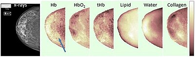
Example of breast constituents' concentrations maps through optical mammography (right cranio-caudal view). The blue arrow points to the lesion. Hb stands for deoxy-hemoglobin, HbO
140:
are non-invasive and they are used especially on young women, who are usually characterized by dense breasts, but the images interpretation depends on the operator's experience.
1821:
Taroni, Paola; Quarto, Giovanna; Pifferi, Antonio; Abbate, Francesca; Balestreri, Nicola; Menna, Simona; Cassano, Enrico; Cubeddu, Rinaldo; Batra, Surinder K. (1 June 2015).
800:
The use of multiple laser sources allows to investigate the breast constituents' concentrations of interest, by selecting some specific wavelengths. Detectors are usually
495:
729:
558:
381:
251:
224:
702:
980:
Taroni, Paola; Paganoni, Anna Maria; Ieva, Francesca; Pifferi, Antonio; Quarto, Giovanna; Abbate, Francesca; Cassano, Enrico; Cubeddu, Rinaldo (16 January 2017).
1823:"Breast Tissue Composition and Its Dependence on Demographic Risk Factors for Breast Cancer: Non-Invasive Assessment by Time Domain Diffuse Optical Spectroscopy"
1158:"Evaluation of Breast Tumor Response to Neoadjuvant Chemotherapy with Tomographic Diffuse Optical Spectroscopy: Case Studies of Tumor Region-of-Interest Changes"
3560:
3244:
535:
515:
339:
3374:
2909:
Roblyer, D.; Ueda, S.; Cerussi, A.; Tanamai, W.; Durkin, A.; Mehta, R.; Hsiang, D.; Butler, J. A.; McLaren, C.; Chen, W.-P.; Tromberg, B. (18 August 2011).
3386:
1883:
Provenzano, Paolo P; Inman, David R; Eliceiri, Kevin W; Knittel, Justin G; Yan, Long; Rueden, Curtis T; White, John G; Keely, Patricia J (28 April 2008).
3714:
2986:
940:
353:
3822:
392:
3352:
2091:"A prospective pilot clinical trial evaluating the utility of a dynamic near-infrared imaging device for characterizing suspicious breast lesions"
1361:"Monitoring the response to neoadjuvant chemotherapy in patients with breast cancer using ultrasound scattering coefficient: A preliminary report"
839:
809:
181:
3357:
950:
864:
851:
816:
1764:
Wang, Xin; Pogue, Brian W.; Jiang, Shudong; Song, Xiaomei; Paulsen, Keith D.; Kogel, Christine; Poplack, Steven P.; Wells, Wendy A. (2005).
253:
denotes the probability per unit length that a scattering event occurs. However, many studies refer to the reduced scattering coefficient
2911:"Optical imaging of breast cancer oxyhemoglobin flare correlates with neoadjuvant chemotherapy response one day after starting treatment"
2862:"Functional Imaging Using Diffuse Optical Spectroscopy of Neoadjuvant Chemotherapy Response in Women with Locally Advanced Breast Cancer"
3198:
1523:
Kaplan, Stuart S. (December 2001). "Clinical
Utility of Bilateral Whole-Breast US in the Evaluation of Women with Dense Breast Tissue".
3750:
3629:
3369:
3278:
3208:
2486:"Non-ionizing near-infrared radiation transillumination spectroscopy for breast tissue density and assessment of breast cancer risk"
2035:
1285:
609:
2153:
Ferocino, Edoardo; Martinenghi, Edoardo; Dalla Mora, Alberto; Pifferi, Antonio; Cubeddu, Rinaldo; Taroni, Paola (23 January 2018).
907:
depends on the characteristics of the tumour and the patient's condition. One of the possible strategies is the administration of
149:
allows the early evaluation of the metabolic changes of the tumour, but it is very expensive and requires the administration of a
3193:
3188:
3071:
781:: injection and collection occur on opposite sides of the breast. The breast is usually compressed between plane parallel plates.
3273:
3256:
2697:"Differentiation of benign and malignant breast tumors by in-vivo three-dimensional parallel-plate diffuse optical tomography"
812:
in the case of a time-resolved optical mammograph, or a filter for frequency modulation in the case of frequency-domain ones.
3634:
3481:
3381:
3364:
2860:
Soliman, H.; Gunasekara, A.; Rycroft, M.; Zubovits, J.; Dent, R.; Spayne, J.; Yaffe, M. J.; Czarnota, G. J. (20 April 2010).
1683:
Taroni, Paola (2012). "Diffuse optical imaging and spectroscopy of the breast: A brief outline of history and perspectives".
1558:
Hylton, Nola (10 March 2005). "Magnetic
Resonance Imaging of the Breast: Opportunities to Improve Breast Cancer Management".
197:
880:
A denser breast is more likely to develop breast cancer. A dense breast is characterized by a meaningful amount of fibrous
3760:
3614:
3213:
3088:
3080:
2979:
2752:
Zhu, Quing; Cronin, Edward B.; Currier, Allen A.; Vine, Hugh S.; Huang, Minming; Chen, NanGuang; Xu, Chen (October 2005).
982:"Non-invasive optical estimate of tissue composition to differentiate malignant from benign breast lesions: A pilot study"
561:
115:
466:
Experimental breast constituent's normalized absorption spectra. Hb stands for deoxy-hemoglobin, HbO2 for oxy-hemoglobin.
3871:
3805:
3737:
3727:
3705:
3476:
3444:
3075:
146:
78:
risk assessment, lesion characterization, therapy monitoring and prediction of therapy outcome. It is an application of
2803:"Shrink pattern of breast cancer after neoadjuvant chemotherapy and its correlation with clinical pathological factors"
2249:
Bevilacqua, Frédéric; Berger, Andrew J.; Cerussi, Albert E.; Jakubowski, Dorota; Tromberg, Bruce J. (1 December 2000).
3535:
3503:
3486:
2018:
Matcher, Stephen J. (25 October 2016). "Signal
Quantification and Localization in Tissue Near-Infrared Spectroscopy".
3609:
3344:
3084:
945:
834:
828:
71:
31:
2535:"The Association between Breast Tissue Optical Content and Mammographic Density in Pre- and Post-Menopausal Women"
226:
represents the probability per unit length that an absorption event takes place, while the scattering coefficient
3850:
3518:
3508:
462:
256:
3915:
3889:
3840:
3619:
3498:
3471:
3232:
3017:
2972:
904:
2754:"Benign versus Malignant Breast Masses: Optical Differentiation with US-guided Optical Imaging Reconstruction"
815:
Based on the number and position of sources and detectors, an optical mammograph can produce bidimensional or
344:
Light propagation through highly diffusive media is typically described through the heuristic approach of the
567:
3910:
3788:
3624:
3565:
3550:
3439:
3261:
3237:
1278:
Light propagation through biological tissue and other diffusive media : theory, solutions, and software
925:
125:
79:
3845:
3659:
3528:
3459:
3266:
1210:
Cerussi, A.; Hsiang, D.; Shah, N.; Mehta, R.; Durkin, A.; Butler, J.; Tromberg, B. J. (28 February 2007).
1105:"Estimate of tissue composition in malignant and benign breast lesions by time-domain optical mammography"
755:. In particular, collagen has been recognized as an independent risk factor for developing breast cancer.
82:, which studies light propagation in strongly diffusive media, such as biological tissues, working in the
2251:"Broadband absorption spectroscopy in turbid media by combined frequency-domain and steady-state methods"
3523:
3454:
3320:
3203:
3122:
3029:
2533:
Blackmore, Kristina M.; Knight, Julia A.; Walter, Jane; Lilge, Lothar; Ho, Yuan-Soon (15 January 2015).
1766:"Approximation of Mie scattering parameters in near-infrared tomography of normal breast tissue in vivo"
2337:"Non–invasive measurements of breast tissue optical properties using frequency–domain photon migration"
3866:
3555:
3493:
3315:
2922:
2708:
2660:
2546:
2497:
2448:
2401:
2348:
2262:
2212:
1984:
1834:
1777:
1730:
1469:
1314:
1223:
1066:
993:
805:
801:
1934:
Taroni, Paola; Pifferi, Antonio; Torricelli, Alessandro; Comelli, Daniela; Cubeddu, Rinaldo (2003).
3812:
3690:
3513:
3249:
2801:
Wang, Shushu; Zhang, Yi; Yang, Xinhua; Fan, Linjun; Qi, Xiaowei; Chen, Qingqiu; Jiang, Jun (2013).
2435:
Moesta, KT; Fantini, S; Jess, H; Totkas, S; Franceschini, MA; Kaschke, M; Schlag, PM (April 1998).
908:
1212:"Predicting response to breast cancer neoadjuvant chemotherapy using diffuse optical spectroscopy"
785:
Whatever the chosen approach is, any optical mammograph must comprehend some essential elements:
473:
3800:
3649:
3644:
3545:
3428:
3283:
3034:
2891:
2842:
2629:
2582:
2313:
2130:
1803:
1505:
1400:
1029:
930:
345:
133:
1055:"Noninvasive assessment of breast cancer risk using time-resolved diffuse optical spectroscopy"
3639:
3604:
3599:
3540:
3288:
3137:
2950:
2883:
2834:
2783:
2734:
2676:
2621:
2574:
2515:
2466:
2417:
2374:
2278:
2228:
2184:
2122:
2071:
2031:
2020:
Handbook of
Optical Biomedical Diagnostics, Second Edition, Volume 1: Light-Tissue Interaction
2000:
1957:
1916:
1862:
1795:
1746:
1700:
1662:
1631:"Imaging in breast cancer: Single-photon computed tomography and positron-emission tomography"
1611:
1575:
1540:
1497:
1435:
1392:
1340:
1281:
1251:
1189:
1134:
1084:
1021:
881:
707:
543:
366:
229:
202:
107:
3577:
3466:
3325:
3097:
3049:
2940:
2930:
2873:
2824:
2814:
2773:
2765:
2724:
2716:
2668:
2613:
2564:
2554:
2505:
2456:
2409:
2364:
2356:
2305:
2270:
2220:
2174:
2166:
2112:
2102:
2063:
2023:
1992:
1947:
1906:
1896:
1852:
1842:
1785:
1738:
1692:
1652:
1642:
1603:
1567:
1532:
1487:
1477:
1427:
1382:
1372:
1330:
1322:
1241:
1231:
1179:
1169:
1124:
1116:
1074:
1011:
1001:
794:
601:
193:
680:
3675:
3127:
3093:
3066:
3061:
2995:
936:
Radiative transfer equation and diffusion theory for photon transport in biological tissue
317:
rather than the simple scattering coefficient, in order to take into account the medium's
172:
Radiative transfer equation and diffusion theory for photon transport in biological tissue
150:
2926:
2712:
2664:
2550:
2501:
2452:
2405:
2352:
2341:
Philosophical
Transactions of the Royal Society of London. Series B: Biological Sciences
2266:
2216:
1988:
1838:
1781:
1734:
1473:
1318:
1303:"Computer modeling of the spatial resolution properties of a dedicated breast CT system"
1227:
1070:
997:
3817:
2945:
2910:
2829:
2802:
2778:
2753:
2729:
2696:
2569:
2534:
2369:
2336:
2179:
2154:
2117:
2090:
1911:
1884:
1857:
1822:
1721:
Jacques, Steven L (7 June 2013). "Optical properties of biological tissues: a review".
1657:
1630:
1387:
1360:
1335:
1302:
1246:
1211:
1184:
1157:
1129:
1104:
1016:
981:
920:
731:
at different wavelengths, the breast constituents’ concentrations can be extrapolated.
520:
500:
384:
324:
177:
99:
63:
17:
2672:
2067:
1996:
1742:
1276:
Martelli, Fabrizio; Del Bianco, Samuele; Ismaelli, Andrea; Zaccanti, Giovanni (2010).
3904:
3449:
3418:
3406:
3401:
3159:
3054:
1456:
Grosenick, Dirk; Rinneberg, Herbert; Cubeddu, Rinaldo; Taroni, Paola (11 July 2016).
1404:
892:
129:
75:
2633:
2586:
2317:
2304:. Vol. 5340. International Society for Optics and Photonics. pp. 104–112.
1807:
1509:
1033:
3832:
3685:
3680:
3587:
3411:
3396:
3330:
2895:
2846:
2437:"Contrast features of breast cancer in frequency-domain laser scanning mammography"
2134:
192:
annihilation, whereas the latter depends on the microscopic inhomogeneities of its
2878:
2861:
2089:
Xu, Ronald X; Young, Donn C; Mao, Jimmy J; Povoski, Stephen P (18 December 2007).
2559:
1847:
1431:
3755:
3310:
3142:
3132:
3112:
3102:
3044:
3039:
3008:
103:
2250:
1607:
3391:
3154:
3149:
3117:
2769:
2617:
1536:
1482:
1457:
1174:
740:
360:
318:
188:. The former is related to the chemical composition of the medium and induces
185:
137:
111:
2819:
537:
refer to the size of the scattering centres and their density, respectively.
453:{\displaystyle \mu '_{s}=a\left({\frac {\lambda }{\lambda _{0}}}\right)^{-b}}
3718:
3654:
3107:
2935:
1571:
1236:
2954:
2887:
2838:
2787:
2738:
2680:
2625:
2578:
2519:
2470:
2421:
2360:
2282:
2232:
2188:
2126:
2075:
2004:
1961:
1920:
1901:
1866:
1799:
1750:
1704:
1666:
1615:
1579:
1544:
1501:
1439:
1396:
1377:
1344:
1255:
1193:
1138:
1088:
1025:
180:, which means that light attenuation during propagation is due not only to
2378:
2027:
3741:
2484:
Simick, Michelle K.; Jong, Roberta; Wilson, Brian; Lilge, Lothar (2004).
2413:
2274:
2224:
2170:
1492:
1120:
790:
752:
87:
74:. It combines in a single non-invasive tool the capability to implement
3770:
3173:
1696:
2720:
2510:
2485:
2309:
1936:"In vivo absorption and scattering spectroscopy of biological tissues"
1790:
1765:
1326:
1079:
1054:
1006:
3765:
2461:
2436:
2155:"High throughput detection chain for time domain optical mammography"
1952:
1935:
189:
67:
2107:
1885:"Collagen density promotes mammary tumor initiation and progression"
1647:
3792:
3003:
2964:
786:
748:
744:
461:
153:. For this reason, its application is not frequently recommended.
145:
of the high costs and the elevated duration of the exam. Finally,
121:
83:
1418:
Marshall, Eliot (18 February 2010). "Brawling Over
Mammography".
49:
investigation of the breast composition through spectral analysis
1301:
Yang, Kai; Kwan, Alexander L. C.; Boone, John M. (15 May 2007).
2968:
668:{\displaystyle \mu _{a}=\sum _{i}\epsilon _{i}(\lambda )C_{i}}
141:
2302:
Commercial and
Biomedical Applications of Ultrafast Lasers IV
704:
is the concentration of the i breast constituent. Measuring
842:
is fundamental to cope with the low-level output signal.
341:, which is the average cosine of the angular deflection.
540:
Regarding the absorption coefficient, the relation with
359:
The relation between reduced scattering coefficient and
321:. The medium's anisotropy is represented by the factor
808:. Finally, the signal processor could be a device for
196:
and determines deviations in photon's trajectory. The
710:
683:
612:
570:
546:
523:
503:
476:
395:
369:
327:
259:
232:
205:
900:
Therapy monitoring and prediction of therapy outcome
3859:
3831:
3787:
3736:
3713:
3703:
3668:
3585:
3576:
3427:
3343:
3303:
3222:
3181:
3172:
3016:
3002:
1458:"Review of optical breast imaging and spectroscopy"
45:
24:
2244:
2242:
1878:
1876:
723:
696:
667:
592:
552:
529:
509:
489:
452:
375:
333:
309:
245:
218:
2645:
2643:
1451:
1449:
2148:
2146:
2144:
1629:Bénard, François; Turcotte, Éric (12 May 2005).
2915:Proceedings of the National Academy of Sciences
1716:
1714:
1271:
1269:
1267:
1265:
1216:Proceedings of the National Academy of Sciences
94:Comparison with conventional imaging techniques
2329:
2327:
2294:
2292:
1678:
1676:
1205:
1203:
90:spectral range, between 600 and 1100 nm.
2980:
2049:
2047:
1047:
1045:
1043:
975:
973:
971:
969:
40:for oxy-hemoglobin, tHb for total hemoglobin.
8:
2598:
2596:
1940:Photochemical & Photobiological Sciences
1150:
1148:
3710:
3592:
3582:
3178:
3022:
3013:
2987:
2973:
2965:
310:{\displaystyle \mu _{s}^{'}=\mu _{s}(1-g)}
2944:
2934:
2877:
2828:
2818:
2777:
2728:
2568:
2558:
2509:
2460:
2368:
2178:
2116:
2106:
1951:
1910:
1900:
1856:
1846:
1789:
1656:
1646:
1491:
1481:
1386:
1376:
1334:
1245:
1235:
1183:
1173:
1128:
1078:
1015:
1005:
941:Near-infrared window in biological tissue
715:
709:
688:
682:
659:
640:
630:
617:
611:
575:
569:
545:
522:
502:
481:
475:
441:
429:
420:
400:
394:
368:
326:
286:
269:
264:
258:
237:
231:
210:
204:
3823:Orthogonal polarization spectral imaging
2695:Rosen, Mark A.; Yodh, Arjun G. (2009).
965:
735:Breast constituents' absorption spectra
593:{\displaystyle \epsilon _{i}(\lambda )}
840:Time-correlated single photon counting
810:Time-correlated single photon counting
66:that enables the investigation of the
21:
951:Computed tomography laser mammography
7:
2606:Breast Cancer Research and Treatment
132:and it is harmful due to the use of
166:Photon migration in diffusive media
3630:Sestamibi parathyroid scintigraphy
2807:World Journal of Surgical Oncology
14:
739:The main breast constituents are
734:
176:Biological tissues are diffusive
124:mammography is widely spread for
3885:
3884:
497:is the reference wavelength and
30:
3387:Cholangiopancreatography (MRCP)
2653:Physics in Medicine and Biology
1977:Physics in Medicine and Biology
1723:Physics in Medicine and Biology
600:, that in combination with the
3635:Radioactive iodine uptake test
652:
646:
587:
581:
560:is mediated by the so-called “
304:
292:
1:
3615:Radionuclide ventriculography
3089:Lower gastrointestinal series
3081:Upper gastrointestinal series
2879:10.1158/1078-0432.CCR-09-1510
2068:10.1016/s1076-6332(03)80169-1
876:Breast cancer risk assessment
3806:Optical coherence tomography
3728:Myocardial perfusion imaging
3316:Dental panoramic radiography
2701:Journal of Biomedical Optics
2560:10.1371/journal.pone.0115851
2490:Journal of Biomedical Optics
2441:Journal of Biomedical Optics
1848:10.1371/journal.pone.0128941
1770:Journal of Biomedical Optics
1560:Journal of Clinical Oncology
1462:Journal of Biomedical Optics
1432:10.1126/science.327.5968.936
1059:Journal of Biomedical Optics
490:{\displaystyle \lambda _{0}}
2673:10.1088/0031-9155/50/11/001
1997:10.1088/0031-9155/47/16/302
1743:10.1088/0031-9155/58/11/R37
819:breast constituents' maps.
98:Currently, the most common
56:Diffuse optical mammography
25:Diffuse optical mammography
3932:
3610:Ventilation/perfusion scan
3085:Small-bowel follow-through
1608:10.1016/j.ejca.2007.06.007
1596:European Journal of Cancer
1365:Journal of Ultrasonography
946:Time-domain diffuse optics
829:Time-domain diffuse optics
826:
348:, sided by the so-called “
346:radiative transport theory
169:
15:
3880:
3851:Dynamic angiothermography
3595:
3519:Abdominal ultrasonography
3025:
2770:10.1148/radiol.2371041236
2618:10.1007/s10549-007-9582-z
2159:Biomedical Optics Express
1685:Photochem. Photobiol. Sci
1537:10.1148/radiol.2213010364
1483:10.1117/1.JBO.21.9.091311
1175:10.1148/radiol.2522081202
1109:Biomedical Optics Express
100:breast imaging techniques
29:
3841:Non-contact thermography
3620:Radionuclide angiography
3472:Doppler echocardiography
2866:Clinical Cancer Research
2820:10.1186/1477-7819-11-166
905:Breast cancer management
763:Possible implementations
741:oxy and deoxy-hemoglobin
724:{\displaystyle \mu _{a}}
553:{\displaystyle \lambda }
376:{\displaystyle \lambda }
246:{\displaystyle \mu _{s}}
219:{\displaystyle \mu _{a}}
3625:Radioisotope renography
2936:10.1073/pnas.1013103108
1572:10.1200/JCO.2005.12.002
1237:10.1073/pnas.0611058104
926:Diffuse optical imaging
888:Lesion characterization
350:diffusion approximation
3660:Gastric emptying study
2361:10.1098/rstb.1997.0047
2095:Breast Cancer Research
1902:10.1186/1741-7015-6-11
1635:Breast Cancer Research
1378:10.15557/JoU.2019.0013
1065:(6): 060501–060501–3.
871:Potential applications
725:
698:
669:
594:
562:extinction coefficient
554:
531:
511:
491:
467:
454:
377:
335:
311:
247:
220:
198:absorption coefficient
3321:X-ray motion analysis
3204:X-ray microtomography
3123:Hysterosalpingography
3030:Pneumoencephalography
2028:10.1117/3.2219603.ch9
1313:(6Part1): 2059–2069.
806:avalanche photodiodes
802:photomultiplier tubes
726:
699:
697:{\displaystyle C_{i}}
670:
595:
555:
532:
512:
492:
465:
455:
378:
336:
312:
248:
221:
3846:Contact thermography
3556:Emergency ultrasound
3494:Transcranial Doppler
3245:Abdominal and pelvis
2414:10.1364/AO.38.002927
2275:10.1364/AO.39.006498
2225:10.1364/AO.46.003628
2171:10.1364/BOE.9.000755
2022:. pp. 585–687.
1121:10.1364/BOE.5.003684
708:
681:
610:
568:
544:
521:
501:
474:
393:
367:
325:
257:
230:
203:
70:composition through
3813:Confocal microscopy
3691:Indium-111 WBC scan
3514:Echoencephalography
3250:Virtual colonoscopy
2927:2011PNAS..10814626R
2921:(35): 14626–14631.
2713:2009JBO....14b4020C
2665:2005PMB....50.2429G
2551:2015PLoSO..1015851B
2502:2004JBO.....9..794S
2453:1998JBO.....3..129M
2406:1999ApOpt..38.2927G
2353:1997RSPTB.352..661T
2267:2000ApOpt..39.6498B
2217:2007ApOpt..46.3628E
1989:2002PMB....47.2847D
1839:2015PLoSO..1028941T
1782:2005JBO....10e1704W
1735:2013PMB....58R..37J
1474:2016JBO....21i1311G
1319:2007MedPh..34.2059Y
1228:2007PNAS..104.4014C
1071:2010JBO....15f0501T
998:2017NatSR...740683T
909:neoadjuvant therapy
408:
383:) derives from the
278:
60:optical mammography
3801:Optical tomography
3650:Dacryoscintigraphy
3645:Immunoscintigraphy
3284:Whole body imaging
3035:Dental radiography
2056:Academic Radiology
1697:10.1039/c1pp05230f
986:Scientific Reports
931:Optical tomography
721:
694:
665:
635:
590:
550:
527:
507:
487:
468:
450:
396:
373:
354:therapeutic window
331:
307:
260:
243:
216:
161:Physical mechanism
151:radioactive tracer
134:ionizing radiation
3898:
3897:
3860:Target conditions
3783:
3782:
3779:
3778:
3699:
3698:
3640:Bone scintigraphy
3605:Scintimammography
3600:Cholescintigraphy
3445:contrast-enhanced
3339:
3338:
3299:
3298:
3289:Full-body CT scan
3189:General operation
3168:
3167:
3138:Angiocardiography
2721:10.1117/1.3103325
2659:(11): 2429–2449.
2511:10.1117/1.1758269
2347:(1354): 661–668.
2310:10.1117/12.529143
2261:(34): 6498–6907.
1983:(16): 2847–2861.
1791:10.1117/1.2098607
1602:(13): 1905–1917.
1426:(5968): 936–938.
1327:10.1118/1.2737263
1222:(10): 4014–4019.
1115:(10): 3684–3698.
1080:10.1117/1.3506043
1007:10.1038/srep40683
817:three-dimensional
626:
530:{\displaystyle a}
510:{\displaystyle b}
435:
334:{\displaystyle g}
104:X-ray mammography
72:spectral analysis
64:imaging technique
62:, is an emerging
53:
52:
3923:
3888:
3887:
3711:
3593:
3583:
3467:Echocardiography
3326:Hounsfield scale
3179:
3098:Cholecystography
3023:
3014:
2989:
2982:
2975:
2966:
2959:
2958:
2948:
2938:
2906:
2900:
2899:
2881:
2872:(9): 2605–2614.
2857:
2851:
2850:
2832:
2822:
2798:
2792:
2791:
2781:
2749:
2743:
2742:
2732:
2691:
2685:
2684:
2647:
2638:
2637:
2600:
2591:
2590:
2572:
2562:
2530:
2524:
2523:
2513:
2481:
2475:
2474:
2464:
2462:10.1117/1.429869
2432:
2426:
2425:
2389:
2383:
2382:
2372:
2331:
2322:
2321:
2296:
2287:
2286:
2246:
2237:
2236:
2199:
2193:
2192:
2182:
2150:
2139:
2138:
2120:
2110:
2086:
2080:
2079:
2051:
2042:
2041:
2015:
2009:
2008:
1972:
1966:
1965:
1955:
1953:10.1039/B209651J
1931:
1925:
1924:
1914:
1904:
1880:
1871:
1870:
1860:
1850:
1818:
1812:
1811:
1793:
1761:
1755:
1754:
1718:
1709:
1708:
1680:
1671:
1670:
1660:
1650:
1626:
1620:
1619:
1590:
1584:
1583:
1566:(8): 1678–1684.
1555:
1549:
1548:
1520:
1514:
1513:
1495:
1485:
1453:
1444:
1443:
1415:
1409:
1408:
1390:
1380:
1355:
1349:
1348:
1338:
1298:
1292:
1291:
1273:
1260:
1259:
1249:
1239:
1207:
1198:
1197:
1187:
1177:
1152:
1143:
1142:
1132:
1099:
1093:
1092:
1082:
1049:
1038:
1037:
1019:
1009:
977:
852:frequency-domain
846:Frequency domain
795:signal processor
730:
728:
727:
722:
720:
719:
703:
701:
700:
695:
693:
692:
674:
672:
671:
666:
664:
663:
645:
644:
634:
622:
621:
602:Lambert-Beer law
599:
597:
596:
591:
580:
579:
559:
557:
556:
551:
536:
534:
533:
528:
516:
514:
513:
508:
496:
494:
493:
488:
486:
485:
459:
457:
456:
451:
449:
448:
440:
436:
434:
433:
421:
404:
382:
380:
379:
374:
340:
338:
337:
332:
316:
314:
313:
308:
291:
290:
277:
276:
268:
252:
250:
249:
244:
242:
241:
225:
223:
222:
217:
215:
214:
194:refractive index
126:breast screening
34:
22:
3931:
3930:
3926:
3925:
3924:
3922:
3921:
3920:
3916:Optical imaging
3901:
3900:
3899:
3894:
3876:
3855:
3827:
3775:
3761:PET mammography
3732:
3695:
3681:Gallium-67 scan
3676:Octreotide scan
3664:
3572:
3423:
3335:
3295:
3218:
3199:High-resolution
3164:
3128:Skeletal survey
3094:Cholangiography
3007:
2998:
2996:Medical imaging
2993:
2963:
2962:
2908:
2907:
2903:
2859:
2858:
2854:
2800:
2799:
2795:
2751:
2750:
2746:
2693:
2692:
2688:
2649:
2648:
2641:
2602:
2601:
2594:
2545:(1): e0115851.
2532:
2531:
2527:
2483:
2482:
2478:
2434:
2433:
2429:
2400:(13): 2927–43.
2391:
2390:
2386:
2333:
2332:
2325:
2298:
2297:
2290:
2248:
2247:
2240:
2211:(17): 3628–38.
2201:
2200:
2196:
2152:
2151:
2142:
2108:10.1186/bcr1837
2088:
2087:
2083:
2053:
2052:
2045:
2038:
2017:
2016:
2012:
1974:
1973:
1969:
1933:
1932:
1928:
1882:
1881:
1874:
1833:(6): e0128941.
1820:
1819:
1815:
1763:
1762:
1758:
1729:(11): R37–R61.
1720:
1719:
1712:
1682:
1681:
1674:
1648:10.1186/bcr1201
1628:
1627:
1623:
1592:
1591:
1587:
1557:
1556:
1552:
1522:
1521:
1517:
1455:
1454:
1447:
1417:
1416:
1412:
1357:
1356:
1352:
1307:Medical Physics
1300:
1299:
1295:
1288:
1275:
1274:
1263:
1209:
1208:
1201:
1154:
1153:
1146:
1101:
1100:
1096:
1051:
1050:
1041:
979:
978:
967:
962:
956:
917:
902:
890:
878:
873:
865:continuous wave
861:
859:Continuous wave
848:
831:
825:
765:
737:
711:
706:
705:
684:
679:
678:
655:
636:
613:
608:
607:
571:
566:
565:
542:
541:
519:
518:
499:
498:
477:
472:
471:
425:
416:
415:
391:
390:
365:
364:
323:
322:
282:
270:
255:
254:
233:
228:
227:
206:
201:
200:
174:
168:
163:
96:
41:
39:
20:
12:
11:
5:
3929:
3927:
3919:
3918:
3913:
3911:Breast imaging
3903:
3902:
3896:
3895:
3893:
3892:
3881:
3878:
3877:
3875:
3874:
3869:
3863:
3861:
3857:
3856:
3854:
3853:
3848:
3843:
3837:
3835:
3829:
3828:
3826:
3825:
3820:
3818:Endomicroscopy
3815:
3810:
3809:
3808:
3797:
3795:
3785:
3784:
3781:
3780:
3777:
3776:
3774:
3773:
3768:
3763:
3758:
3753:
3747:
3745:
3734:
3733:
3731:
3730:
3724:
3722:
3708:
3701:
3700:
3697:
3696:
3694:
3693:
3688:
3683:
3678:
3672:
3670:
3666:
3665:
3663:
3662:
3657:
3652:
3647:
3642:
3637:
3632:
3627:
3622:
3617:
3612:
3607:
3602:
3596:
3590:
3580:
3574:
3573:
3571:
3570:
3569:
3568:
3563:
3553:
3548:
3543:
3538:
3533:
3532:
3531:
3526:
3516:
3511:
3506:
3501:
3496:
3491:
3490:
3489:
3484:
3479:
3474:
3464:
3463:
3462:
3457:
3452:
3447:
3442:
3433:
3431:
3425:
3424:
3422:
3421:
3416:
3415:
3414:
3409:
3404:
3394:
3389:
3384:
3379:
3378:
3377:
3367:
3362:
3361:
3360:
3349:
3347:
3341:
3340:
3337:
3336:
3334:
3333:
3328:
3323:
3318:
3313:
3307:
3305:
3301:
3300:
3297:
3296:
3294:
3293:
3292:
3291:
3281:
3276:
3271:
3270:
3269:
3264:
3254:
3253:
3252:
3242:
3241:
3240:
3235:
3226:
3224:
3220:
3219:
3217:
3216:
3211:
3206:
3201:
3196:
3191:
3185:
3183:
3176:
3170:
3169:
3166:
3165:
3163:
3162:
3157:
3152:
3147:
3146:
3145:
3140:
3130:
3125:
3120:
3115:
3110:
3105:
3100:
3091:
3078:
3069:
3064:
3059:
3058:
3057:
3047:
3042:
3037:
3032:
3026:
3020:
3011:
3000:
2999:
2994:
2992:
2991:
2984:
2977:
2969:
2961:
2960:
2901:
2852:
2793:
2744:
2686:
2639:
2592:
2525:
2496:(4): 794–803.
2476:
2427:
2394:Applied Optics
2384:
2323:
2288:
2255:Applied Optics
2238:
2205:Applied Optics
2194:
2165:(2): 755–770.
2140:
2081:
2062:(2): 186–194.
2043:
2036:
2010:
1967:
1946:(2): 124–129.
1926:
1872:
1813:
1756:
1710:
1691:(2): 241–250.
1672:
1621:
1585:
1550:
1531:(3): 641–649.
1515:
1445:
1410:
1350:
1293:
1286:
1261:
1199:
1168:(2): 551–560.
1144:
1094:
1039:
964:
963:
961:
958:
954:
953:
948:
943:
938:
933:
928:
923:
921:Breast imaging
916:
913:
901:
898:
889:
886:
877:
874:
872:
869:
860:
857:
847:
844:
824:
821:
783:
782:
776:
764:
761:
736:
733:
718:
714:
691:
687:
662:
658:
654:
651:
648:
643:
639:
633:
629:
625:
620:
616:
589:
586:
583:
578:
574:
549:
526:
506:
484:
480:
447:
444:
439:
432:
428:
424:
419:
414:
411:
407:
403:
399:
372:
330:
306:
303:
300:
297:
294:
289:
285:
281:
275:
272:
267:
263:
240:
236:
213:
209:
184:, but also to
167:
164:
162:
159:
95:
92:
80:diffuse optics
51:
50:
47:
43:
42:
37:
35:
27:
26:
18:Breast imaging
16:Main article:
13:
10:
9:
6:
4:
3:
2:
3928:
3917:
3914:
3912:
3909:
3908:
3906:
3891:
3883:
3882:
3879:
3873:
3870:
3868:
3865:
3864:
3862:
3858:
3852:
3849:
3847:
3844:
3842:
3839:
3838:
3836:
3834:
3830:
3824:
3821:
3819:
3816:
3814:
3811:
3807:
3804:
3803:
3802:
3799:
3798:
3796:
3794:
3790:
3786:
3772:
3769:
3767:
3764:
3762:
3759:
3757:
3754:
3752:
3749:
3748:
3746:
3743:
3739:
3735:
3729:
3726:
3725:
3723:
3720:
3716:
3712:
3709:
3707:
3702:
3692:
3689:
3687:
3686:Ga-68-DOTATOC
3684:
3682:
3679:
3677:
3674:
3673:
3671:
3667:
3661:
3658:
3656:
3653:
3651:
3648:
3646:
3643:
3641:
3638:
3636:
3633:
3631:
3628:
3626:
3623:
3621:
3618:
3616:
3613:
3611:
3608:
3606:
3603:
3601:
3598:
3597:
3594:
3591:
3589:
3584:
3581:
3579:
3575:
3567:
3564:
3562:
3559:
3558:
3557:
3554:
3552:
3549:
3547:
3544:
3542:
3539:
3537:
3534:
3530:
3527:
3525:
3522:
3521:
3520:
3517:
3515:
3512:
3510:
3507:
3505:
3502:
3500:
3499:Intravascular
3497:
3495:
3492:
3488:
3485:
3483:
3480:
3478:
3475:
3473:
3470:
3469:
3468:
3465:
3461:
3458:
3456:
3453:
3451:
3448:
3446:
3443:
3441:
3438:
3437:
3435:
3434:
3432:
3430:
3426:
3420:
3419:Synthetic MRI
3417:
3413:
3410:
3408:
3405:
3403:
3400:
3399:
3398:
3395:
3393:
3390:
3388:
3385:
3383:
3380:
3376:
3373:
3372:
3371:
3368:
3366:
3363:
3359:
3356:
3355:
3354:
3351:
3350:
3348:
3346:
3342:
3332:
3329:
3327:
3324:
3322:
3319:
3317:
3314:
3312:
3309:
3308:
3306:
3302:
3290:
3287:
3286:
3285:
3282:
3280:
3277:
3275:
3272:
3268:
3265:
3263:
3260:
3259:
3258:
3255:
3251:
3248:
3247:
3246:
3243:
3239:
3236:
3234:
3231:
3230:
3228:
3227:
3225:
3221:
3215:
3212:
3210:
3209:Electron beam
3207:
3205:
3202:
3200:
3197:
3195:
3192:
3190:
3187:
3186:
3184:
3180:
3177:
3175:
3171:
3161:
3160:Orbital x-ray
3158:
3156:
3153:
3151:
3148:
3144:
3141:
3139:
3136:
3135:
3134:
3131:
3129:
3126:
3124:
3121:
3119:
3116:
3114:
3111:
3109:
3106:
3104:
3101:
3099:
3095:
3092:
3090:
3086:
3082:
3079:
3077:
3073:
3070:
3068:
3065:
3063:
3060:
3056:
3055:Bronchography
3053:
3052:
3051:
3048:
3046:
3043:
3041:
3038:
3036:
3033:
3031:
3028:
3027:
3024:
3021:
3019:
3015:
3012:
3010:
3005:
3001:
2997:
2990:
2985:
2983:
2978:
2976:
2971:
2970:
2967:
2956:
2952:
2947:
2942:
2937:
2932:
2928:
2924:
2920:
2916:
2912:
2905:
2902:
2897:
2893:
2889:
2885:
2880:
2875:
2871:
2867:
2863:
2856:
2853:
2848:
2844:
2840:
2836:
2831:
2826:
2821:
2816:
2812:
2808:
2804:
2797:
2794:
2789:
2785:
2780:
2775:
2771:
2767:
2763:
2759:
2755:
2748:
2745:
2740:
2736:
2731:
2726:
2722:
2718:
2714:
2710:
2707:(2): 024020.
2706:
2702:
2698:
2690:
2687:
2682:
2678:
2674:
2670:
2666:
2662:
2658:
2654:
2646:
2644:
2640:
2635:
2631:
2627:
2623:
2619:
2615:
2611:
2607:
2599:
2597:
2593:
2588:
2584:
2580:
2576:
2571:
2566:
2561:
2556:
2552:
2548:
2544:
2540:
2536:
2529:
2526:
2521:
2517:
2512:
2507:
2503:
2499:
2495:
2491:
2487:
2480:
2477:
2472:
2468:
2463:
2458:
2454:
2450:
2447:(2): 129–36.
2446:
2442:
2438:
2431:
2428:
2423:
2419:
2415:
2411:
2407:
2403:
2399:
2395:
2388:
2385:
2380:
2376:
2371:
2366:
2362:
2358:
2354:
2350:
2346:
2342:
2338:
2330:
2328:
2324:
2319:
2315:
2311:
2307:
2303:
2295:
2293:
2289:
2284:
2280:
2276:
2272:
2268:
2264:
2260:
2256:
2252:
2245:
2243:
2239:
2234:
2230:
2226:
2222:
2218:
2214:
2210:
2206:
2198:
2195:
2190:
2186:
2181:
2176:
2172:
2168:
2164:
2160:
2156:
2149:
2147:
2145:
2141:
2136:
2132:
2128:
2124:
2119:
2114:
2109:
2104:
2100:
2096:
2092:
2085:
2082:
2077:
2073:
2069:
2065:
2061:
2057:
2050:
2048:
2044:
2039:
2037:9781628419092
2033:
2029:
2025:
2021:
2014:
2011:
2006:
2002:
1998:
1994:
1990:
1986:
1982:
1978:
1971:
1968:
1963:
1959:
1954:
1949:
1945:
1941:
1937:
1930:
1927:
1922:
1918:
1913:
1908:
1903:
1898:
1894:
1890:
1886:
1879:
1877:
1873:
1868:
1864:
1859:
1854:
1849:
1844:
1840:
1836:
1832:
1828:
1824:
1817:
1814:
1809:
1805:
1801:
1797:
1792:
1787:
1783:
1779:
1776:(5): 051704.
1775:
1771:
1767:
1760:
1757:
1752:
1748:
1744:
1740:
1736:
1732:
1728:
1724:
1717:
1715:
1711:
1706:
1702:
1698:
1694:
1690:
1686:
1679:
1677:
1673:
1668:
1664:
1659:
1654:
1649:
1644:
1641:(4): 153–62.
1640:
1636:
1632:
1625:
1622:
1617:
1613:
1609:
1605:
1601:
1597:
1589:
1586:
1581:
1577:
1573:
1569:
1565:
1561:
1554:
1551:
1546:
1542:
1538:
1534:
1530:
1526:
1519:
1516:
1511:
1507:
1503:
1499:
1494:
1493:11311/1013563
1489:
1484:
1479:
1475:
1471:
1468:(9): 091311.
1467:
1463:
1459:
1452:
1450:
1446:
1441:
1437:
1433:
1429:
1425:
1421:
1414:
1411:
1406:
1402:
1398:
1394:
1389:
1384:
1379:
1374:
1371:(77): 89–97.
1370:
1366:
1362:
1354:
1351:
1346:
1342:
1337:
1332:
1328:
1324:
1320:
1316:
1312:
1308:
1304:
1297:
1294:
1289:
1287:9780819476586
1283:
1279:
1272:
1270:
1268:
1266:
1262:
1257:
1253:
1248:
1243:
1238:
1233:
1229:
1225:
1221:
1217:
1213:
1206:
1204:
1200:
1195:
1191:
1186:
1181:
1176:
1171:
1167:
1163:
1159:
1151:
1149:
1145:
1140:
1136:
1131:
1126:
1122:
1118:
1114:
1110:
1106:
1098:
1095:
1090:
1086:
1081:
1076:
1072:
1068:
1064:
1060:
1056:
1048:
1046:
1044:
1040:
1035:
1031:
1027:
1023:
1018:
1013:
1008:
1003:
999:
995:
991:
987:
983:
976:
974:
972:
970:
966:
959:
957:
952:
949:
947:
944:
942:
939:
937:
934:
932:
929:
927:
924:
922:
919:
918:
914:
912:
910:
906:
899:
897:
894:
887:
885:
883:
875:
870:
868:
866:
858:
856:
853:
845:
843:
841:
836:
830:
822:
820:
818:
813:
811:
807:
803:
798:
796:
792:
788:
780:
779:Transmittance
777:
774:
771:
770:
769:
762:
760:
756:
754:
750:
746:
742:
732:
716:
712:
689:
685:
675:
660:
656:
649:
641:
637:
631:
627:
623:
618:
614:
605:
603:
584:
576:
572:
563:
547:
538:
524:
504:
482:
478:
464:
460:
445:
442:
437:
430:
426:
422:
417:
412:
409:
405:
401:
397:
388:
386:
370:
362:
357:
355:
351:
347:
342:
328:
320:
301:
298:
295:
287:
283:
279:
273:
271:
265:
261:
238:
234:
211:
207:
199:
195:
191:
187:
183:
179:
173:
165:
160:
158:
154:
152:
148:
143:
139:
135:
131:
130:dense breasts
127:
123:
119:
117:
113:
109:
105:
101:
93:
91:
89:
88:near-infrared
85:
81:
77:
76:breast cancer
73:
69:
65:
61:
57:
48:
44:
33:
28:
23:
19:
3867:Acute stroke
3833:Thermography
3588:scintigraphy
3578:Radionuclide
3566:pre-hospital
3412:Tractography
3331:Radiodensity
3233:calcium scan
3194:Quantitative
2918:
2914:
2904:
2869:
2865:
2855:
2810:
2806:
2796:
2764:(1): 57–66.
2761:
2757:
2747:
2704:
2700:
2689:
2656:
2652:
2609:
2605:
2542:
2538:
2528:
2493:
2489:
2479:
2444:
2440:
2430:
2397:
2393:
2387:
2344:
2340:
2301:
2258:
2254:
2208:
2204:
2197:
2162:
2158:
2098:
2094:
2084:
2059:
2055:
2019:
2013:
1980:
1976:
1970:
1943:
1939:
1929:
1892:
1889:BMC Medicine
1888:
1830:
1826:
1816:
1773:
1769:
1759:
1726:
1722:
1688:
1684:
1638:
1634:
1624:
1599:
1595:
1588:
1563:
1559:
1553:
1528:
1524:
1518:
1465:
1461:
1423:
1419:
1413:
1368:
1364:
1353:
1310:
1306:
1296:
1277:
1219:
1215:
1165:
1161:
1112:
1108:
1097:
1062:
1058:
992:(1): 40683.
989:
985:
955:
903:
891:
879:
862:
849:
832:
814:
799:
784:
778:
772:
766:
757:
738:
676:
606:
539:
469:
389:
358:
343:
175:
155:
120:
97:
59:
58:, or simply
55:
54:
3756:Cardiac PET
3529:renal tract
3504:Gynecologic
3436:Techniques
3407:restriction
3382:Angiography
3365:Neurography
3311:Fluoroscopy
3257:Angiography
3238:angiography
3182:Techniques:
3143:Aortography
3133:Angiography
3113:Cystography
3103:Mammography
3045:Myelography
3040:Sialography
3009:radiography
2612:(1): 9–22.
835:time-domain
823:Time domain
789:sources, a
773:Reflectance
138:Ultrasounds
108:ultrasounds
3905:Categories
3669:Full body:
3455:endoscopic
3429:Ultrasound
3358:functional
3155:Lymphogram
3150:Venography
3118:Arthrogram
2813:(1): 166.
2101:(6): R88.
960:References
827:See also:
385:Mie theory
361:wavelength
319:anisotropy
186:scattering
182:absorption
170:See also:
3872:Pregnancy
3751:Brain PET
3719:gamma ray
3655:DMSA scan
3509:Obstetric
3402:diffusion
3397:Sequences
3375:perfusion
3267:Pulmonary
3214:Cone beam
3108:Pyelogram
2758:Radiology
1895:(1): 11.
1594:cancer".
1525:Radiology
1405:198295706
1162:Radiology
713:μ
650:λ
638:ϵ
628:∑
615:μ
585:λ
573:ϵ
548:λ
479:λ
443:−
427:λ
423:λ
398:μ
371:λ
299:−
284:μ
262:μ
235:μ
208:μ
3890:Category
3742:positron
3262:Coronary
2955:21852577
2888:20406836
2839:23883300
2788:16183924
2739:19405750
2681:15901947
2634:10705543
2626:17468951
2587:15113061
2579:25590139
2539:PLOS ONE
2520:15250768
2471:23015049
2422:18319875
2318:17283884
2283:18354663
2233:17514325
2189:29552410
2127:18088411
2076:11918371
2005:12222850
1962:12664972
1921:18442412
1867:26029912
1827:PLOS ONE
1808:45813277
1800:16292956
1751:23666068
1705:22094324
1667:15987467
1616:17681781
1580:15755976
1545:11719658
1510:42000848
1502:27403837
1440:20167758
1397:31355579
1345:17654909
1280:. SPIE.
1256:17360469
1194:19508985
1139:25360382
1089:21198142
1034:33523292
1026:28091596
915:See also
791:detector
753:collagen
406:′
274:′
3789:Optical
3771:PET-MRI
3551:Carotid
3546:Scrotal
3440:doppler
3370:Cardiac
3279:Thyroid
3223:Targets
3174:CT scan
2946:3167535
2923:Bibcode
2896:1275542
2847:6217814
2830:3728037
2779:1533766
2730:2782703
2709:Bibcode
2661:Bibcode
2570:4295879
2547:Bibcode
2498:Bibcode
2449:Bibcode
2402:Bibcode
2379:9232853
2370:1691955
2349:Bibcode
2263:Bibcode
2213:Bibcode
2180:5854076
2135:3323560
2118:2246191
1985:Bibcode
1912:2386807
1858:4452361
1835:Bibcode
1778:Bibcode
1731:Bibcode
1658:1175073
1470:Bibcode
1420:Science
1388:6750328
1336:2838398
1315:Bibcode
1247:1805697
1224:Bibcode
1185:2753781
1130:4206334
1067:Bibcode
1017:5238417
994:Bibcode
893:Tumours
46:Purpose
3766:PET-CT
3541:Breast
3536:Rectal
3460:duplex
3392:Breast
3229:Heart
2953:
2943:
2894:
2886:
2845:
2837:
2827:
2786:
2776:
2737:
2727:
2679:
2632:
2624:
2585:
2577:
2567:
2518:
2469:
2420:
2377:
2367:
2316:
2281:
2231:
2187:
2177:
2133:
2125:
2115:
2074:
2034:
2003:
1960:
1919:
1909:
1865:
1855:
1806:
1798:
1749:
1703:
1665:
1655:
1614:
1578:
1543:
1508:
1500:
1438:
1403:
1395:
1385:
1343:
1333:
1284:
1254:
1244:
1192:
1182:
1137:
1127:
1087:
1032:
1024:
1014:
882:tissue
749:lipids
677:where
604:gives
470:where
190:photon
68:breast
3793:Laser
3715:SPECT
3524:renal
3353:Brain
3304:Other
3004:X-ray
2892:S2CID
2843:S2CID
2630:S2CID
2583:S2CID
2314:S2CID
2131:S2CID
1804:S2CID
1506:S2CID
1401:S2CID
1030:S2CID
787:laser
745:water
178:media
122:X-ray
3561:FAST
3274:Head
2951:PMID
2884:PMID
2835:PMID
2784:PMID
2735:PMID
2677:PMID
2622:PMID
2575:PMID
2516:PMID
2467:PMID
2418:PMID
2375:PMID
2279:PMID
2229:PMID
2185:PMID
2123:PMID
2072:PMID
2032:ISBN
2001:PMID
1958:PMID
1917:PMID
1863:PMID
1796:PMID
1747:PMID
1701:PMID
1663:PMID
1612:PMID
1576:PMID
1541:PMID
1498:PMID
1436:PMID
1393:PMID
1341:PMID
1282:ISBN
1252:PMID
1190:PMID
1135:PMID
1085:PMID
1022:PMID
793:, a
751:and
517:and
114:and
102:are
86:and
3738:PET
3706:ECT
3704:3D/
3586:2D/
3487:ICE
3482:TEE
3477:TTE
3345:MRI
3076:DXR
3072:DXA
3067:KUB
3062:AXR
3050:CXR
2941:PMC
2931:doi
2919:108
2874:doi
2825:PMC
2815:doi
2774:PMC
2766:doi
2762:237
2725:PMC
2717:doi
2669:doi
2614:doi
2610:108
2565:PMC
2555:doi
2506:doi
2457:doi
2410:doi
2365:PMC
2357:doi
2345:352
2306:doi
2271:doi
2221:doi
2175:PMC
2167:doi
2113:PMC
2103:doi
2064:doi
2024:doi
1993:doi
1948:doi
1907:PMC
1897:doi
1853:PMC
1843:doi
1786:doi
1739:doi
1693:doi
1653:PMC
1643:doi
1604:doi
1568:doi
1533:doi
1529:221
1488:hdl
1478:doi
1428:doi
1424:327
1383:PMC
1373:doi
1331:PMC
1323:doi
1242:PMC
1232:doi
1220:104
1180:PMC
1170:doi
1166:252
1125:PMC
1117:doi
1075:doi
1012:PMC
1002:doi
863:In
850:In
833:In
804:or
147:PET
142:MRI
116:PET
112:MRI
84:red
3907::
3744:):
3721:):
3450:3D
3018:2D
2949:.
2939:.
2929:.
2917:.
2913:.
2890:.
2882:.
2870:16
2868:.
2864:.
2841:.
2833:.
2823:.
2811:11
2809:.
2805:.
2782:.
2772:.
2760:.
2756:.
2733:.
2723:.
2715:.
2705:14
2703:.
2699:.
2675:.
2667:.
2657:50
2655:.
2642:^
2628:.
2620:.
2608:.
2595:^
2581:.
2573:.
2563:.
2553:.
2543:10
2541:.
2537:.
2514:.
2504:.
2492:.
2488:.
2465:.
2455:.
2443:.
2439:.
2416:.
2408:.
2398:38
2396:.
2373:.
2363:.
2355:.
2343:.
2339:.
2326:^
2312:.
2291:^
2277:.
2269:.
2259:39
2257:.
2253:.
2241:^
2227:.
2219:.
2209:46
2207:.
2183:.
2173:.
2161:.
2157:.
2143:^
2129:.
2121:.
2111:.
2097:.
2093:.
2070:.
2058:.
2046:^
2030:.
1999:.
1991:.
1981:47
1979:.
1956:.
1942:.
1938:.
1915:.
1905:.
1891:.
1887:.
1875:^
1861:.
1851:.
1841:.
1831:10
1829:.
1825:.
1802:.
1794:.
1784:.
1774:10
1772:.
1768:.
1745:.
1737:.
1727:58
1725:.
1713:^
1699:.
1689:11
1687:.
1675:^
1661:.
1651:.
1637:.
1633:.
1610:.
1600:43
1598:.
1574:.
1564:23
1562:.
1539:.
1527:.
1504:.
1496:.
1486:.
1476:.
1466:21
1464:.
1460:.
1448:^
1434:.
1422:.
1399:.
1391:.
1381:.
1369:19
1367:.
1363:.
1339:.
1329:.
1321:.
1311:34
1309:.
1305:.
1264:^
1250:.
1240:.
1230:.
1218:.
1214:.
1202:^
1188:.
1178:.
1164:.
1160:.
1147:^
1133:.
1123:.
1111:.
1107:.
1083:.
1073:.
1063:15
1061:.
1057:.
1042:^
1028:.
1020:.
1010:.
1000:.
988:.
984:.
968:^
797:.
747:,
743:,
564:”
387::
136:.
118:.
110:,
106:,
3791:/
3740:(
3717:(
3096:/
3087:/
3083:/
3074:/
3006:/
2988:e
2981:t
2974:v
2957:.
2933::
2925::
2898:.
2876::
2849:.
2817::
2790:.
2768::
2741:.
2719::
2711::
2683:.
2671::
2663::
2636:.
2616::
2589:.
2557::
2549::
2522:.
2508::
2500::
2494:9
2473:.
2459::
2451::
2445:3
2424:.
2412::
2404::
2381:.
2359::
2351::
2320:.
2308::
2285:.
2273::
2265::
2235:.
2223::
2215::
2191:.
2169::
2163:9
2137:.
2105::
2099:9
2078:.
2066::
2060:9
2040:.
2026::
2007:.
1995::
1987::
1964:.
1950::
1944:2
1923:.
1899::
1893:6
1869:.
1845::
1837::
1810:.
1788::
1780::
1753:.
1741::
1733::
1707:.
1695::
1669:.
1645::
1639:7
1618:.
1606::
1582:.
1570::
1547:.
1535::
1512:.
1490::
1480::
1472::
1442:.
1430::
1407:.
1375::
1347:.
1325::
1317::
1290:.
1258:.
1234::
1226::
1196:.
1172::
1141:.
1119::
1113:5
1091:.
1077::
1069::
1036:.
1004::
996::
990:7
717:a
690:i
686:C
661:i
657:C
653:)
647:(
642:i
632:i
624:=
619:a
588:)
582:(
577:i
525:a
505:b
483:0
446:b
438:)
431:0
418:(
413:a
410:=
402:s
363:(
329:g
305:)
302:g
296:1
293:(
288:s
280:=
266:s
239:s
212:a
38:2
Text is available under the Creative Commons Attribution-ShareAlike License. Additional terms may apply.