392:
416:
40:
404:
380:
potentially address biomechanical errors that cause the inflammation and microtears in the tendon. Some FHL injuries can be treated through rest, physical therapy, splints, and anti-inflammatory medication. However, more serious or chronic injuries may require surgery. If surgery is indicated, tears in the FHL will be repaired, and debris will be removed from the area. It is worth noting that an os trigonum may cause similar symptoms to the ones caused by FHL tendinitis or tenosynovitis. A radiograph should be taken to rule out this condition.
1401:
52:
433:
368:
impingement can be found using this method. A diagnostic ultrasound can also be used to diagnose FHL injuries, as it shows the muscle in movement and potential areas of impingement. Conservatively, an FHL injury can be evaluated by determining if movements caused by the FHL muscle cause pain along the inner ankle or under the big toe.
367:
Common injuries associated with the FHL tendon are tenosynovitis, tendinopathies, and muscle strains. Because the FHL muscle is small, injuries associated with this muscle and its tendon are often overlooked. An MRI can be used to evaluate the cause and condition of the FHL tendon. Tears and areas of
379:
Most FHL injuries can be managed through conservative treatment. Rest is usually the first indicated intervention for minor FHL injuries. Ice and ultrasound therapy can also help with the inflammation and pain. Physical therapy exercises and stretches can help rehabilitate the muscle and tendon and
375:
Hallux saltans is a condition that develops as a result of overusing the FHL muscle. With this condition, a nodule develops along the FHL tendon which may produce a popping effect during contraction because it drags along surrounding tissues. If left untreated and continually irritated, stenosis of
345:
Usually a slip runs to the flexor digitorum and frequently an additional slip runs from the flexor digitorum to the flexor hallucis. Peroneocalcaneus internus, rare, arises below or outside the flexor hallucis from the back of the fibula, passes over the sustentaculum tali with the flexor hallucis
371:
After passing through the tarsal tunnel, the flexor hallucis longus tendon must curve around a bony landmark called the sustentaculum tali. Friction at this site is likely to cause pain on the posteromedial aspect of the ankle. While commonly referred to as "dancer's tendinitis," FHL tendinitis
1421:
372:
occurs commonly in ballet dancers, gymnasts, and runners. Due to their excessive use of toe flexion, which results in ten times their body weight being applied to this small muscle and tendon, inflammation and irritation is common at the site of the sustentaculum tali.
358:
Similar to the flexor digitorum longus and tibialis posterior muscles, the flexor hallucis longus muscle functions to plantar flex and invert the foot. However, it is unique in that it also functions to flex the great toe and helps supinate the ankle.
391:
578:
415:
472:
302:
This tendon lies in a groove which crosses the posterior surface of the lower end of the tibia, between the medial and lateral tubercles of the posterior surface of the
268:. The tibialis posterior is the most powerful of these deep muscles. All three muscles are innervated by the tibial nerve which comprises half of the sciatic nerve.
403:
216:
276:
The flexor hallucis longus is situated on the fibular side of the leg. It arises from the inferior two-thirds of the posterior surface of the body of the
551:
1312:
326:. The grooves on the talus and calcaneus, which contain the tendon of the muscle, are converted by tendinous fibers into distinct canals, lined by a
571:
333:
As the tendon passes forward in the sole of the foot, it is situated above, and crosses from the lateral to the medial side of the tendon of the
497:
1367:
1362:
1273:
1199:
951:
192:
1194:
956:
1431:
564:
376:
the tendon may occur, resulting in the big toe becoming stiff and relatively immobile. This condition is known as Hallux
Rigidus.
299:
on the medial side of the foot and end in a tendon which occupies nearly the whole length of the posterior surface of the muscle.
1242:
1013:
39:
1237:
1008:
155:
1268:
1104:
793:
56:
The mucous sheaths of the tendons around the ankle. Medial aspect. (Tendon of flexor hallucis longus labeled at bottom left.)
1324:
1302:
743:
734:
211:
1391:
1028:
257:
995:
986:
851:
828:
667:
1372:
1329:
1130:
223:
128:
80:
67:
1109:
841:
771:
756:
710:
705:
1307:
1285:
1263:
1003:
783:
700:
690:
1062:
871:
766:
334:
261:
107:
1400:
1290:
1204:
866:
861:
856:
816:
315:
199:
187:
1426:
811:
806:
761:
679:
442:
281:
1052:
1047:
1357:
1172:
545:
150:
1184:
616:
612:
587:
307:
289:
265:
1138:
1018:
961:
715:
447:
518:
1148:
1143:
1116:
1074:
946:
836:
751:
285:
1422:
Knowledge (XXG) articles incorporating text from the 20th edition of Gray's
Anatomy (1918)
1067:
922:
917:
846:
776:
695:
660:
650:
645:
256:
and is responsible for flexing that toe. The FHL is one of the three deep muscles of the
138:
103:
1405:
1349:
912:
895:
655:
621:
17:
1415:
1094:
1057:
900:
438:
319:
296:
86:
907:
599:
556:
280:, with the exception of 2.5 cm at its lowest part; from the lower part of the
249:
120:
934:
303:
295:
The fibers pass obliquely downward and backward, where it passes through the
204:
981:
801:
607:
591:
347:
323:
311:
253:
51:
633:
229:
314:; in the sole of the foot it runs forward between the two heads of the
134:
1341:
1160:
883:
462:
Aids to the
Examination of the Peripheral Nervous System, 5th edition
277:
97:
90:
72:
729:
327:
167:
142:
115:
1222:
560:
1389:
1340:
1251:
1230:
1221:
1181:
1159:
1129:
1083:
1036:
1027:
994:
980:
931:
882:
827:
792:
742:
728:
676:
632:
598:
210:
198:
186:
178:
166:
161:
149:
127:
114:
96:
79:
66:
61:
32:
284:; from an intermuscular septum between it and the
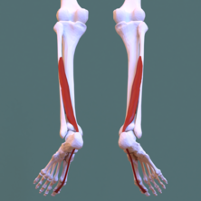
397:Animation. Flexor hallucis longus shown in red.
421:Muscles of the sole of the foot. Second layer.
337:, to which it is connected by a fibrous slip.
288:, laterally, and from the fascia covering the
27:One of the three deep muscles in the lower leg
572:
8:
318:, and is inserted into the base of the last
1227:
1165:
1033:
991:
888:
739:
638:
579:
565:
557:
44:Flexor hallucis longus. Seen from behind.
1396:
455:
387:
437:This article incorporates text in the
409:Right leg seen from back. Deep layer.
227:
29:
952:Lateral intermuscular septum of thigh
248:) attaches to the plantar surface of
7:
957:Medial intermuscular septum of thigh
548:at the SUNY Downstate Medical Center
519:"Flexor Hallucis Longus Dysfunction"
25:
1399:
431:
414:
402:
390:
258:posterior compartment of the leg
85:Plantar surface; base of distal
75:, posterior aspect of middle 1/3
50:
38:
306:, and the under surface of the
173:musculus flexor hallucis longus
156:Extensor hallucis longus muscle
1:
242:flexor hallucis longus muscle
33:Flexor hallucis longus muscle
1139:Fibularis (peroneus) muscles
1019:Fibularis (peroneus) tertius
1313:Flexor digiti minimi brevis
445:of the 20th edition of
1448:
224:Anatomical terms of muscle
1432:Muscles of the lower limb
1243:Extensor digitorum brevis
1168:
1014:Extensor digitorum longus
891:
641:
473:"Peripheral Nerve Injury"
222:
133:Flexes all joints of the
49:
37:
1238:Extensor hallucis brevis
1009:Extensor hallucis longus
546:Anatomy photo:15:st-0404
498:"Flexor Hallucis Longus"
106:(peroneal branch of the
1269:Flexor digitorum brevis
1105:Flexor digitorum longus
477:www.hopkinsmedicine.org
335:flexor digitorum longus
262:flexor digitorum longus
260:, the others being the
108:posterior tibial artery
1303:Flexor hallucis brevis
1274:Abductor digiti minimi
1100:Flexor hallucis longus
316:flexor hallucis brevis
18:Flexor hallucis longus
680:Lateral rotator group
346:and inserts into the
282:interosseous membrane
668:Tensor fasciae latae
363:Injury and treatment
102:(Muscular branch of
1185:Intermuscular septa
1330:Plantar interossei
1110:Tibialis posterior
842:External obturator
772:Vastus intermedius
711:External obturator
706:Internal obturator
588:Muscles of the hip
308:sustentaculum tali
290:tibialis posterior
266:tibialis posterior
1387:
1386:
1383:
1382:
1368:Superior extensor
1363:Inferior extensor
1325:Dorsal interossei
1308:Adductor hallucis
1286:Quadratus plantae
1264:Abductor hallucis
1217:
1216:
1213:
1212:
1125:
1124:
1004:Tibialis anterior
976:
975:
972:
971:
962:Cribriform fascia
784:Articularis genus
724:
723:
701:Superior gemellus
696:Inferior gemellus
691:Quadratus femoris
384:Additional images
238:
237:
233:
16:(Redirected from
1439:
1404:
1403:
1395:
1291:Lumbrical muscle
1228:
1188:
1166:
1088:
1063:Accessory soleus
1041:
1034:
992:
947:Iliotibial tract
938:
889:
767:Vastus lateralis
740:
684:
639:
581:
574:
567:
558:
534:
533:
531:
529:
515:
509:
508:
506:
504:
494:
488:
487:
485:
483:
469:
463:
460:
435:
434:
418:
406:
394:
286:peroneus muscles
230:edit on Wikidata
54:
42:
30:
21:
1447:
1446:
1442:
1441:
1440:
1438:
1437:
1436:
1412:
1411:
1410:
1398:
1390:
1388:
1379:
1336:
1247:
1209:
1182:
1177:
1155:
1121:
1084:
1079:
1068:Achilles tendon
1037:
1023:
985:
968:
932:
927:
923:Muscular lacuna
918:Adductor hiatus
878:
823:
817:Semimembranosus
788:
777:Vastus medialis
733:
720:
677:
672:
646:Gluteal muscles
628:
594:
585:
542:
537:
527:
525:
517:
516:
512:
502:
500:
496:
495:
491:
481:
479:
471:
470:
466:
461:
457:
432:
429:
422:
419:
410:
407:
398:
395:
386:
365:
356:
343:
274:
234:
139:plantar flexion
104:peroneal artery
57:
45:
28:
23:
22:
15:
12:
11:
5:
1445:
1443:
1435:
1434:
1429:
1424:
1414:
1413:
1409:
1408:
1385:
1384:
1381:
1380:
1378:
1377:
1376:
1375:
1370:
1365:
1360:
1352:
1350:Plantar fascia
1346:
1344:
1338:
1337:
1335:
1334:
1333:
1332:
1327:
1317:
1316:
1315:
1310:
1305:
1295:
1294:
1293:
1288:
1278:
1277:
1276:
1271:
1266:
1255:
1253:
1249:
1248:
1246:
1245:
1240:
1234:
1232:
1225:
1219:
1218:
1215:
1214:
1211:
1210:
1208:
1207:
1202:
1197:
1191:
1189:
1179:
1178:
1176:
1175:
1169:
1163:
1157:
1156:
1154:
1153:
1152:
1151:
1146:
1135:
1133:
1127:
1126:
1123:
1122:
1120:
1119:
1114:
1113:
1112:
1107:
1102:
1091:
1089:
1081:
1080:
1078:
1077:
1072:
1071:
1070:
1065:
1060:
1055:
1044:
1042:
1031:
1025:
1024:
1022:
1021:
1016:
1011:
1006:
1000:
998:
989:
978:
977:
974:
973:
970:
969:
967:
966:
965:
964:
959:
954:
949:
941:
939:
929:
928:
926:
925:
920:
915:
913:Adductor canal
910:
905:
904:
903:
896:Femoral sheath
892:
886:
880:
879:
877:
876:
875:
874:
869:
864:
859:
849:
844:
839:
833:
831:
825:
824:
822:
821:
820:
819:
814:
812:Semitendinosus
809:
807:Biceps femoris
798:
796:
790:
789:
787:
786:
781:
780:
779:
774:
769:
764:
762:Rectus femoris
754:
748:
746:
737:
726:
725:
722:
721:
719:
718:
713:
708:
703:
698:
693:
687:
685:
674:
673:
671:
670:
665:
664:
663:
658:
653:
642:
636:
630:
629:
627:
626:
625:
624:
619:
604:
602:
596:
595:
586:
584:
583:
576:
569:
561:
555:
554:
549:
541:
540:External links
538:
536:
535:
510:
489:
464:
454:
448:Gray's Anatomy
428:
425:
424:
423:
420:
413:
411:
408:
401:
399:
396:
389:
385:
382:
364:
361:
355:
352:
342:
339:
273:
270:
236:
235:
226:
220:
219:
214:
208:
207:
202:
196:
195:
190:
184:
183:
180:
176:
175:
170:
164:
163:
159:
158:
153:
147:
146:
131:
125:
124:
118:
112:
111:
100:
94:
93:
83:
77:
76:
70:
64:
63:
59:
58:
55:
47:
46:
43:
35:
34:
26:
24:
14:
13:
10:
9:
6:
4:
3:
2:
1444:
1433:
1430:
1428:
1425:
1423:
1420:
1419:
1417:
1407:
1402:
1397:
1393:
1374:
1371:
1369:
1366:
1364:
1361:
1359:
1356:
1355:
1353:
1351:
1348:
1347:
1345:
1343:
1339:
1331:
1328:
1326:
1323:
1322:
1321:
1318:
1314:
1311:
1309:
1306:
1304:
1301:
1300:
1299:
1296:
1292:
1289:
1287:
1284:
1283:
1282:
1279:
1275:
1272:
1270:
1267:
1265:
1262:
1261:
1260:
1257:
1256:
1254:
1250:
1244:
1241:
1239:
1236:
1235:
1233:
1229:
1226:
1224:
1220:
1206:
1203:
1201:
1198:
1196:
1193:
1192:
1190:
1187:
1186:
1180:
1174:
1173:Pes anserinus
1171:
1170:
1167:
1164:
1162:
1158:
1150:
1147:
1145:
1142:
1141:
1140:
1137:
1136:
1134:
1132:
1128:
1118:
1115:
1111:
1108:
1106:
1103:
1101:
1098:
1097:
1096:
1095:tarsal tunnel
1093:
1092:
1090:
1087:
1082:
1076:
1073:
1069:
1066:
1064:
1061:
1059:
1056:
1054:
1053:Gastrocnemius
1051:
1050:
1049:
1048:Triceps surae
1046:
1045:
1043:
1040:
1035:
1032:
1030:
1026:
1020:
1017:
1015:
1012:
1010:
1007:
1005:
1002:
1001:
999:
997:
993:
990:
988:
983:
979:
963:
960:
958:
955:
953:
950:
948:
945:
944:
943:
942:
940:
937:
936:
930:
924:
921:
919:
916:
914:
911:
909:
906:
902:
901:Femoral canal
899:
898:
897:
894:
893:
890:
887:
885:
881:
873:
870:
868:
865:
863:
860:
858:
855:
854:
853:
850:
848:
845:
843:
840:
838:
835:
834:
832:
830:
826:
818:
815:
813:
810:
808:
805:
804:
803:
800:
799:
797:
795:
791:
785:
782:
778:
775:
773:
770:
768:
765:
763:
760:
759:
758:
755:
753:
750:
749:
747:
745:
741:
738:
736:
731:
727:
717:
714:
712:
709:
707:
704:
702:
699:
697:
694:
692:
689:
688:
686:
683:
681:
675:
669:
666:
662:
659:
657:
654:
652:
649:
648:
647:
644:
643:
640:
637:
635:
631:
623:
620:
618:
614:
611:
610:
609:
606:
605:
603:
601:
597:
593:
589:
582:
577:
575:
570:
568:
563:
562:
559:
553:
550:
547:
544:
543:
539:
524:
523:Massage Today
520:
514:
511:
499:
493:
490:
478:
474:
468:
465:
459:
456:
453:
452:
449:
446:
444:
440:
439:public domain
426:
417:
412:
405:
400:
393:
388:
383:
381:
377:
373:
369:
362:
360:
353:
351:
349:
340:
338:
336:
331:
329:
325:
321:
317:
313:
309:
305:
300:
298:
297:tarsal tunnel
293:
291:
287:
283:
279:
271:
269:
267:
263:
259:
255:
251:
247:
243:
231:
225:
221:
218:
215:
213:
209:
206:
203:
201:
197:
194:
191:
189:
185:
181:
177:
174:
171:
169:
165:
160:
157:
154:
152:
148:
144:
140:
136:
132:
130:
126:
122:
119:
117:
113:
109:
105:
101:
99:
95:
92:
88:
84:
82:
78:
74:
71:
69:
65:
60:
53:
48:
41:
36:
31:
19:
1427:Calf muscles
1319:
1297:
1280:
1258:
1183:
1099:
1085:
1038:
987:compartments
933:
908:Femoral ring
735:compartments
678:
600:Iliac region
526:. Retrieved
522:
513:
501:. Retrieved
492:
480:. Retrieved
476:
467:
458:
450:
436:
430:
378:
374:
370:
366:
357:
344:
332:
301:
294:
292:, medially.
275:
245:
241:
239:
193:A04.7.02.053
172:
123:, S2 and S3
121:Tibial nerve
1354:retinacula
1039:Superficial
935:Fascia lata
617:Psoas minor
613:Psoas major
162:Identifiers
1416:Categories
1205:Transverse
757:Quadriceps
716:Piriformis
528:20 October
503:20 October
482:20 October
427:References
179:Acronym(s)
151:Antagonist
1320:4th layer
1298:3rd layer
1281:2nd layer
1259:1st layer
1200:Posterior
1117:Popliteus
1075:Plantaris
1029:Posterior
837:Pectineus
802:Hamstring
794:Posterior
752:Sartorius
608:Iliopsoas
592:human leg
552:PTCentral
348:calcaneum
341:Variation
324:great toe
312:calcaneus
272:Structure
254:great toe
81:Insertion
1358:Peroneal
1195:Anterior
996:Anterior
852:Adductor
847:Gracilis
744:Anterior
634:Buttocks
443:page 485
354:Function
330:sheath.
264:and the
1406:Anatomy
1252:Plantar
1131:Lateral
872:Minimus
661:Minimus
651:Maximus
622:Iliacus
322:of the
320:phalanx
310:of the
252:of the
250:phalanx
141:of the
135:big toe
129:Actions
87:phalanx
62:Details
1392:Portal
1373:Flexor
1342:Fascia
1231:Dorsal
1161:Fascia
1149:Brevis
1144:Longus
1058:Soleus
884:Fascia
867:Magnus
862:Brevis
857:Longus
829:Medial
656:Medius
451:(1918)
328:mucous
278:fibula
98:Artery
91:hallux
73:Fibula
68:Origin
730:Thigh
441:from
304:talus
228:[
217:22593
168:Latin
145:joint
143:ankle
116:Nerve
1223:Foot
1086:Deep
590:and
530:2020
505:2020
484:2020
240:The
205:2668
188:TA98
982:Leg
246:FHL
212:FMA
200:TA2
182:FHL
89:of
1418::
521:.
475:.
350:.
137:,
1394::
984:/
732:/
682::
615:/
580:e
573:t
566:v
532:.
507:.
486:.
244:(
232:]
110:)
20:)
Text is available under the Creative Commons Attribution-ShareAlike License. Additional terms may apply.