38:
513:
464:
479:
498:
211:
46:
390:. Staining by basic dyes occurs only from solutions that are less acidic than hemalum, and it is prevented by prior chemical or enzymatic extraction of nucleic acids. There is evidence to indicate that co-ordinate bonds, similar to those that hold aluminium and hematein together, bind the hemalum complex to DNA and to carboxy groups of proteins in the nuclear
148:
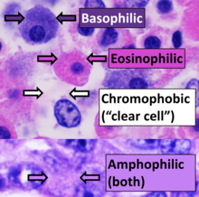
pink, with other structures taking on different shades, hues, and combinations of these colors. Hence a pathologist can easily differentiate between the nuclear and cytoplasmic parts of a cell, and additionally, the overall patterns of coloration from the stain show the general layout and
449:
397:
The structures do not have to be acidic or basic to be called basophilic and eosinophilic; the terminology is based on the affinity of cellular components for the dyes. Other colors, e.g. yellow and brown, can be present in the sample; they are caused by intrinsic pigments such as
382:(a combination of aluminum ions and hematein) is ordinarily due to binding of the dye-metal complex to DNA, but nuclear staining can be obtained after extraction of DNA from tissue sections. The mechanism is different from that of nuclear staining by basic (cationic) dyes such as
478:
246:
and cut into thin slices. The slices are affixed to microscope slides at which point the wax is removed with a solvent and the tissue slices attached to the slides are rehydrated and are ready for staining. Alternatively, H&E stain is the most used stain in
1307:
Lillie RD, Pizzolato P, Donaldson PT (1976) Nuclear stains with soluble metachrome mordant lake dyes. The effect of chemical endgroup blocking reactions and the artificial introduction of acid groups into tissues. Histochemistry 49:
222:
for producing H&E stained slides, some of which may be specific to a certain laboratory. Although there is no standard procedure, the results by convention are reasonably consistent in that cell nuclei are stained blue and the
463:
191:
H&E staining does not always provide enough contrast to differentiate all tissues, cellular structures, or the distribution of chemical substances, and in these cases more specific stains and methods are used.
512:
176:
The H&E staining procedure is the principal stain in histology in part because it can be done quickly, is not expensive, and stains tissues in such a way that a considerable amount of
72:
995:
1684:
1314:
Puchtler H, Meloan SN, Waldrop FS (1986) Application of current chemical concepts to metal-haematein and -brazilein stains. Histochemistry 85: 353–364.
1149:"Hematoxylin: Mesoamerica's Gift to Histopathology. Palo de Campeche (Logwood Tree), Pirates' Most Desired Treasure, and Irreplaceable Tissue Stain"
497:
834:"Traditional staining for routine diagnostic pathology including the role of tannic acid. 1. Value and limitations of the hematoxylin-eosin stain"
1324:
491:
patient. Cell nuclei (blue-purple), red blood cells (bright red), other cell bodies and extracellular material (pink), and air spaces (white).
37:
1761:
1339:
1258:
1029:
1201:"Does progressive nuclear staining with hemalum (alum hematoxylin) involve DNA, and what is the nature of the dye-chromatin complex?"
448:
1353:
218:
There are many ways to prepare the hematoxylin solutions (formulation) used in the H&E procedure, in addition, there are many
1825:
1385:
150:
1487:
1830:
188:
the tissue or slight inconsistencies in laboratory protocol, and these factors contribute to its routine use in histology.
1579:
1716:
231:
are stained pink. Histology laboratories may also adjust the amount or type of staining for a particular pathologist.
149:
distribution of cells and provides a general overview of a tissue sample's structure. Thus, pattern recognition, both
1746:
1620:
418:
also require silver stain. Hydrophobic structures also tend to remain clear; these are usually rich in fats, e.g.
1766:
1840:
1629:
469:
165:
1554:
362:
form of hematoxylin, is the active colorant (when combined with a mordant), the stain is still referred to as
1835:
31:
326:(substances that are stained by eosin) structures are generally composed of intracellular or extracellular
1845:
1789:
1354:
Rosen Lab, Department of
Molecular and Cellular Biology, Baylor College of Medicine) Step by step protocol
1304:
Kiernan JA (2008) Histological and
Histochemical Methods: Theory and Practice. 4th ed. Bloxham, UK: Scion.
1721:
1596:
989:
1658:
1624:
1348:
1639:
1378:
1358:
518:
315:
228:
185:
141:
122:
104:
1820:
1414:
177:
154:
1756:
1228:
1126:
1079:
977:
921:
861:
765:
659:
575:
219:
184:
conditions. The results from H&E staining are not overly dependent on the chemical used to
1850:
1773:
1608:
1482:
1477:
1254:
1220:
1170:
1118:
1071:
1025:
969:
913:
853:
757:
703:
651:
609:
567:
158:
99:
1336:
1736:
1634:
1311:
Llewellyn BD (2009) Nuclear staining with alum-hematoxylin. Biotech. Histochem. 84: 159–177.
1283:
1212:
1160:
1110:
1063:
961:
903:
845:
749:
693:
643:
559:
210:
1616:
1371:
1343:
431:
415:
181:
1644:
1572:
1563:
1537:
1274:
Kahr, Bart; Lovell, Scott; Subramony, Anand (1998). "The progress of logwood extract".
387:
343:
299:
738:"The wonderful colors of the hematoxylin-eosin stain in diagnostic surgical pathology"
1814:
1731:
1706:
1674:
1558:
1549:
1533:
1457:
1130:
965:
1363:
1232:
1083:
925:
865:
794:
Stevens, Alan (1982). "The
Haematoxylins". In Bancroft, John; Stevens, Alan (eds.).
769:
663:
579:
1751:
1711:
1694:
1469:
1022:
Histology : a text and atlas : with correlated cell and molecular biology
981:
411:
403:
335:
323:
303:
295:
279:
259:
248:
239:
205:
137:
1216:
892:"Progress in the Development of Microscopical Techniques for Diagnostic Pathology"
472:(DCIS) in breast tissue, cell nuclei (blue-purple), extracellular material (pink).
164:
This stain combination was introduced in 1877 by chemist
Nicolaus Wissozky at the
45:
849:
1799:
1741:
1726:
1589:
1432:
1406:
129:
110:
57:
266:, often followed by a rinse in a weak acid solution to remove excess staining (
1794:
1649:
1584:
1528:
1510:
1114:
698:
681:
647:
563:
1165:
1148:
917:
753:
1505:
1452:
1442:
908:
891:
488:
419:
407:
391:
339:
331:
311:
307:
243:
224:
201:
145:
95:
1224:
1200:
1174:
1122:
1098:
1052:"Standardization of biological dyes and stains: pitfalls and possibilities"
973:
950:"Hematoxylin and eosin tissue stain in Mohs micrographic surgery: a review"
949:
857:
833:
761:
737:
707:
655:
631:
613:
597:
571:
255:(a microtome that cuts frozen tissue), fixed in alcohol, and then stained.
1075:
1051:
64:, cell nuclei stained blue-purple and extracellular material stained pink.
1567:
1542:
1520:
1447:
1437:
1394:
383:
371:
359:
355:
319:
275:
252:
235:
91:
17:
1699:
1679:
1497:
1067:
399:
375:
327:
283:
263:
1142:
1140:
503:
423:
118:
114:
49:
1287:
370:) is often considered to "resemble" a basic, positively charged, or
366:. Hematoxylin, when combined with a mordant (most commonly aluminum
1424:
238:) and fixed, they are typically dehydrated and embedded in melted
209:
133:
61:
44:
36:
378:(negatively charged) and acidic stain. The staining of nuclei by
1402:
682:"Why microscopy will remain a cornerstone of surgical pathology"
522:
484:
454:
427:
367:
1367:
214:
Rack of slides being removed from a bath of hematoxylin stain.
53:
302:
blue or dark-purple, along with a few other tissues, such as
789:
787:
785:
783:
781:
779:
278:
water. After the application of haematoxylin, the tissue is
525:, cell nuclei (blue-purple), extracellular material (pink).
1244:
1242:
625:
623:
598:"Our debt to the logwood tree: the history of hematoxylin"
1015:
1013:
1011:
1009:
1007:
1005:
180:
is revealed, and can be used to diagnose a wide range of
1253:(Fourth ed.). W. B. Saunders Company. p. 600.
550:
Titford, M. (2005). "The long history of hematoxylin".
128:
H&E is the combination of two histological stains:
1337:
943:
941:
939:
937:
935:
338:
are examples of eosinophilic structures. Most of the
258:
The H&E staining method involves application of
1782:
1667:
1607:
1519:
1496:
1468:
1423:
1401:
1349:
Hematoxylin & Eosin (H&E) Staining
Protocol
798:(2nd ed.). Longman Group Limited. p. 109.
827:
825:
796:The Theory and Practice of Histological Techniques
823:
821:
819:
817:
815:
813:
811:
809:
807:
805:
1194:
1192:
1190:
1188:
1186:
1184:
1045:
1043:
1041:
457:, cell nuclei (blue-purple), bone matrix (pink).
251:in which tissues are typically frozen, cut on a
1024:(7th ed.). Wolters Kluwer. pp. 984p.
885:
883:
881:
879:
877:
875:
545:
543:
632:"Dyes from a twenty-first century perspective"
506:, cell nuclei (blue-purple), cell body (pink).
30:"H&E" redirects here. For other uses, see
1379:
1249:Leeson, Thomas S.; Leeson, C. Roland (1981).
948:Larson K, Ho HH, Anumolu PL, Chen TM (2011).
675:
673:
591:
589:
41:Main types of staining seen on H&E stain.
8:
1020:Ross, Michael H.; Pawlina, Wojciech (2016).
994:: CS1 maint: multiple names: authors list (
731:
729:
727:
725:
723:
721:
719:
717:
234:After tissues have been collected (often as
1386:
1372:
1364:
1325:SIGMA-ALDRICH H&E Informational Primer
1164:
907:
697:
1099:"Nuclear staining with alum hematoxylin"
1147:Ortiz-Hidalgo C, Pina-Oviedo S (2019).
539:
444:
1685:Jaswant Singh–Bhattacharji (JSB) stain
987:
342:is eosinophilic and is rendered pink.
242:, the resulting block is mounted on a
140:a purplish blue, and eosin stains the
125:is likely to be stained with H&E.
98:. It is the most widely used stain in
7:
314:and some other structures including
161:), provides histologic information.
438:Examples of H&E stained tissues
414:, if they have to be well visible.
294:Hematoxylin principally colors the
202:Histology § Sample preparation
155:by software that aids those experts
1762:Grocott's methenamine silver stain
322:in up to five shades of pink. The
25:
1395:Microbial and histological stains
90:) is one of the principal tissue
966:10.1111/j.1524-4725.2011.02051.x
511:
496:
477:
462:
447:
552:Biotechnic & Histochemistry
262:mixed with a metallic salt, or
630:Dapson RW, Horobin RW (2009).
136:. The hematoxylin stains cell
1:
1217:10.1080/10520295.2017.1399466
850:10.1080/10520290310001633725
76:haematoxylin and eosin stain
346:are stained intensely red.
310:material. Eosin stains the
151:by expert humans themselves
69:Hematoxylin and eosin stain
1867:
1488:Periodic acid–Schiff stain
896:Journal of Histotechnology
282:with eosin (most commonly
199:
29:
1115:10.1080/10520290903052899
890:Titford, Michael (2009).
699:10.1038/labinvest.3700551
648:10.1080/10520290902908802
564:10.1080/10520290500138372
166:Kazan Imperial University
27:Histological stain method
1630:Light Green SF yellowish
1621:Masson's trichrome stain
1580:Auramine–rhodamine stain
1166:10.1177/1066896918787652
754:10.1177/1066896913517939
470:Ductal carcinoma in situ
32:H&E (disambiguation)
1826:Microbiology techniques
909:10.1179/his.2009.32.1.9
82:; often abbreviated as
80:hematoxylin-eosin stain
1747:Schaeffer–Fulton stain
1717:Gömöri trichrome stain
406:need to be stained by
215:
65:
42:
1831:Laboratory techniques
1722:Luxol fast blue stain
1597:Auramine phenol stain
1097:Llewellyn BD (2009).
213:
196:Method of application
48:
40:
1767:Warthin–Starry stain
1640:Phosphomolybdic acid
832:Wittekind D (2003).
519:Basal cell carcinoma
316:extracellular matrix
229:extracellular matrix
220:laboratory protocols
142:extracellular matrix
123:histological section
109:For example, when a
1783:Tissue stainability
1555:Ziehl–Neelsen stain
1415:Perls Prussian blue
1199:Kiernan JA (2018).
1050:Schulte EK (1991).
374:stain. Eosin is an
178:microscopic anatomy
1757:Bielschowsky stain
1659:Van Gieson's stain
1625:Lillie's trichrome
1342:2023-06-02 at the
1068:10.1007/BF00266958
216:
66:
43:
1808:
1807:
1609:Connective tissue
1205:Biotech Histochem
1153:Int J Surg Pathol
1103:Biotech Histochem
838:Biotech Histochem
742:Int J Surg Pathol
636:Biotech Histochem
159:digital pathology
102:and is often the
100:medical diagnosis
16:(Redirected from
1858:
1635:Biebrich scarlet
1388:
1381:
1374:
1365:
1292:
1291:
1271:
1265:
1264:
1246:
1237:
1236:
1196:
1179:
1178:
1168:
1144:
1135:
1134:
1094:
1088:
1087:
1047:
1036:
1035:
1017:
1000:
999:
993:
985:
945:
930:
929:
911:
887:
870:
869:
829:
800:
799:
791:
774:
773:
736:Chan JK (2014).
733:
712:
711:
701:
680:Rosai J (2007).
677:
668:
667:
627:
618:
617:
596:Smith C (2006).
593:
584:
583:
547:
515:
500:
481:
466:
451:
416:Reticular fibers
21:
1866:
1865:
1861:
1860:
1859:
1857:
1856:
1855:
1841:Histotechnology
1811:
1810:
1809:
1804:
1778:
1663:
1617:trichrome stain
1603:
1515:
1492:
1464:
1419:
1397:
1392:
1344:Wayback Machine
1333:
1321:
1301:
1299:Further reading
1296:
1295:
1288:10.1002/chir.12
1273:
1272:
1268:
1261:
1248:
1247:
1240:
1198:
1197:
1182:
1146:
1145:
1138:
1096:
1095:
1091:
1049:
1048:
1039:
1032:
1019:
1018:
1003:
986:
947:
946:
933:
889:
888:
873:
831:
830:
803:
793:
792:
777:
735:
734:
715:
679:
678:
671:
629:
628:
621:
608:(5): 18, 20–2.
602:MLO Med Lab Obs
595:
594:
587:
549:
548:
541:
536:
531:
530:
529:
526:
516:
507:
501:
492:
482:
473:
467:
458:
452:
440:
432:Golgi apparatus
352:
344:Red blood cells
292:
270:), followed by
268:differentiation
208:
200:Main articles:
198:
182:histopathologic
174:
117:of a suspected
56:) stained with
35:
28:
23:
22:
15:
12:
11:
5:
1864:
1862:
1854:
1853:
1848:
1843:
1838:
1836:Histopathology
1833:
1828:
1823:
1813:
1812:
1806:
1805:
1803:
1802:
1797:
1792:
1786:
1784:
1780:
1779:
1777:
1776:
1774:Wright's stain
1771:
1770:
1769:
1764:
1759:
1749:
1744:
1739:
1734:
1729:
1724:
1719:
1714:
1709:
1704:
1703:
1702:
1697:
1687:
1682:
1677:
1671:
1669:
1665:
1664:
1662:
1661:
1655:
1654:
1653:
1652:
1647:
1645:Fast Green FCF
1642:
1637:
1632:
1613:
1611:
1605:
1604:
1602:
1601:
1600:
1599:
1594:
1593:
1592:
1587:
1577:
1576:
1575:
1573:Methylene blue
1570:
1564:Carbol fuchsin
1547:
1546:
1545:
1540:
1538:Gentian violet
1525:
1523:
1517:
1516:
1514:
1513:
1508:
1502:
1500:
1494:
1493:
1491:
1490:
1485:
1480:
1474:
1472:
1466:
1465:
1463:
1462:
1461:
1460:
1455:
1450:
1445:
1440:
1429:
1427:
1421:
1420:
1418:
1417:
1411:
1409:
1399:
1398:
1393:
1391:
1390:
1383:
1376:
1368:
1362:
1361:
1356:
1351:
1346:
1332:
1329:
1328:
1327:
1320:
1319:External links
1317:
1316:
1315:
1312:
1309:
1305:
1300:
1297:
1294:
1293:
1282:(1–2): 66–77.
1266:
1260:978-0721657042
1259:
1238:
1211:(2): 133–148.
1180:
1136:
1089:
1056:Histochemistry
1037:
1031:978-1451187427
1030:
1001:
960:(8): 1089–99.
931:
871:
801:
775:
713:
669:
619:
585:
538:
537:
535:
532:
528:
527:
517:
510:
508:
502:
495:
493:
487:taken from an
483:
476:
474:
468:
461:
459:
453:
446:
443:
442:
441:
439:
436:
426:around neuron
388:toluidine blue
351:
350:Mode of action
348:
336:Mallory bodies
291:
288:
280:counterstained
197:
194:
173:
170:
26:
24:
14:
13:
10:
9:
6:
4:
3:
2:
1863:
1852:
1849:
1847:
1846:Staining dyes
1844:
1842:
1839:
1837:
1834:
1832:
1829:
1827:
1824:
1822:
1819:
1818:
1816:
1801:
1798:
1796:
1793:
1791:
1788:
1787:
1785:
1781:
1775:
1772:
1768:
1765:
1763:
1760:
1758:
1755:
1754:
1753:
1750:
1748:
1745:
1743:
1740:
1738:
1737:Movat's stain
1735:
1733:
1732:Moeller stain
1730:
1728:
1725:
1723:
1720:
1718:
1715:
1713:
1710:
1708:
1707:Janus Green B
1705:
1701:
1698:
1696:
1693:
1692:
1691:
1690:H&E stain
1688:
1686:
1683:
1681:
1678:
1676:
1675:Cresyl violet
1673:
1672:
1670:
1666:
1660:
1657:
1656:
1651:
1648:
1646:
1643:
1641:
1638:
1636:
1633:
1631:
1628:
1627:
1626:
1622:
1618:
1615:
1614:
1612:
1610:
1606:
1598:
1595:
1591:
1588:
1586:
1583:
1582:
1581:
1578:
1574:
1571:
1569:
1565:
1562:
1561:
1560:
1559:Kinyoun stain
1556:
1553:
1552:
1551:
1548:
1544:
1541:
1539:
1535:
1534:Methyl violet
1532:
1531:
1530:
1527:
1526:
1524:
1522:
1518:
1512:
1509:
1507:
1504:
1503:
1501:
1499:
1495:
1489:
1486:
1484:
1481:
1479:
1476:
1475:
1473:
1471:
1470:Carbohydrates
1467:
1459:
1458:Sudan Black B
1456:
1454:
1451:
1449:
1446:
1444:
1441:
1439:
1436:
1435:
1434:
1431:
1430:
1428:
1426:
1422:
1416:
1413:
1412:
1410:
1408:
1404:
1400:
1396:
1389:
1384:
1382:
1377:
1375:
1370:
1369:
1366:
1360:
1359:the H-E Stain
1357:
1355:
1352:
1350:
1347:
1345:
1341:
1338:
1335:
1334:
1330:
1326:
1323:
1322:
1318:
1313:
1310:
1306:
1303:
1302:
1298:
1289:
1285:
1281:
1277:
1270:
1267:
1262:
1256:
1252:
1245:
1243:
1239:
1234:
1230:
1226:
1222:
1218:
1214:
1210:
1206:
1202:
1195:
1193:
1191:
1189:
1187:
1185:
1181:
1176:
1172:
1167:
1162:
1158:
1154:
1150:
1143:
1141:
1137:
1132:
1128:
1124:
1120:
1116:
1112:
1109:(4): 159–77.
1108:
1104:
1100:
1093:
1090:
1085:
1081:
1077:
1073:
1069:
1065:
1062:(4): 319–28.
1061:
1057:
1053:
1046:
1044:
1042:
1038:
1033:
1027:
1023:
1016:
1014:
1012:
1010:
1008:
1006:
1002:
997:
991:
983:
979:
975:
971:
967:
963:
959:
955:
954:Dermatol Surg
951:
944:
942:
940:
938:
936:
932:
927:
923:
919:
915:
910:
905:
901:
897:
893:
886:
884:
882:
880:
878:
876:
872:
867:
863:
859:
855:
851:
847:
844:(5): 261–70.
843:
839:
835:
828:
826:
824:
822:
820:
818:
816:
814:
812:
810:
808:
806:
802:
797:
790:
788:
786:
784:
782:
780:
776:
771:
767:
763:
759:
755:
751:
747:
743:
739:
732:
730:
728:
726:
724:
722:
720:
718:
714:
709:
705:
700:
695:
691:
687:
683:
676:
674:
670:
665:
661:
657:
653:
649:
645:
641:
637:
633:
626:
624:
620:
615:
611:
607:
603:
599:
592:
590:
586:
581:
577:
573:
569:
565:
561:
557:
553:
546:
544:
540:
533:
524:
520:
514:
509:
505:
504:Muscle tissue
499:
494:
490:
486:
480:
475:
471:
465:
460:
456:
450:
445:
437:
435:
433:
429:
425:
421:
417:
413:
412:silver stains
409:
405:
404:Basal laminae
401:
395:
393:
389:
385:
381:
377:
373:
369:
365:
361:
357:
349:
347:
345:
341:
337:
333:
329:
325:
321:
317:
313:
309:
306:granules and
305:
301:
297:
289:
287:
285:
281:
277:
273:
269:
265:
261:
256:
254:
250:
245:
241:
237:
232:
230:
226:
221:
212:
207:
203:
195:
193:
189:
187:
183:
179:
171:
169:
167:
162:
160:
156:
152:
147:
143:
139:
135:
131:
126:
124:
120:
116:
112:
108:
106:
105:gold standard
101:
97:
93:
89:
85:
84:H&E stain
81:
77:
74:
70:
63:
59:
55:
52:(part of the
51:
47:
39:
33:
19:
1800:Chromophobic
1752:Silver stain
1712:Giemsa stain
1695:Haematoxylin
1689:
1279:
1275:
1269:
1250:
1208:
1204:
1156:
1152:
1106:
1102:
1092:
1059:
1055:
1021:
990:cite journal
957:
953:
899:
895:
841:
837:
795:
748:(1): 12–32.
745:
741:
692:(5): 403–8.
689:
685:
642:(4): 135–7.
639:
635:
605:
601:
558:(2): 73–80.
555:
551:
396:
379:
363:
353:
324:eosinophilic
304:keratohyalin
293:
271:
267:
260:haematoxylin
257:
249:Mohs surgery
240:paraffin wax
233:
217:
206:Haematoxylin
190:
175:
163:
127:
103:
87:
83:
79:
75:
68:
67:
1790:Acidophilic
1742:Neutral red
1727:Methyl blue
1590:Rhodamine B
1483:Mucicarmine
1478:Alcian blue
1433:Sudan stain
1407:hemosiderin
1159:(1): 4–14.
902:(1): 9–19.
485:Lung tissue
434:membranes.
364:hematoxylin
332:Lewy bodies
168:in Russia.
130:hematoxylin
113:looks at a
111:pathologist
58:hematoxylin
1821:Microscopy
1815:Categories
1795:Basophilic
1650:Sirius Red
1585:Auramine O
1529:Gram stain
1511:Thioflavin
686:Lab Invest
534:References
420:adipocytes
274:in mildly
1550:Acid-fast
1506:Congo red
1453:Oil Red O
1443:Sudan III
1276:Chirality
1251:Histology
1131:205713596
918:0147-8885
489:emphysema
408:PAS stain
392:chromatin
354:Although
340:cytoplasm
312:cytoplasm
308:calcified
244:microtome
225:cytoplasm
146:cytoplasm
96:histology
1851:Staining
1568:Fuchsine
1543:Safranin
1521:Bacteria
1448:Sudan IV
1438:Sudan II
1340:Archived
1331:Protocol
1233:13481905
1225:29320873
1175:30001639
1123:19579146
1084:29628388
974:21635628
926:26801839
866:10563849
858:14989644
770:26847314
762:24406626
708:17401434
664:28563610
656:19384743
614:16761865
580:20338201
572:16195172
410:or some
384:thionine
372:cationic
360:oxidized
356:hematein
328:proteins
320:collagen
318:such as
276:alkaline
253:cryostat
236:biopsies
94:used in
88:HE stain
18:HE stain
1700:Eosin Y
1680:Cyanine
1498:Amyloid
1076:1708749
982:2538853
521:of the
400:melanin
380:hemalum
376:anionic
290:Results
284:eosin Y
264:mordant
1425:Lipids
1308:23–35.
1257:
1231:
1223:
1173:
1129:
1121:
1082:
1074:
1028:
980:
972:
924:
916:
864:
856:
768:
760:
706:
662:
654:
612:
578:
570:
430:, and
424:myelin
330:. The
296:nuclei
272:bluing
204:, and
138:nuclei
121:, the
119:cancer
115:biopsy
92:stains
50:Retina
1668:Other
1229:S2CID
1127:S2CID
1080:S2CID
978:S2CID
922:S2CID
862:S2CID
766:S2CID
660:S2CID
576:S2CID
428:axons
358:, an
300:cells
134:eosin
62:eosin
1403:Iron
1255:ISBN
1221:PMID
1171:PMID
1119:PMID
1072:PMID
1026:ISBN
996:link
970:PMID
914:ISSN
854:PMID
758:PMID
704:PMID
652:PMID
610:PMID
568:PMID
523:skin
455:Bone
368:alum
334:and
227:and
172:Uses
157:(in
153:and
144:and
132:and
60:and
1284:doi
1213:doi
1161:doi
1111:doi
1064:doi
962:doi
904:doi
846:doi
750:doi
694:doi
644:doi
560:doi
386:or
298:of
286:).
186:fix
86:or
78:or
54:eye
1817::
1619::
1280:10
1278:.
1241:^
1227:.
1219:.
1209:93
1207:.
1203:.
1183:^
1169:.
1157:27
1155:.
1151:.
1139:^
1125:.
1117:.
1107:84
1105:.
1101:.
1078:.
1070:.
1060:95
1058:.
1054:.
1040:^
1004:^
992:}}
988:{{
976:.
968:.
958:37
956:.
952:.
934:^
920:.
912:.
900:32
898:.
894:.
874:^
860:.
852:.
842:78
840:.
836:.
804:^
778:^
764:.
756:.
746:22
744:.
740:.
716:^
702:.
690:87
688:.
684:.
672:^
658:.
650:.
640:84
638:.
634:.
622:^
606:38
604:.
600:.
588:^
574:.
566:.
556:80
554:.
542:^
422:,
402:.
394:.
73:or
1623:/
1566:/
1557:/
1536:/
1405:/
1387:e
1380:t
1373:v
1290:.
1286::
1263:.
1235:.
1215::
1177:.
1163::
1133:.
1113::
1086:.
1066::
1034:.
998:)
984:.
964::
928:.
906::
868:.
848::
772:.
752::
710:.
696::
666:.
646::
616:.
582:.
562::
107:.
71:(
34:.
20:)
Text is available under the Creative Commons Attribution-ShareAlike License. Additional terms may apply.