1060:, or PAP staining, was developed to replace fine needle aspiration cytology (FNAC) in hopes of decreasing staining times and cost without compromising quality. This stain is a frequently used method for examining cell samples from a variety of tissue types in various organs. PAP staining has endured several modifications in order to become a “suitable alternative” for FNAC. This transition stemmed from the appreciation of wet fixed smears by scientists preserving the structures of the nuclei opposed to the opaque appearance of air dried Romanowsky smears. This led to the creation of a hybrid stain of wet fixed and air dried known as the ultrafast papanicolaou stain. This modification includes the use of nasal saline to rehydrate cells to increase cell transparency and is paired with the use of alcoholic formalin to enhance colors of the nuclei. The papanicolaou stain is now used in place of cytological staining in all organ types due to its increase in morphological quality, decreased staining time, and decreased cost. It is frequently used to stain
955:. Prior to the development of more efficient methods, this stain was performed using the Wirtz method with heat fixation and counterstain. Through the use of malachite green and a diluted ratio of carbol fuchsin, fixing bacteria in osmic acid was a great way to ensure no blending of dyes. However, newly revised staining methods have significantly decreased the time it takes to create these stains. This revision included substitution of carbol fuchsin with aqueous Safranin paired with a newly diluted 5% formula of malachite green. This new and improved composition of stains was performed in the same way as before with the use of heat fixation, rinsing, and blotting dry for later examination. Upon examination, all endospore forming bacteria will be stained green accompanied by all other cells appearing red.
1900:
1132:). PAS is commonly used on liver tissue where glycogen deposits are made which is done in efforts to distinguish different types of glycogen storage diseases. PAS is important because it can detect glycogen granules found in tumors of the ovaries and pancreas of the endocrine system, as well as in the bladder and kidneys of the renal system. Basement membranes can also show up in a PAS stain and can be important when diagnosing renal disease. Due to the high volume of carbohydrates within the cell wall of hyphae and yeast forms of fungi, the Periodic acid -Schiff stain can help locate these species inside tissue samples of the human body.
570:(an aqueous suspension of carbon particles). After drying, the microorganisms may be viewed in bright field microscopy as lighter inclusions well-contrasted against the dark environment surrounding them. Negative staining is able to stain the background instead of the organisms because the cell wall of microorganisms typically has a negative charge which repels the negatively charged stain. The dyes used in negative staining are acidic. Note: negative staining is a mild technique that may not destroy the microorganisms, and is therefore unsuitable for studying pathogens.
1402:
helps open the spore's membrane so the dye can enter. The main purpose of this stain is to show germination of bacterial spores. If the process of germination is taking place, then the spore will turn green in color due to malachite green and the surrounding cell will be red from the safranin. This stain can also help determine the orientation of the spore within the bacterial cell; whether it being terminal (at the tip), subterminal (within the cell), or central (completely in the middle of the cell).
1468:
1255:
1443:(AO) is a nucleic acid selective fluorescent cationic dye useful for cell cycle determination. It is cell-permeable, and interacts with DNA and RNA by intercalation or electrostatic attractions. When bound to DNA, it is very similar spectrally to fluorescein. Like fluorescein, it is also useful as a non-specific stain for backlighting conventionally stained cells on the surface of a solid sample of tissue (fluorescence backlighted staining).
27:
1099:
2001:, and other biological tissue materials. It is mostly used in a .5-2% ph form making it neutral and is paired with water to make an aqueous solution. Phosphotungstic acid is filled with electron dense matter that stains the background surrounding the specimen dark and the specimen itself light. This process is not the normal positive technique for staining where the specimen is dark and the background remains light.
1005:
2016:. It dissolves in fats, and is reduced by organic materials to elemental osmium, an easily visible black substance. Because it is a heavy metal that absorbs electrons, it is perhaps the most common stain used for morphology in biological electron microscopy. It is also used for the staining of various polymers for the study of their morphology by TEM.
1419:(CHP) staining allows for an easy, direct way to stain denatured collagens of any type (Type I, II, IV, etc.) regardless if they were damaged or degraded via enzymatic, mechanical, chemical, or thermal means. They work by refolding into the collagen triple helix with the available single strands in the tissue. CHPs can be visualized by a simple
1625:. Acid fuchsin is used as the nuclear and cytoplasmic stain in Mallory's trichrome method. Acid fuchsin stains cytoplasm in some variants of Masson's trichrome. In Van Gieson's picro-fuchsine, acid fuchsin imparts its red colour to collagen fibres. Acid fuchsin is also a traditional stain for mitochondria (Altmann's method).
592:
Differential staining uses multiple stains per slide. Based on the stains being used, organisms with different properties will appear different colors allowing for categorization of multiple specimens. Differential staining can also be used to color different organelles within one organism which can be seen in
1710:. When starch is mixed with iodine in solution, an intensely dark blue colour develops, representing a starch/iodine complex. Starch is a substance common to most plant cells and so a weak iodine solution will stain starch present in the cells. Iodine is one component in the staining technique known as
923:
organisms appear red or pink due to their counterstain. Due to the presence of higher lipid content, after alcohol-treatment, the porosity of the cell wall increases, hence the CVI complex (crystal violet – iodine) can pass through. Thus, the primary stain is not retained. In addition, in contrast to
1887:
in cartilage and mast cells, and components of lignin and plastids in plant tissues. Safranine should not be confused with saffron, an expensive natural dye that is used in some methods to impart a yellow colour to collagen, to contrast with blue and red colours imparted by other dyes to nuclei and
1580:(also known as eosin Y ws or eosin yellowish); it has a very slightly yellowish cast. The other eosin compound is eosin B (eosin bluish or imperial red); it has a very faint bluish cast. The two dyes are interchangeable, and the use of one or the other is more a matter of preference and tradition.
1401:
The Wirtz-Conklin stain is a special technique designed for staining true endospores with the use of malachite green dye as the primary stain and safranin as the counterstain. Once stained, they do not decolourize. The addition of heat during the staining process is a huge contributing factor. Heat
591:
Simple
Staining is a technique that only uses one type of stain on a slide at a time. Because only one stain is being used, the specimens (for positive stains) or background (for negative stains) will be one color. Therefore, simple stains are typically used for viewing only one organism per slide.
265:
staining). However, these stains are eventually toxic to the organism, some more so than others. Partly due to their toxic interaction inside a living cell, when supravital stains enter a living cell, they might produce a characteristic pattern of staining different from the staining of an already
1856:
is a fluorescent intercalating agent that can be used to stain cells. Propidium iodide is used as a DNA stain in flow cytometry to evaluate cell viability or DNA content in cell cycle analysis, or in microscopy to visualise the nucleus and other DNA-containing organelles. Propidium Iodide cannot
582:
is used for both negative and positive staining alike, the type of chromophore used in this technique is a positively charged ion instead of a negative one. The negatively charged cell wall of many microorganisms attracts the positively charged chromophore which causes the specimen to absorb the
1431:
Different stains react or concentrate in different parts of a cell or tissue, and these properties are used to advantage to reveal specific parts or areas. Some of the most common biological stains are listed below. Unless otherwise marked, all of these dyes may be used with fixed cells and
911:
counterstain to (mark all bacteria). Gram status, helps divide specimens of bacteria into two groups, generally representative of their underlying phylogeny. This characteristic, in combination with other techniques makes it a useful tool in clinical microbiology laboratories, where it can be
1458:
and an intense brown color to mast cells. One default of this stain is that it blots out any other structure surrounding it and makes the quality of the contrast low. It has to be paired with other stains in order to be useful. Some complementing stains used alongside
Bismark brown are
207:) or position within a cell or tissue can be readily seen and studied. The usual purpose is to reveal cytological details that might otherwise not be apparent; however, staining can also reveal where certain chemicals or specific chemical reactions are taking place within cells or tissues.
819:
The smear is first treated with chloroform to remove fats . Smear applied with
Alberts stain which contains cationic dyes such as toluidine blue and malachite green. Toluidine blue preferentially stains granules while malachite green stains cytoplasm.
538:
308:
are used to view live organisms and can be made using water and certain stains. The liquid is added to the slide before the addition of the organism and a coverslip is placed over the specimen in the water and stain to help contain it within the
945:, which make bacteria very difficult to kill. Bacterial spores have proven to be difficult to stain as they are not permeable to aqueous dye reagents. Endospore staining is particularly useful for identifying endospore-forming bacterial
522:), and found to meet or exceed certain standards of purity, dye content and performance in staining techniques ensuring more accurately performed experiments and more reliable results. These standards are published in the commission's journal
215:
staining involves colouring cells or structures that have been removed from their biological context. Certain stains are often combined to reveal more details and features than a single stain alone. Combined with specific protocols for
2028:
is very volatile and extremely toxic. It is a strong oxidizing agent as the osmium has an oxidation number of +8. It aggressively oxidizes many materials, leaving behind a deposit of non-volatile osmium in a lower oxidation state.
1739:
in cervical and vaginal tissues during "Pap smear" follow up examinations in preparation for biopsy. The acetic acid causes the abnormal cells to blanch white, while the normal tissues stain a mahogany brown from the iodine.
236:
While ex vivo, many cells continue to live and metabolize until they are "fixed". Some staining methods are based on this property. Those stains excluded by the living cells but taken up by the already dead cells are called
1553:. DAPI binds with A=T rich repeats of chromosomes. DAPI is also not visible with regular transmission microscopy. It may be used in living or fixed cells. DAPI-stained cells are especially appropriate for cell counting.
1149:
is (as the name implies) a three-colour staining protocol. The recipe has evolved from Masson's original technique for different specific applications, but all are well-suited to distinguish cells from surrounding
1576:. Eosin may also be used as a counterstain in some variants of Gram staining, and in many other protocols. There are actually two very closely related compounds commonly referred to as eosin. Most often used is
1857:
cross the membrane of live cells, making it useful to differentiate necrotic, apoptotic and healthy cells. PI also binds to RNA, necessitating treatment with nucleases to distinguish between RNA and DNA staining
493:
usually involves attaching the samples to a glass microscope slide for observation and analysis. In some cases, cells may be grown directly on a slide. For samples of loose cells (as with a blood smear or a
799:
Smear is treated for hydrolysis to release purines from DNA, purines to cause shift form furanose to aldehyde. Aldehyde groups are available to react with schiff's reagent to form addition compounds.
765:
Smear treated with C.P.C. which dissociates to form positively charged cetyl pyridinium and negatively charged chloride ions. Positively charged ions are adsorbed on negatively charged cell wall
1667:. Often used in fluorescence microscopy for DNA staining, Hoechst stains appear yellow when dissolved in aqueous solutions and emit blue light under UV excitation. There are two major types of
3091:
is an independent non-profit company that has been testing dyes since the early 1920s and issuing
Certificates of approval for batches of dyes that meet internationally recognized standards.
3596:
1043:, colouring them bright red. In a skillfully made H&E preparation the red blood cells are almost orange, and collagen and cytoplasm (especially muscle) acquire different shades of pink.
583:
stain giving it the color of the stain being used. Positive staining is more commonly used than negative staining in microbiology. The different types of positive staining are listed below.
837:
Lipids are stained with fat soluble dyes like Sudan black. On application of Sudan black-B dyes move into lipids and are retained there while cytoplasm is counter stained with safranin.
1830:
globules inside cells, staining them red. Nile red can be used with living cells. It fluoresces strongly when partitioned into lipids, but practically not at all in aqueous solution.
1211:). This stain develops varying colors for all cell structures (“Romanowsky-Giemsa effect) and thus was used in staining neutrophil polymorphs and cell nuclei. Common variants include
529:
Some vendors sell stains "certified" by themselves rather than by the
Biological Stain Commission. Such products may or may not be suitable for diagnostic and other applications.
2168:
2784:
Sellors JW, Sankaranarayanan R (eds.). "Chapter 4: An introduction to colposcopy: indications for colposcopy, instrumentation, principles and documentation of results".
857:
Polysaccharide is oxidized with periodate to form polyaldehyde which reacts with Schiff's reagents to red color, while cytoplasm is counter stained with malachite green
1594:
and stains DNA, providing a fluorescent red-orange stain. Although it will not stain healthy cells, it can be used to identify cells that are in the final stages of
1610:(AO) in viable cell counting. This EB/AO combined stain causes live cells to fluoresce green whilst apoptotic cells retain the distinctive red-orange fluorescence.
1636:(hematoxylin in North America) is a nuclear stain. Used with a mordant, haematoxylin stains nuclei blue-violet or brown. It is most often used with eosin in the
3589:
228:
Crystal violet stains both Gram positive and Gram negative organisms. Treatment with alcohol removes the crystal violet colour from gram negative organisms only.
924:
most Gram-positive bacteria, Gram-negative bacteria have only a few layers of peptidoglycan and a secondary cell membrane made primarily of lipopolysaccharide.
2805:
Prieto D, Aparicio G, Morande PE, Zolessi FR (September 2014). "A fast, low cost, and highly efficient fluorescent DNA labeling method using methyl green".
266:
fixed cell (e.g. "reticulocyte" look versus diffuse "polychromasia"). To achieve desired effects, the stains are used in very dilute solutions ranging from
3448:
802:
To demonstrate the presence of DNA in cell. But for detection of the DNA, RNA should be selectively destroyed by acid hydrolysis without affecting DNA
3582:
2262:
Penney DP, Powers JM, Frank M, Willis C, Churukian C (2002). "Analysis and testing of biological stains--the
Biological Stain Commission Procedures".
2040:
is equally volatile and even more aggressive than osmium tetraoxide and able to stain even materials that resist the osmium stain, e.g. polyethylene.
526:. Many dyes are inconsistent in composition from one supplier to another. The use of BSC-certified stains eliminates a source of unexpected results.
4153:
1297:
203:
or intravital staining) is the process of dyeing living tissues. By causing certain cells or structures to take on contrasting colours, their form (
3110:
3106:
2669:
Corey L (March 1986). "Laboratory diagnosis of herpes simplex virus infections. Principles guiding the development of rapid diagnostic tests".
164:. Light microscopes are used for viewing stained samples at high magnification, typically using bright-field or epi-fluorescence illumination.
1899:
4075:
3525:
3042:
3000:
2981:
2768:
2527:
2359:
2309:
2246:
1781:
is used to stain animal cells, such as human cheek cells, to make their nuclei more observable. Also used to stain blood films in cytology.
1770:
is used commonly with bright-field, as well as fluorescence microscopes to dye the chromatin of cells so that they are more easily viewed.
1258:
167:
Staining is not limited to only biological materials, since it can also be used to study the structure of other materials; for example, the
3059:
676:
Primary stain: Crystal violet applied to film then treated with iodine (mordant), alcohol (decolourizer) and counter stained with safranin
1728:
or Lugol's iodine (IKI) is a brown solution that turns black in the presence of starches and can be used as a cell stain, making the cell
3889:
321:, which may itself consist of several steps, aims to preserve the shape of the cells or tissue involved as much as possible. Sometimes
2785:
1242:(white blood cells) can be readily distinguished. All are also suited to examination of blood to detect blood-borne parasites such as
748:
Capsules can be observed as clear zones surrounding cells of capsulated bacteria and are used to demonstrate the presence of capsules.
2180:
1691:
substitution on the terminal hydroxyl group (i.e. an ethylether group) making it more hydrophobic for easier plasma membrane passage
3023:
325:
is used to kill, adhere, and alter the specimen so it accepts stains. Most chemical fixatives (chemicals causing fixation) generate
220:
and sample preparation, scientists and physicians can use these standard techniques as consistent, repeatable diagnostic tools. A
4043:
3149:
1971:
1891:
The incorrect spelling "safranin" is in common use. The -ine ending is appropriate for safranine O because this dye is an amine.
951:
300:
The preparatory steps involved depend on the type of analysis planned. Some or all of the following procedures may be required.
3251:
1093:
4148:
3951:
3725:
2626:
Bezrukov AV (2017-01-02). "Romanowsky staining, the
Romanowsky effect and thoughts on the question of scientific priority".
1602:. Consequently, ethidium bromide is often used as a marker for apoptosis in cells populations and to locate bands of DNA in
523:
1679:. The two compounds are functionally similar, but with a little difference in structure. Hoechst 33258 contains a terminal
782:
Mordant acts to thicken flagella before staining and increases visibility microscopically when stained with
Leifson stain
3343:
3946:
3909:
3894:
1591:
1416:
1411:
3480:
498:) the sample can be directly applied to a slide. For larger pieces of tissue, thin sections (slices) are made using a
2144:
1683:
group and is thus more soluble in aqueous solution, however this characteristics reduces its ability to penetrate the
515:
3510:
3384:
1141:
578:
Unlike negative staining, positive staining uses basic dyes to color the specimen against a bright background. While
3941:
2704:
Wells J (1988). "A Technique for
Staining the Superficial Cells of Plucked Hair Follicles and Other Solid Tissues".
2602:
1845:. It dissolves in fats, and is reduced by organic materials to elemental osmium, an easily visible black substance.
3834:
1039:
stains cytoplasm, connective tissue and other extracellular substances pink or red. Eosin is strongly absorbed by
975:
3990:
1722:
in Gram's staining, iodine enhances the entrance of the dye through the pores present in the cell wall/membrane.
20:
3931:
3530:
1751:(also known as diamond green B or victoria green B) can be used as a blue-green counterstain to safranin in the
716:
Primary stain
Malachite green heat fixed to penetrate spores; vegetative cells are counterstained with Safranin
3980:
3747:
3639:
3614:
3393:
1497:
1077:
172:
3779:
3318:
970:
964:
3904:
3899:
3879:
3859:
3829:
1420:
224:
is stain that makes cells or structures more visible, when not completely visible with the principal stain.
699:
Separate non-decolorized acid fast bacteria that are not decolorized from colorized non-acid fast bacteria
4143:
4015:
3914:
3619:
3553:
2096:
2092:
2068:
1935:
4023:
3936:
2112:
3972:
3606:
3485:
3360:
1193:
258:
3422:
3388:
1146:
361:
are chemical agents which have power of making dyes to stain materials which otherwise are unstainable
2302:
Conn's Biological Stains: A Handbook of Dyes, Stains and Fluorochromes for Use in Biology and Medicine
3839:
3665:
3403:
3142:
2447:
2391:"Impact of Reporting Gram Stain Results From Blood Cultures on the Selection of Antimicrobial Agents"
2162:
2088:
1982:
1325:
1313:
1113:
353:
to increase their mechanical strength and stability and to make them easier to cut into thin slices.
317:
217:
204:
3706:
3078:
4097:
4038:
4028:
3956:
3742:
3178:
2037:
1603:
1599:
1508:
1238:
samples. They are preferred over H&E for inspection of blood cells because different types of
1192:
is considered a polychrome staining effect and is based on a combination of eosin plus (chemically
891:
is used to determine gram status to classifying bacteria broadly based on the composition of their
514:. This means that samples of the manufacturer's batch have been tested by an independent body, the
367:
a) Basic mordant: React with acidic dyes e.g. alum, ferrous sulfate, cetylpyridinium chloride etc.
161:
2603:"Periodic Acid-Schiff (PAS): Diagnostic Applications - LabCE.com, Laboratory Continuing Education"
4107:
3754:
3520:
2909:
2840:
2651:
2064:
2044:
1277:
1057:
1052:
938:
933:
696:
Film stained with hot Z.N.C.F. decolourised (acid-alcohol) and counter stain with methylene blue
593:
254:
98:
1500:
nonspecifically stains proteins a strong blue colour. It is often used in gel electrophoresis.
840:
To detect the presence of lipids in cell wall, cell membrane or fat globules (PHB in cytoplasm)
3884:
2926:
1485:, while Carmine alum is a nuclear stain. Carmine stains require the use of a mordant, usually
1467:
1254:
4138:
4128:
4092:
3924:
3701:
3537:
3372:
3246:
3241:
3038:
3019:
2996:
2977:
2901:
2832:
2764:
2686:
2643:
2584:
2533:
2523:
2463:
2420:
2412:
2355:
2305:
2279:
2242:
2120:
2108:
1884:
1838:
1725:
1329:
1239:
1216:
1212:
1151:
549:
542:
333:
and other substances within the sample, increasing their rigidity. Common fixatives include
232:
as counterstain is used to colour the gram negative organisms that got decolorised by alcohol.
144:) dye to a substrate to qualify or quantify the presence of a specific compound. Staining and
106:
3100:
1208:
4102:
4000:
3500:
3398:
2893:
2822:
2814:
2791:
2739:
2678:
2635:
2574:
2564:
2515:
2455:
2402:
2271:
2116:
2080:
2009:
1951:
1853:
1588:
1451:
1378:
1189:
1183:
1081:
489:
250:
246:
62:
35:
1336:
reduce silver solution to metallic silver after being exposed to the stain that contains a
3874:
3774:
3764:
3644:
3380:
3135:
2132:
2056:
1748:
1736:
1684:
1607:
1459:
Hematoxylin and Toluidine blue which provide better contrast within the histology sample.
1440:
979:
that do not stain with the standard laboratory staining procedures such as Gram staining.
823:
The granules show the typical monochromatism nature, this is used to demonstrate granules
176:
145:
26:
2724:
2451:
1564:
is most often used as a counterstain to haematoxylin, imparting a pink or red colour to
1098:
3854:
3675:
3670:
3408:
3336:
3327:
3301:
3012:
2579:
2552:
2438:
Schaeffer AB, Fulton MD (February 1933). "A Simplified Method of Staining Endospores".
2192:
2189:- a type of in vivo stain that creates contrast in the x-ray part of the light spectrum
2128:
2124:
2104:
2052:
2048:
1998:
1986:
1778:
1573:
1523:
1337:
1321:
1293:
1220:
1200:
1040:
987:
983:
896:
559:
200:
149:
110:
78:
70:
3574:
2682:
2325:
4133:
4122:
4068:
4005:
3799:
3711:
3685:
3680:
3660:
3495:
3470:
3438:
3322:
3313:
3297:
3221:
2100:
2084:
1819:
1752:
1711:
1668:
1652:
1622:
1569:
1511:
stains the acidic components of the neuronal cytoplasm a violet colour, specifically
1382:
1317:
920:
888:
883:
744:
Bacterial suspension smeared along with Congo red and the Maneval's stain is applied
482:
326:
322:
310:
86:
3127:
3116:
2943:
2913:
2844:
2655:
3849:
3794:
3769:
3515:
3475:
3458:
3233:
2787:
Colposcopy and treatment of cervical intraepithelial neoplasia: a beginners' manual
2186:
2150:
2076:
2072:
1879:(or Safranine O) is a red cationic dye. It binds to nuclei (DNA) and other tissue
1767:
1729:
1715:
1633:
1341:
1269:
1224:
1171:
1129:
1125:
1117:
1102:
1065:
1032:
350:
334:
262:
221:
157:
141:
125:
102:
31:
3453:
2884:
Kiernan JA (2001). "Classification and naming of dyes, stains and fluorochromes".
2639:
1904:
1868:
is a protein specific fluorescent stain commonly used in fluorescence microscopy.
1637:
1024:
999:
370:
b) Acidic mordant : React with basic dyes e.g. picric acid, tannic acid etc.
16:
Technique used to enhance visual contrast of specimens observed under a microscope
2519:
292:(Howey, 2000). Note that many stains may be used in both living and fixed cells.
4087:
3804:
3563:
3505:
3490:
3353:
3196:
3170:
2459:
2174:
1959:
1927:
1916:
1789:
1688:
1546:
1542:
1390:
1358:
1353:
1262:
1235:
1106:
1013:
1004:
579:
554:
A simple staining method for bacteria that is usually successful, even when the
346:
242:
238:
3073:
2897:
562:. This can be achieved by smearing the sample onto the slide and then applying
537:
4063:
3759:
3558:
3413:
3348:
3292:
3274:
2818:
2725:"Modified bismarck brown staining for demonstration of soft tissue mast cells"
1947:
1880:
1823:
1793:
1512:
913:
478:
148:
can serve similar purposes. Biological staining is also used to mark cells in
114:
82:
46:
2744:
2537:
2416:
2407:
2390:
1735:
Used with common vinegar (acetic acid), Lugol's solution is used to identify
1572:, and some extracellular structures. It also imparts a strong red colour to
753:
Bacteria: Purple capsule, bacterial cell, stands out against dark background
3985:
3844:
3269:
3216:
3206:
2156:
1931:
1876:
1865:
1804:
1756:
1703:
1641:
1595:
1565:
1527:
1454:(also Bismarck brown Y or Manchester brown) imparts a yellow colour to acid
1386:
1374:
1366:
1167:
1061:
1028:
942:
892:
679:
Characterizes bacteria in one of two groups, Gram positive or Gram negative
567:
499:
495:
386:
Table represents Indirect Staining Techniques and mordants applied in each:
304:
118:
90:
58:
2905:
2836:
2647:
2588:
2467:
2424:
2283:
1796:
red. It is usually used as a counterstain in combination with other dyes.
2690:
2569:
1934:. This may also be used for more generalized staining properties, such as
249:
for eukaryotic cells). Those that enter and stain living cells are called
3961:
3869:
3809:
3786:
3331:
3306:
3284:
3211:
3201:
3088:
2553:"Modified ultrafast Papanicolaou staining technique: A comparative study"
2060:
1815:
1680:
1618:
1486:
1482:
1370:
1310:
1285:
1159:
1121:
1069:
946:
908:
563:
342:
229:
211:
168:
66:
3121:
1807:(or Nile blue A) stains nuclei blue. It may be used with living cells.
1640:(haematoxylin and eosin) staining, one of the most common procedures in
1432:
tissues; vital dyes (suitable for use with living organisms) are noted.
4080:
3634:
3463:
3443:
3261:
2827:
1922:
Positive affinity for a specific stain may be designated by the suffix
1719:
1577:
1478:
1471:
1385:
are often used. Sudan staining is often used to determine the level of
1281:
1243:
1204:
1155:
1073:
904:
742:
Smear stained with Hiss stain following treatment with copper sulphate
357:
338:
330:
194:
153:
133:
94:
74:
2275:
1818:(also known as Nile blue oxazone) is formed by boiling Nile blue with
1361:
utilizes Sudan dyes to stain sudanophilic substances, often including
3919:
3101:
Vital Staining for Protozoa and Related Temporary Mounting Techniques
1939:
1707:
1699:
1273:
900:
45:
is a technique used to enhance contrast in samples, generally at the
4033:
3995:
3864:
3188:
2013:
1994:
1990:
1970:
As in light microscopy, stains can be used to enhance contrast in
1943:
1898:
1842:
1827:
1561:
1466:
1455:
1362:
1306:
1253:
1231:
1196:
1097:
1036:
1017:
1003:
536:
137:
50:
25:
2241:. Vol. I. Jones & Bartlett Learning. pp. 248, 249.
1919:
were so named because of their ability to absorb a violet stain.
1822:. This produces a mix of Nile red and Nile blue. Nile red is a
860:
Detects the accumulation of polysaccharide granules in the cells
3166:
2354:(4th ed.). Baltimore: Williams & Wilkins. p. 412.
1538:
1163:
1009:
3578:
3131:
2514:. Essentials in Cytopathology. Vol. 12. pp. 143–189.
1974:. Electron-dense compounds of heavy metals are typically used.
3094:
2761:
An efficient method for counting DAPI-stained cells using Fiji
2043:
Other chemicals used in electron microscopy staining include:
1664:
1550:
1289:
510:
Most of the dyes commonly used in microscopy are available as
129:
54:
2487:
2485:
2483:
2481:
2479:
2477:
1280:. This kind of staining is important in the demonstration of
982:
This stain is performed through the use of both red coloured
719:
Detects the presence of endospores in six genera of bacteria
1530:
purple. Crystal violet is the stain used in Gram staining.
485:, and allows larger dye molecules into the cell's interior.
3018:(5th ed.). Baltimore: Johns Hopkins University Press.
2993:
Histological and Histochemical Methods. Theory and Practice
1958:
when staining with either acid or basic dyes. In contrast,
2944:"Negative Staining | Central Microscopy Research Facility"
2147:: Third-party quality control and certification of stains
1549:
light and showing strong blue fluorescence when bound to
1328:, thus precipitating silver chromate in some cells (see
1292:. It is used to show both substances inside and outside
97:
at the microscopic level. Stains may be used to define
1755:
for bacteria. It can also be used to directly stain
1659:-benzimidazole derivative compound that binds to the
986:
that stains the bacteria and a counter stain such as
2389:
Stone, Rebecca B.; Steele, John C. H. (2009-07-01).
645:
Organisms are stained in the color of applied stain
4056:
4014:
3970:
3820:
3735:
3724:
3694:
3653:
3627:
3613:
3546:
3431:
3371:
3283:
3260:
3232:
3187:
3165:
2169:
Ruthenium(II) tris(bathophenanthroline disulfonate)
1008:Microscopic view of a histologic specimen of human
639:Used to highlight microbes and illustrate cellular
3011:
2494:The Theory and Practice of Histological Techniques
502:; these slices can then be mounted and inspected.
477:involves treatment of cells with (usually) a mild
1834:Osmium tetroxide (formal name: osmium tetraoxide)
1621:may be used to stain collagen, smooth muscle, or
1606:. The stain may also be used in conjunction with
3122:Frequently asked questions in staining exercises
2326:"Vendors List - The Biological Stain Commission"
2295:
2293:
2165:: the use of antisera to label specific antigens
1526:, when combined with a suitable mordant, stains
2976:(5th ed.). London: Churchill-Livingstone.
2790:. The World Health Organization. Archived from
1888:cytoplasm in animal (including human) tissues.
973:is an acid-fast stain used to stain species of
941:is used to identify the presence or absence of
2974:Theory and Practice of Histological Techniques
2671:Diagnostic Microbiology and Infectious Disease
3590:
3143:
2505:
2503:
1962:tissues do not take up coloured dye readily.
1158:and muscle fibers, blue or green staining of
665:Organism is stained, the background is black
364:Mordants are classified into two categories:
8:
2931:Saunders Comprehensive Veterinary Dictionary
912:important in early selection of appropriate
3097:Reference for dyes and staining techniques.
3732:
3624:
3597:
3583:
3575:
3150:
3136:
3128:
2859:Botanical Microtechnique and Cytochemistry
1926:. For example, tissues that stain with an
1116:is a histology special stain used to mark
816:Metachromatic granules (Alberts's method)
2826:
2743:
2578:
2568:
2406:
1309:silver solution to metallic silver after
826:Granules: Bluish black, Cytoplasm: Green
1911:Tissues which take up stains are called
1316:. This method was discovered by Italian
1298:temperature gradient gel electrophoresis
635:e.g. Methylene blue, Safranin°≤×←→ etc.
603:
384:
2872:Principles of Biological Microtechnique
2204:
2012:is used in optical microscopy to stain
1841:is used in optical microscopy to stain
834:Intracellular lipids (Burdon's method)
751:Capsule: Light violet/pale mauve color
349:. Pieces of tissue may be embedded in
128:, it involves adding a class-specific (
3449:Jaswant Singh–Bhattacharji (JSB) stain
2927:thefreedictionary.com > amphophilic
2510:Gill GW (2013). "Papanicolaou Stain".
2496:(2nd ed.). Longman Group Limited.
2395:American Journal of Clinical Pathology
1598:– such cells have much more permeable
1515:bodies. Often used in brain research.
1481:is an intensely red dye used to stain
682:Gram positive appears purple in color
555:
3037:. New York: Oxford University Press.
796:Nuclear material (Feulgen technique)
788:Flagella: Red Vegetative cells: Blue
656:Smear mixed with Nigrosin and spread
447:Loeffler's mordant (20%Tannic acid )
34:specimen, sandwiched between a glass
7:
3117:Photomicrographs of Histology Stains
2972:Bancroft JD, Gamble M, eds. (2002).
2492:Bancroft J, Stevens A, eds. (1982).
2232:
2230:
2214:
2212:
2210:
2208:
1064:specimens. It uses a combination of
735:A: Hiss method (Positive technique)
693:Acid fast (Ziehl-Neelsen technique)
684:Gram negative appears pink in color
394:Name of Indirect Staining Technique
382:Staining with the aid of a mordant.
3890:Oxidative/fermentation glucose test
3035:Plant Microtechnique and Microscopy
3010:Presnell JK, Schreibman MP (1997).
2300:Horobin R, Kiernan J, eds. (2002).
1737:pre-cancerous and cancerous changes
1203:(containing its oxidation products
919:On most Gram-stained preparations,
854:Polysaccharide (Hotch kuss method)
738:B: Manevals's technique (Negative)
113:populations (classifying different
3526:Grocott's methenamine silver stain
3014:Humason's Animal tissue Techniques
1296:. Silver staining is also used in
1031:to examine thin tissue sections.
805:Nuclear material- pinkish purple,
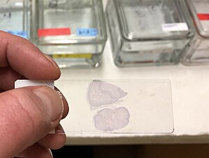
785:Demonstrates presence of flagella
14:
4016:Antibiotic susceptibility testing
3823:biochemical and immunologic tests
3159:Microbial and histological stains
2183:: separation of protein molecules
1474:staining of a parasitic flatworm.
467:Fontana's mordant(5%Tannic acid)
465:Fontana's mordant(5%Tannic acid)
61:(microscopic study of biological
4044:Minimum inhibitory concentration
3736:Manual testing: basic techniques
2551:Thakur M, Guttikonda VR (2017).
1972:transmission electron microscopy
1166:, light red or pink staining of
994:Haematoxylin and eosin (H&E)
4154:Biological techniques and tools
3089:The Biological Stain commission
2886:Biotechnic & Histochemistry
2807:Histochemistry and Cell Biology
2628:Biotechnic & Histochemistry
2376:Elementary Microbiology Vol - I
2264:Biotechnic & Histochemistry
2177:: stains that do not kill cells
1905:hematoxylin and eosin (H&E)
1903:Main staining types when using
1259:Gömöri methenamine silver stain
1035:stains cell nuclei blue, while
1025:Haematoxylin and eosin staining
771:Cell wall: Red Cytoplasm: Blue
524:Biotechnic & Histochemistry
445:Tannic acid in Leifson's stain
3952:Novobiocin susceptibility test
3942:Bacitracin susceptibility test
3124:at Sridhar Rao P.N's home page
2874:. pp. 329 ff. London: Methuen.
2861:. Iowa State University Press.
2857:Berlyn GP, Miksche JP (1976).
1340:. An example of this would be
1320:, by using a reaction between
768:Stains cell wall of bacterium
633:Smear stain with single dye .
1:
3835:Amino acid decarboxylase test
2683:10.1016/s0732-8893(86)80049-9
2640:10.1080/10520295.2016.1250285
2330:biologicalstaincommission.org
1826:stain; it will accumulate in
605:Types of staining techniques
376:Carried out without mordant.
3947:Optochin susceptibility test
3910:Sulfide indole motility test
3895:Phenylalanine deaminase test
3107:Speaking of Fixation: Part 1
3079:Resources in other libraries
2723:Tomov N, Dimitrov N (2017).
2520:10.1007/978-1-4614-4933-1_10
2239:Fundamentals of Microbiology
1687:. Hoechst 33342 contains an
1417:Collagen hybridizing peptide
1412:Collagen hybridizing peptide
1406:Collagen hybridizing peptide
1154:. Most recipes produce red
779:Flagella (Leifson's method)
713:Endospore (Dornor's method)
481:. This treatment dissolves
175:or the domain structures of
101:(highlighting, for example,
89:that focus on the study and
2460:10.1126/science.77.1990.194
2145:Biological Stain Commission
843:Lipid granules: Deep blue,
566:(a black synthetic dye) or
516:Biological Stain Commission
4172:
3252:Periodic acid–Schiff stain
2933:, 3 ed. 2007 Elsevier, Inc
2898:10.1080/bih.76.5-6.261.278
2732:Trakia Journal of Sciences
1938:for tissues that stain by
1792:(or toluylene red) stains
1753:Gimenez staining technique
1545:nuclear stain, excited by
1409:
1351:
1181:
1139:
1094:Periodic acid–Schiff stain
1091:
1050:
997:
976:Mycobacterium tuberculosis
962:
931:
881:
762:Cell wall (Dyar's method)
587:Simple versus differential
558:methods fail, is to use a
547:
18:
3991:Polymerase chain reaction
3074:Resources in your library
2819:10.1007/s00418-014-1215-0
173:semi-crystalline polymers
121:within individual cells.
21:Staining (disambiguation)
3981:Analytical profile index
3394:Light Green SF yellowish
3385:Masson's trichrome stain
3344:Auramine–rhodamine stain
2745:10.15547/tjs.2017.03.001
2408:10.1309/AJCP9RUV0YGLBVHA
2304:. Taylor & Francis.
1498:Coomassie brilliant blue
1427:Common biological stains
1230:All are used to examine
1142:Masson's trichrome stain
1078:Light Green SF yellowish
641:shapes and arrangements
397:Name of mordant applied
2948:cmrf.research.uiowa.edu
2237:Pommerville JC (2017).
1895:Stainability of tissues
1421:fluorescence microscope
1265:(illustrated in black).
702:Acid fast bacteria:Red
57:are frequently used in
3915:Triple sugar iron test
3511:Schaeffer–Fulton stain
3481:Gömöri trichrome stain
2995:. Banbury, UK: Scion.
2677:(3 Suppl): 111S–119S.
2159:: the study of tissues
2097:potassium ferrocyanide
2093:potassium ferricyanide
2069:lanthanum(III) nitrate
1930:may be referred to as
1908:
1475:
1284:(for example type III
1266:
1219:, May-Grunwald stain,
1110:
1027:is frequently used in
1021:
903:(as a mordant), and a
845:Cytoplasm: Light pink
724:Vegetative cells: Red
662:Study cell morphology
545:
441:b.) Loeffler's method
69:(microscopic study of
39:
4149:Scientific techniques
3973:point-of-care testing
3654:Cultures by body site
3607:clinical microbiology
3486:Luxol fast blue stain
3361:Auramine phenol stain
2570:10.4103/JOC.JOC_23_16
1942:stains (most notably
1902:
1470:
1257:
1101:
1058:Papanicolaou staining
1007:
952:Clostridium difficile
899:to stain cell walls,
895:. Gram staining uses
807:Cytoplasm- colorless
540:
458:a.) Fontana's method
438:a.) Leifson's method
259:brilliant cresyl blue
29:
3932:Voges–Proskauer test
3840:Bile solubility test
3695:Cultures by organism
3628:Isolation techniques
3531:Warthin–Starry stain
3404:Phosphomolybdic acid
2163:Immunohistochemistry
2153:: the study of cells
2089:phosphomolybdic acid
1983:Phosphotungstic acid
1978:Phosphotungstic acid
1706:as an indicator for
1326:potassium dichromate
1114:Periodic acid-Schiff
1012:tissue stained with
863:Polysaccharide: Red
704:Non acid fast: Blue
630:Simple (Monochrome)
512:BSC-certified stains
461:b.) Becker's method
456:Spirochete Staining
418:a.) Ringer's method
19:For other uses, see
4039:McFarland standards
4029:Disk diffusion test
4024:Beta-lactamase test
3957:Lancefield grouping
3937:X and V factor test
3905:Salt tolerance test
3780:Ziehl–Neelsen stain
3743:Colonial morphology
3547:Tissue stainability
3319:Ziehl–Neelsen stain
3179:Perls Prussian blue
2991:Kiernan JA (2015).
2794:on 31 January 2019.
2759:Levenfus I (2011).
2557:Journal of Cytology
2452:1933Sci....77..194S
2352:Staining Procedures
2113:sodium chloroaurate
2038:Ruthenium tetroxide
2033:Ruthenium tetroxide
1966:Electron microscopy
1604:gel electrophoresis
1278:histologic sections
1199:) and demethylated
1105:showing the fungus
971:Ziehl–Neelsen stain
965:Ziehl–Neelsen stain
613:Staining Technique
606:
416:Cell Wall Staining
387:
162:gel electrophoresis
146:fluorescent tagging
4108:Inoculation needle
3521:Bielschowsky stain
3423:Van Gieson's stain
3389:Lillie's trichrome
3113:– by M. Halit Umar
2065:indium trichloride
2045:ammonium molybdate
1909:
1885:glycosaminoglycans
1476:
1267:
1147:Masson's trichrome
1111:
1053:Papanicolaou stain
1022:
939:Endospore staining
934:Endospore staining
722:Endospores: Green
653:Negative (Relief)
604:
594:endospore staining
546:
436:Flagella Staining
421:b.) Dyar's method
385:
380:Indirect Staining:
255:New Methylene Blue
99:biological tissues
40:
4116:
4115:
4093:Biosafety cabinet
4052:
4051:
3900:Reverse CAMP test
3720:
3719:
3702:Bacterial culture
3572:
3571:
3373:Connective tissue
3060:Library resources
3044:978-0-19-508956-1
3033:Ruzin SE (1999).
3002:978-1-907904-32-5
2983:978-0-443-06435-7
2870:Baker JR (1958).
2770:978-3-640-86284-9
2529:978-1-4614-4932-4
2361:978-0-683-01707-6
2311:978-1-85996-099-8
2276:10.1080/714028210
2248:978-1-284-10095-2
2219:Parker N (2012).
2121:thiosemicarbazide
2109:silver proteinate
1950:when staining in
1839:Osmium tetraoxide
1334:rgyrophilic cells
1304:Argentaffin cells
1190:Romanowsky stains
1152:connective tissue
870:
869:
865:Cytoplasm: Green
574:Positive staining
556:positive staining
550:Negative staining
543:negative staining
533:Negative staining
472:
471:
251:supravital stains
107:connective tissue
4161:
4103:Inoculation loop
4001:Rapid strep test
3733:
3625:
3599:
3592:
3585:
3576:
3399:Biebrich scarlet
3152:
3145:
3138:
3129:
3048:
3029:
3017:
3006:
2987:
2958:
2957:
2955:
2954:
2940:
2934:
2924:
2918:
2917:
2881:
2875:
2868:
2862:
2855:
2849:
2848:
2830:
2802:
2796:
2795:
2781:
2775:
2774:
2763:. Munich: Grin.
2756:
2750:
2749:
2747:
2729:
2720:
2714:
2713:
2706:Stain Technology
2701:
2695:
2694:
2666:
2660:
2659:
2623:
2617:
2616:
2614:
2613:
2599:
2593:
2592:
2582:
2572:
2548:
2542:
2541:
2507:
2498:
2497:
2489:
2472:
2471:
2435:
2429:
2428:
2410:
2386:
2380:
2379:
2372:
2366:
2365:
2350:Clark G (1981).
2347:
2341:
2340:
2338:
2336:
2322:
2316:
2315:
2297:
2288:
2287:
2259:
2253:
2252:
2234:
2225:
2224:
2216:
2171:, a protein dye.
2117:thallium nitrate
2081:lead(II) nitrate
2027:
2026:
2025:
2010:Osmium tetroxide
2005:Osmium tetroxide
1854:Propidium iodide
1849:Propidium iodide
1726:Lugol's solution
1589:Ethidium bromide
1584:Ethidium bromide
1379:Osmium tetroxide
1184:Romanowsky stain
1082:Bismarck Brown Y
1080:, and sometimes
607:
475:Permeabilization
425:10% Tannic acid
405:Gram's Staining
388:
374:Direct Staining:
291:
290:
287:
284:
278:
277:
274:
271:
247:propidium iodide
177:block copolymers
36:microscope slide
4171:
4170:
4164:
4163:
4162:
4160:
4159:
4158:
4119:
4118:
4117:
4112:
4048:
4010:
3966:
3875:Methyl red test
3822:
3821:Manual testing:
3816:
3775:India ink stain
3765:Acid-fast stain
3728:
3716:
3690:
3666:Genital culture
3649:
3645:Selective media
3617:
3609:
3603:
3573:
3568:
3542:
3427:
3381:trichrome stain
3367:
3279:
3256:
3228:
3183:
3161:
3156:
3085:
3084:
3083:
3068:
3067:
3063:
3056:
3051:
3045:
3032:
3026:
3009:
3003:
2990:
2984:
2971:
2967:
2965:Further reading
2962:
2961:
2952:
2950:
2942:
2941:
2937:
2925:
2921:
2892:(5–6): 261–78.
2883:
2882:
2878:
2869:
2865:
2856:
2852:
2804:
2803:
2799:
2783:
2782:
2778:
2771:
2758:
2757:
2753:
2727:
2722:
2721:
2717:
2703:
2702:
2698:
2668:
2667:
2663:
2625:
2624:
2620:
2611:
2609:
2601:
2600:
2596:
2550:
2549:
2545:
2530:
2512:Cytopreparation
2509:
2508:
2501:
2491:
2490:
2475:
2437:
2436:
2432:
2388:
2387:
2383:
2374:
2373:
2369:
2362:
2349:
2348:
2344:
2334:
2332:
2324:
2323:
2319:
2312:
2299:
2298:
2291:
2270:(5–6): 237–75.
2261:
2260:
2256:
2249:
2236:
2235:
2228:
2218:
2217:
2206:
2201:
2141:
2133:vanadyl sulfate
2057:ferric chloride
2035:
2024:
2021:
2020:
2019:
2017:
2007:
1999:polysaccharides
1980:
1968:
1897:
1874:
1863:
1851:
1836:
1813:
1802:
1794:Nissl substance
1787:
1776:
1765:
1749:Malachite green
1746:
1744:Malachite green
1697:
1685:plasma membrane
1650:
1631:
1616:
1608:acridine orange
1586:
1574:red blood cells
1559:
1536:
1521:
1506:
1495:
1465:
1449:
1441:Acridine orange
1438:
1436:Acridine orange
1429:
1414:
1408:
1399:
1356:
1350:
1270:Silver staining
1252:
1186:
1180:
1144:
1138:
1096:
1090:
1055:
1049:
1041:red blood cells
1002:
996:
967:
961:
936:
930:
886:
880:
875:
658:into thin film
602:
589:
576:
552:
535:
508:
506:Standardization
298:
288:
285:
282:
280:
275:
272:
269:
267:
191:
24:
17:
12:
11:
5:
4169:
4168:
4165:
4157:
4156:
4151:
4146:
4141:
4136:
4131:
4121:
4120:
4114:
4113:
4111:
4110:
4105:
4100:
4095:
4090:
4085:
4084:
4083:
4073:
4072:
4071:
4060:
4058:
4054:
4053:
4050:
4049:
4047:
4046:
4041:
4036:
4031:
4026:
4020:
4018:
4012:
4011:
4009:
4008:
4003:
3998:
3993:
3988:
3983:
3977:
3975:
3971:Automated and
3968:
3967:
3965:
3964:
3959:
3954:
3949:
3944:
3939:
3934:
3929:
3928:
3927:
3917:
3912:
3907:
3902:
3897:
3892:
3887:
3882:
3877:
3872:
3867:
3862:
3857:
3855:Coagulase test
3852:
3847:
3842:
3837:
3832:
3826:
3824:
3818:
3817:
3815:
3814:
3813:
3812:
3807:
3802:
3797:
3789:
3784:
3783:
3782:
3777:
3772:
3767:
3762:
3752:
3751:
3750:
3739:
3737:
3730:
3726:Identification
3722:
3721:
3718:
3717:
3715:
3714:
3709:
3707:Fungal culture
3704:
3698:
3696:
3692:
3691:
3689:
3688:
3683:
3678:
3676:Throat culture
3673:
3671:Sputum culture
3668:
3663:
3657:
3655:
3651:
3650:
3648:
3647:
3642:
3637:
3631:
3629:
3622:
3611:
3610:
3605:Techniques in
3604:
3602:
3601:
3594:
3587:
3579:
3570:
3569:
3567:
3566:
3561:
3556:
3550:
3548:
3544:
3543:
3541:
3540:
3538:Wright's stain
3535:
3534:
3533:
3528:
3523:
3513:
3508:
3503:
3498:
3493:
3488:
3483:
3478:
3473:
3468:
3467:
3466:
3461:
3451:
3446:
3441:
3435:
3433:
3429:
3428:
3426:
3425:
3419:
3418:
3417:
3416:
3411:
3409:Fast Green FCF
3406:
3401:
3396:
3377:
3375:
3369:
3368:
3366:
3365:
3364:
3363:
3358:
3357:
3356:
3351:
3341:
3340:
3339:
3337:Methylene blue
3334:
3328:Carbol fuchsin
3311:
3310:
3309:
3304:
3302:Gentian violet
3289:
3287:
3281:
3280:
3278:
3277:
3272:
3266:
3264:
3258:
3257:
3255:
3254:
3249:
3244:
3238:
3236:
3230:
3229:
3227:
3226:
3225:
3224:
3219:
3214:
3209:
3204:
3193:
3191:
3185:
3184:
3182:
3181:
3175:
3173:
3163:
3162:
3157:
3155:
3154:
3147:
3140:
3132:
3126:
3125:
3119:
3114:
3104:
3098:
3092:
3082:
3081:
3076:
3070:
3069:
3058:
3057:
3055:
3054:External links
3052:
3050:
3049:
3043:
3030:
3024:
3007:
3001:
2988:
2982:
2968:
2966:
2963:
2960:
2959:
2935:
2919:
2876:
2863:
2850:
2797:
2776:
2769:
2751:
2738:(3): 195–197.
2715:
2696:
2661:
2618:
2594:
2563:(3): 149–153.
2543:
2528:
2499:
2473:
2430:
2381:
2367:
2360:
2342:
2317:
2310:
2289:
2254:
2247:
2226:
2203:
2202:
2200:
2197:
2196:
2195:
2193:Diaphonization
2190:
2184:
2178:
2172:
2166:
2160:
2154:
2148:
2140:
2137:
2129:uranyl nitrate
2125:uranyl acetate
2105:silver nitrate
2053:carbohydrazide
2049:cadmium iodide
2034:
2031:
2022:
2006:
2003:
1987:negative stain
1979:
1976:
1967:
1964:
1896:
1893:
1873:
1870:
1862:
1859:
1850:
1847:
1835:
1832:
1812:
1809:
1801:
1798:
1786:
1783:
1779:Methylene blue
1775:
1774:Methylene blue
1772:
1764:
1761:
1745:
1742:
1732:more visible.
1696:
1693:
1649:
1648:Hoechst stains
1646:
1630:
1627:
1615:
1612:
1585:
1582:
1570:cell membranes
1558:
1555:
1535:
1532:
1524:Crystal violet
1520:
1519:Crystal violet
1517:
1505:
1502:
1494:
1493:Coomassie blue
1491:
1464:
1461:
1452:Bismarck brown
1448:
1447:Bismarck brown
1445:
1437:
1434:
1428:
1425:
1410:Main article:
1407:
1404:
1398:
1395:
1389:in diagnosing
1359:Sudan staining
1352:Main article:
1349:
1346:
1330:Golgi's method
1322:silver nitrate
1272:is the use of
1261:demonstrating
1251:
1248:
1221:Leishman stain
1217:Jenner's stain
1213:Wright's stain
1201:methylene blue
1182:Main article:
1179:
1176:
1140:Main article:
1137:
1134:
1092:Main article:
1089:
1086:
1051:Main article:
1048:
1045:
998:Main article:
995:
992:
988:methylene blue
984:carbol fuchsin
963:Main article:
960:
957:
932:Main article:
929:
926:
897:crystal violet
882:Main article:
879:
876:
874:
871:
868:
867:
861:
858:
855:
852:
848:
847:
841:
838:
835:
832:
828:
827:
824:
821:
817:
814:
810:
809:
803:
800:
797:
794:
790:
789:
786:
783:
780:
777:
773:
772:
769:
766:
763:
760:
756:
755:
749:
746:
740:
731:
727:
726:
720:
717:
714:
711:
707:
706:
700:
697:
694:
691:
687:
686:
680:
677:
674:
671:
667:
666:
663:
660:
654:
651:
647:
646:
643:
637:
631:
628:
624:
623:
620:
617:
614:
611:
601:
598:
588:
585:
575:
572:
560:negative stain
548:Main article:
534:
531:
507:
504:
483:cell membranes
470:
469:
463:
454:
450:
449:
443:
434:
430:
429:
423:
414:
410:
409:
408:Gram's iodine
406:
403:
399:
398:
395:
392:
327:chemical bonds
297:
294:
234:
233:
201:vital staining
190:
181:
171:structures of
152:, and to flag
150:flow cytometry
79:histopathology
73:), and in the
15:
13:
10:
9:
6:
4:
3:
2:
4167:
4166:
4155:
4152:
4150:
4147:
4145:
4144:Staining dyes
4142:
4140:
4137:
4135:
4132:
4130:
4127:
4126:
4124:
4109:
4106:
4104:
4101:
4099:
4096:
4094:
4091:
4089:
4086:
4082:
4079:
4078:
4077:
4076:Anaerobic jar
4074:
4070:
4069:Growth medium
4067:
4066:
4065:
4062:
4061:
4059:
4055:
4045:
4042:
4040:
4037:
4035:
4032:
4030:
4027:
4025:
4022:
4021:
4019:
4017:
4013:
4007:
4006:Monospot test
4004:
4002:
3999:
3997:
3994:
3992:
3989:
3987:
3984:
3982:
3979:
3978:
3976:
3974:
3969:
3963:
3960:
3958:
3955:
3953:
3950:
3948:
3945:
3943:
3940:
3938:
3935:
3933:
3930:
3926:
3923:
3922:
3921:
3918:
3916:
3913:
3911:
3908:
3906:
3903:
3901:
3898:
3896:
3893:
3891:
3888:
3886:
3883:
3881:
3878:
3876:
3873:
3871:
3868:
3866:
3863:
3861:
3858:
3856:
3853:
3851:
3848:
3846:
3843:
3841:
3838:
3836:
3833:
3831:
3828:
3827:
3825:
3819:
3811:
3808:
3806:
3803:
3801:
3798:
3796:
3793:
3792:
3790:
3788:
3785:
3781:
3778:
3776:
3773:
3771:
3768:
3766:
3763:
3761:
3758:
3757:
3756:
3753:
3749:
3746:
3745:
3744:
3741:
3740:
3738:
3734:
3731:
3727:
3723:
3713:
3712:Viral culture
3710:
3708:
3705:
3703:
3700:
3699:
3697:
3693:
3687:
3686:Wound culture
3684:
3682:
3681:Urine culture
3679:
3677:
3674:
3672:
3669:
3667:
3664:
3662:
3661:Blood culture
3659:
3658:
3656:
3652:
3646:
3643:
3641:
3638:
3636:
3633:
3632:
3630:
3626:
3623:
3621:
3616:
3612:
3608:
3600:
3595:
3593:
3588:
3586:
3581:
3580:
3577:
3565:
3562:
3560:
3557:
3555:
3552:
3551:
3549:
3545:
3539:
3536:
3532:
3529:
3527:
3524:
3522:
3519:
3518:
3517:
3514:
3512:
3509:
3507:
3504:
3502:
3501:Movat's stain
3499:
3497:
3496:Moeller stain
3494:
3492:
3489:
3487:
3484:
3482:
3479:
3477:
3474:
3472:
3471:Janus Green B
3469:
3465:
3462:
3460:
3457:
3456:
3455:
3454:H&E stain
3452:
3450:
3447:
3445:
3442:
3440:
3439:Cresyl violet
3437:
3436:
3434:
3430:
3424:
3421:
3420:
3415:
3412:
3410:
3407:
3405:
3402:
3400:
3397:
3395:
3392:
3391:
3390:
3386:
3382:
3379:
3378:
3376:
3374:
3370:
3362:
3359:
3355:
3352:
3350:
3347:
3346:
3345:
3342:
3338:
3335:
3333:
3329:
3326:
3325:
3324:
3323:Kinyoun stain
3320:
3317:
3316:
3315:
3312:
3308:
3305:
3303:
3299:
3298:Methyl violet
3296:
3295:
3294:
3291:
3290:
3288:
3286:
3282:
3276:
3273:
3271:
3268:
3267:
3265:
3263:
3259:
3253:
3250:
3248:
3245:
3243:
3240:
3239:
3237:
3235:
3234:Carbohydrates
3231:
3223:
3222:Sudan Black B
3220:
3218:
3215:
3213:
3210:
3208:
3205:
3203:
3200:
3199:
3198:
3195:
3194:
3192:
3190:
3186:
3180:
3177:
3176:
3174:
3172:
3168:
3164:
3160:
3153:
3148:
3146:
3141:
3139:
3134:
3133:
3130:
3123:
3120:
3118:
3115:
3112:
3108:
3105:
3103:~ Howey, 2000
3102:
3099:
3096:
3093:
3090:
3087:
3086:
3080:
3077:
3075:
3072:
3071:
3066:
3061:
3053:
3046:
3040:
3036:
3031:
3027:
3025:9780801854019
3021:
3016:
3015:
3008:
3004:
2998:
2994:
2989:
2985:
2979:
2975:
2970:
2969:
2964:
2949:
2945:
2939:
2936:
2932:
2928:
2923:
2920:
2915:
2911:
2907:
2903:
2899:
2895:
2891:
2887:
2880:
2877:
2873:
2867:
2864:
2860:
2854:
2851:
2846:
2842:
2838:
2834:
2829:
2824:
2820:
2816:
2813:(3): 335–45.
2812:
2808:
2801:
2798:
2793:
2789:
2788:
2780:
2777:
2772:
2766:
2762:
2755:
2752:
2746:
2741:
2737:
2733:
2726:
2719:
2716:
2711:
2707:
2700:
2697:
2692:
2688:
2684:
2680:
2676:
2672:
2665:
2662:
2657:
2653:
2649:
2645:
2641:
2637:
2633:
2629:
2622:
2619:
2608:
2604:
2598:
2595:
2590:
2586:
2581:
2576:
2571:
2566:
2562:
2558:
2554:
2547:
2544:
2539:
2535:
2531:
2525:
2521:
2517:
2513:
2506:
2504:
2500:
2495:
2488:
2486:
2484:
2482:
2480:
2478:
2474:
2469:
2465:
2461:
2457:
2453:
2449:
2446:(1990): 194.
2445:
2441:
2434:
2431:
2426:
2422:
2418:
2414:
2409:
2404:
2400:
2396:
2392:
2385:
2382:
2377:
2371:
2368:
2363:
2357:
2353:
2346:
2343:
2331:
2327:
2321:
2318:
2313:
2307:
2303:
2296:
2294:
2290:
2285:
2281:
2277:
2273:
2269:
2265:
2258:
2255:
2250:
2244:
2240:
2233:
2231:
2227:
2222:
2215:
2213:
2211:
2209:
2205:
2198:
2194:
2191:
2188:
2185:
2182:
2179:
2176:
2173:
2170:
2167:
2164:
2161:
2158:
2155:
2152:
2149:
2146:
2143:
2142:
2138:
2136:
2134:
2130:
2126:
2122:
2118:
2114:
2110:
2106:
2102:
2101:ruthenium red
2098:
2094:
2090:
2086:
2085:periodic acid
2082:
2078:
2074:
2070:
2066:
2062:
2058:
2054:
2050:
2046:
2041:
2039:
2032:
2030:
2015:
2011:
2004:
2002:
2000:
1996:
1992:
1988:
1984:
1977:
1975:
1973:
1965:
1963:
1961:
1957:
1953:
1949:
1945:
1941:
1937:
1933:
1929:
1925:
1920:
1918:
1914:
1906:
1901:
1894:
1892:
1889:
1886:
1882:
1878:
1871:
1869:
1867:
1860:
1858:
1855:
1848:
1846:
1844:
1840:
1833:
1831:
1829:
1825:
1821:
1820:sulfuric acid
1817:
1810:
1808:
1806:
1799:
1797:
1795:
1791:
1784:
1782:
1780:
1773:
1771:
1769:
1762:
1760:
1758:
1754:
1750:
1743:
1741:
1738:
1733:
1731:
1727:
1723:
1721:
1717:
1713:
1712:Gram staining
1709:
1705:
1701:
1694:
1692:
1690:
1686:
1682:
1678:
1677:Hoechst 33342
1674:
1673:Hoechst 33258
1670:
1666:
1662:
1658:
1654:
1647:
1645:
1643:
1639:
1638:H&E stain
1635:
1628:
1626:
1624:
1620:
1619:Acid fuchsine
1613:
1611:
1609:
1605:
1601:
1597:
1593:
1590:
1583:
1581:
1579:
1575:
1571:
1567:
1563:
1556:
1554:
1552:
1548:
1544:
1540:
1533:
1531:
1529:
1525:
1518:
1516:
1514:
1510:
1509:Cresyl violet
1504:Cresyl violet
1503:
1501:
1499:
1492:
1490:
1488:
1484:
1480:
1473:
1469:
1462:
1460:
1457:
1453:
1446:
1444:
1442:
1435:
1433:
1426:
1424:
1422:
1418:
1413:
1405:
1403:
1397:Wirtz-Conklin
1396:
1394:
1392:
1388:
1384:
1383:Sudan Black B
1380:
1376:
1372:
1368:
1364:
1360:
1355:
1347:
1345:
1344:or formalin.
1343:
1339:
1335:
1331:
1327:
1323:
1319:
1318:Camillo Golgi
1315:
1312:
1308:
1305:
1301:
1299:
1295:
1291:
1287:
1283:
1279:
1275:
1271:
1264:
1260:
1256:
1249:
1247:
1245:
1241:
1237:
1233:
1228:
1226:
1222:
1218:
1214:
1210:
1206:
1202:
1198:
1195:
1191:
1185:
1177:
1175:
1173:
1169:
1165:
1161:
1157:
1153:
1148:
1143:
1135:
1133:
1131:
1130:proteoglycans
1127:
1123:
1119:
1118:carbohydrates
1115:
1108:
1104:
1100:
1095:
1087:
1085:
1083:
1079:
1075:
1071:
1067:
1063:
1059:
1054:
1046:
1044:
1042:
1038:
1034:
1030:
1026:
1019:
1015:
1011:
1006:
1001:
1000:H&E stain
993:
991:
989:
985:
980:
978:
977:
972:
966:
959:Ziehl-Neelsen
958:
956:
954:
953:
948:
944:
940:
935:
927:
925:
922:
921:Gram-negative
917:
915:
910:
906:
902:
898:
894:
890:
889:Gram staining
885:
884:Gram staining
877:
872:
866:
862:
859:
856:
853:
850:
849:
846:
842:
839:
836:
833:
830:
829:
825:
822:
818:
815:
812:
811:
808:
804:
801:
798:
795:
792:
791:
787:
784:
781:
778:
775:
774:
770:
767:
764:
761:
758:
757:
754:
750:
747:
745:
741:
739:
736:
732:
729:
728:
725:
721:
718:
715:
712:
709:
708:
705:
701:
698:
695:
692:
689:
688:
685:
681:
678:
675:
672:
669:
668:
664:
661:
659:
655:
652:
649:
648:
644:
642:
638:
636:
632:
629:
626:
625:
621:
618:
615:
612:
609:
608:
599:
597:
595:
586:
584:
581:
573:
571:
569:
565:
561:
557:
551:
544:
539:
532:
530:
527:
525:
521:
517:
513:
505:
503:
501:
497:
492:
491:
486:
484:
480:
476:
468:
464:
462:
459:
455:
452:
451:
448:
444:
442:
439:
435:
432:
431:
428:
424:
422:
419:
415:
412:
411:
407:
404:
401:
400:
396:
393:
390:
389:
383:
381:
377:
375:
371:
368:
365:
362:
360:
359:
354:
352:
348:
344:
340:
336:
332:
328:
324:
323:heat fixation
320:
319:
314:
312:
311:field of view
307:
306:
301:
295:
293:
264:
260:
256:
252:
248:
244:
240:
231:
227:
226:
225:
223:
219:
214:
213:
208:
206:
202:
199:(also called
198:
196:
189:
185:
182:
180:
178:
174:
170:
165:
163:
159:
158:nucleic acids
155:
151:
147:
143:
142:carbohydrates
139:
135:
131:
127:
122:
120:
116:
112:
108:
104:
103:muscle fibers
100:
96:
92:
88:
87:cytopathology
84:
80:
76:
72:
68:
64:
60:
56:
52:
48:
44:
37:
33:
28:
22:
3880:Nitrite test
3850:Citrate test
3791:Rapid tests
3770:Giemsa stain
3640:Streak plate
3564:Chromophobic
3516:Silver stain
3476:Giemsa stain
3459:Haematoxylin
3158:
3064:
3034:
3013:
2992:
2973:
2951:. Retrieved
2947:
2938:
2930:
2922:
2889:
2885:
2879:
2871:
2866:
2858:
2853:
2810:
2806:
2800:
2792:the original
2786:
2779:
2760:
2754:
2735:
2731:
2718:
2709:
2705:
2699:
2674:
2670:
2664:
2634:(1): 29–35.
2631:
2627:
2621:
2610:. Retrieved
2606:
2597:
2560:
2556:
2546:
2511:
2493:
2443:
2439:
2433:
2398:
2394:
2384:
2375:
2370:
2351:
2345:
2333:. Retrieved
2329:
2320:
2301:
2267:
2263:
2257:
2238:
2221:Microbiology
2220:
2187:Barium enema
2077:lead citrate
2073:lead acetate
2042:
2036:
2008:
1985:is a common
1981:
1969:
1960:chromophobic
1955:
1923:
1921:
1912:
1910:
1890:
1883:, including
1875:
1864:
1852:
1837:
1814:
1803:
1788:
1777:
1768:Methyl green
1766:
1763:Methyl green
1747:
1734:
1724:
1718:. Used as a
1716:microbiology
1698:
1676:
1672:
1661:minor groove
1660:
1656:
1651:
1634:Haematoxylin
1632:
1629:Haematoxylin
1623:mitochondria
1617:
1614:Acid fuchsin
1592:intercalates
1587:
1560:
1537:
1522:
1507:
1496:
1477:
1450:
1439:
1430:
1415:
1400:
1357:
1342:hydroquinone
1333:
1303:
1302:
1268:
1229:
1225:Giemsa stain
1187:
1170:, and black
1145:
1126:glycoprotein
1112:
1103:PAS diastase
1066:haematoxylin
1056:
1047:Papanicolaou
1033:Haematoxylin
1023:
981:
974:
968:
950:
937:
918:
887:
864:
844:
806:
752:
743:
737:
734:
723:
703:
683:
657:
640:
634:
619:Application
616:Preparation
590:
577:
553:
528:
519:
511:
509:
488:
487:
474:
473:
466:
460:
457:
446:
440:
437:
427:0.34% C.P.C
426:
420:
417:
379:
378:
373:
372:
369:
366:
363:
356:
355:
351:paraffin wax
335:formaldehyde
316:
315:
303:
302:
299:
263:reticulocyte
239:vital stains
235:
222:counterstain
210:
209:
193:
192:
187:
183:
166:
126:biochemistry
123:
42:
41:
32:histological
4088:Durham tube
3920:Urease test
3729:and testing
3554:Acidophilic
3506:Neutral red
3491:Methyl blue
3354:Rhodamine B
3247:Mucicarmine
3242:Alcian blue
3197:Sudan stain
3171:hemosiderin
2828:11336/35891
2223:. OpenStax.
2175:Vital stain
1956:amphophilic
1936:acidophilic
1932:azurophilic
1928:azure stain
1917:Chromosomes
1790:Neutral red
1785:Neutral red
1702:is used in
1566:cytoplasmic
1547:ultraviolet
1543:fluorescent
1391:steatorrhea
1354:Sudan stain
1263:histoplasma
1236:bone marrow
1172:cell nuclei
1107:Histoplasma
1014:hematoxylin
914:antibiotics
580:chromophore
541:Example of
347:picric acid
296:Preparation
243:trypan blue
115:blood cells
47:microscopic
4123:Categories
4064:Agar plate
3860:DNAse test
3760:Gram stain
3559:Basophilic
3414:Sirius Red
3349:Auramine O
3293:Gram stain
3275:Thioflavin
3095:StainsFile
2953:2020-04-16
2612:2020-04-16
2401:(1): 5–6.
2199:References
1954:dyes, and
1948:basophilic
1881:polyanions
1824:lipophilic
1714:, used in
1568:material,
1528:cell walls
1240:leukocytes
1178:Romanowsky
943:endospores
873:Techniques
479:surfactant
305:Wet mounts
205:morphology
119:organelles
83:hematology
77:fields of
30:A stained
4098:Incubator
4057:Equipment
3986:MALDI-TOF
3885:ONPG test
3845:CAMP test
3748:Hemolysis
3615:Isolation
3314:Acid-fast
3270:Congo red
3217:Oil Red O
3207:Sudan III
2607:labce.com
2538:1574-9053
2417:0002-9173
2157:Histology
1913:chromatic
1877:Safranine
1872:Safranine
1866:Rhodamine
1861:Rhodamine
1805:Nile blue
1800:Nile blue
1704:chemistry
1642:histology
1600:membranes
1596:apoptosis
1387:fecal fat
1375:Oil Red O
1367:Sudan III
1338:reductant
1276:to stain
1168:cytoplasm
1062:Pap smear
1029:histology
947:pathogens
928:Endospore
893:cell wall
568:India ink
500:microtome
496:pap smear
345:, and/or
91:diagnoses
59:histology
4139:Pigments
4129:Staining
3962:RPR test
3870:KOH test
3830:ALA test
3800:Catalase
3787:Wet prep
3755:Staining
3332:Fuchsine
3307:Safranin
3285:Bacteria
3212:Sudan IV
3202:Sudan II
3065:Staining
2929:Citing:
2914:32479873
2906:11871748
2845:11094194
2837:24671497
2656:37401579
2648:28098484
2589:28701828
2468:17741261
2425:19864226
2335:25 March
2284:12564600
2151:Cytology
2139:See also
2061:hexamine
1816:Nile red
1811:Nile red
1681:hydroxyl
1487:aluminum
1483:glycogen
1371:Sudan IV
1314:fixation
1311:formalin
1286:collagen
1282:proteins
1160:collagen
1122:glycogen
1070:Orange G
949:such as
909:safranin
733:Capsule
610:Sr. No.
564:nigrosin
490:Mounting
358:Mordants
343:methanol
331:proteins
329:between
318:Fixation
230:Safranin
218:fixation
212:In vitro
197:staining
188:In vitro
169:lamellar
154:proteins
134:proteins
95:diseases
67:cytology
43:Staining
4081:Gas-pak
3795:Oxidase
3635:Asepsis
3620:culture
3464:Eosin Y
3444:Cyanine
3262:Amyloid
2691:3009082
2580:5492752
2448:Bibcode
2440:Science
1991:viruses
1924:-philic
1720:mordant
1669:Hoechst
1653:Hoechst
1578:eosin Y
1479:Carmine
1472:Carmine
1463:Carmine
1244:malaria
1209:azure B
1205:azure A
1194:reduced
1156:keratin
1074:eosin Y
905:fuchsin
622:Result
391:Sr No.
339:ethanol
195:In vivo
184:In vivo
75:medical
63:tissues
49:level.
3805:Indole
3189:Lipids
3111:Part 2
3062:about
3041:
3022:
2999:
2980:
2912:
2904:
2843:
2835:
2767:
2689:
2654:
2646:
2587:
2577:
2536:
2526:
2466:
2423:
2415:
2358:
2308:
2282:
2245:
2131:, and
2014:lipids
1995:nerves
1940:acidic
1843:lipids
1757:spores
1730:nuclei
1708:starch
1700:Iodine
1695:Iodine
1456:mucins
1381:, and
1363:lipids
1307:reduce
1288:) and
1274:silver
1250:Silver
1136:Masson
901:iodine
253:(e.g.
241:(e.g.
138:lipids
117:), or
85:, and
65:), in
51:Stains
4034:Etest
3996:VITEK
3925:rapid
3865:IMViC
3432:Other
2910:S2CID
2841:S2CID
2728:(PDF)
2652:S2CID
1952:basic
1944:eosin
1828:lipid
1689:ethyl
1655:is a
1562:Eosin
1557:Eosin
1541:is a
1513:nissl
1348:Sudan
1294:cells
1232:blood
1197:eosin
1037:eosin
1018:eosin
673:Gram
600:Types
71:cells
4134:Dyes
3618:and
3167:Iron
3109:and
3039:ISBN
3020:ISBN
2997:ISBN
2978:ISBN
2902:PMID
2833:PMID
2765:ISBN
2712:(3).
2687:PMID
2644:PMID
2585:PMID
2534:ISSN
2524:ISBN
2464:PMID
2421:PMID
2413:ISSN
2356:ISBN
2337:2018
2306:ISBN
2280:PMID
2243:ISBN
2181:PAGE
1989:for
1675:and
1539:DAPI
1534:DAPI
1332:). A
1324:and
1223:and
1207:and
1188:The
1164:bone
1162:and
1016:and
1010:lung
878:Gram
453:4.)
433:3.)
413:2.)
402:1.)
261:for
257:and
111:cell
55:dyes
53:and
3810:PYR
2894:doi
2823:hdl
2815:doi
2811:142
2740:doi
2679:doi
2636:doi
2575:PMC
2565:doi
2516:doi
2456:doi
2403:doi
2399:132
2272:doi
2018:OsO
1946:),
1915:.
1665:DNA
1663:of
1657:bis
1551:DNA
1290:DNA
1234:or
1088:PAS
907:or
851:12
831:11
813:10
650:2.
627:1.
520:BSC
289:000
286:500
279:to
276:000
245:or
186:vs
160:in
156:or
130:DNA
124:In
109:),
105:or
93:of
4125::
3383::
2946:.
2908:.
2900:.
2890:76
2888:.
2839:.
2831:.
2821:.
2809:.
2736:15
2734:.
2730:.
2710:63
2708:.
2685:.
2673:.
2650:.
2642:.
2632:92
2630:.
2605:.
2583:.
2573:.
2561:34
2559:.
2555:.
2532:.
2522:.
2502:^
2476:^
2462:.
2454:.
2444:77
2442:.
2419:.
2411:.
2397:.
2393:.
2328:.
2292:^
2278:.
2268:77
2266:.
2229:^
2207:^
2135:.
2127:,
2123:,
2119:,
2115:,
2111:,
2107:,
2103:,
2099:,
2095:,
2091:,
2087:,
2083:,
2079:,
2075:,
2071:,
2067:,
2063:,
2059:,
2055:,
2051:,
2047:,
1997:,
1993:,
1759:.
1671::
1644:.
1489:.
1423:.
1393:.
1377:,
1373:,
1369:,
1365:.
1300:.
1246:.
1227:.
1215:,
1174:.
1128:,
1124:,
1084:.
1076:,
1072:,
1068:,
990:.
969:A
916:.
793:9
776:8
759:7
730:6
710:5
690:4
670:3
596:.
341:,
337:,
313:.
179:.
140:,
136:,
132:,
81:,
3598:e
3591:t
3584:v
3387:/
3330:/
3321:/
3300:/
3169:/
3151:e
3144:t
3137:v
3047:.
3028:.
3005:.
2986:.
2956:.
2916:.
2896::
2847:.
2825::
2817::
2773:.
2748:.
2742::
2693:.
2681::
2675:4
2658:.
2638::
2615:.
2591:.
2567::
2540:.
2518::
2470:.
2458::
2450::
2427:.
2405::
2378:.
2364:.
2339:.
2314:.
2286:.
2274::
2251:.
2023:4
1907:.
1120:(
1109:.
1020:.
518:(
283::
281:1
273:5
270::
268:1
38:.
23:.
Text is available under the Creative Commons Attribution-ShareAlike License. Additional terms may apply.