180:
bone marrow are released, resulting in a grayish blue color of the cells. This color is seen because of the ribosomes still left on the immature blood cells, which are not found on mature red blood cells. These cells still contain a nucleus as well due to the early release, which is not needed in mature blood cells because their only function is to carry oxygen in the blood. The life span of a typical red blood cell is acknowledged to be approximately 120 days, and the time period of a reticulocyte found in the blood to be one day. The percentage of reticulocytes calculated to be in the blood at any given time indicates the rapidity of the red blood cell turnover in a healthy patient. The number of reticulocytes, however, reflects the amount of erythropoiesis that has occurred on any certain day. The absolute number of reticulocytes is referred to as the
123:
111:, a hormone made by the kidneys, controls the production of red blood cells as well as the rate at which they are released from the bone marrow. When these levels of erythropoetin rise, they signal the release of immature red blood cells into the bloodstream and is linked to anemia. Damaged bone marrow can also lead to polychromasia. The most common cause of bone marrow damage is penetration by
46:
218:, who was studying the blood of an anemic patient, discovered granulation in the blood cells that were polychromatic. Later studies were done by other scientists also showed the same results in other forms of anemia. This pattern of granulation was also seen in several types of toxic poisoning, especially
153:
There is a slight correlation between polychromasia and reticulocytosis. It is much easier to test for polychromasia in blood cells than to perform special staining for reticulocytosis. If polychromasia is found in the blood cells, the reticulocyte count is taken to detect further disease or stress.
196:
Polychromasia can be detected through the use of stains that will change the color of the red blood cells that are affected. Under certain conditions, these red blood cells are shown to have an affinity for basic stains, contrary to the usual acid stains used. Polychromatic cells usually stain dark
209:
indicated that certain red blood cells found both in fetal circulation and bone marrow (of a cat) had unusual granulation. These granules are also called
Grawitz granules. In most instances, he found that these granules were connected by a network of sorts. The cells that had this granulation were
179:
is the main source of hematopoiesis. The liver is then used as the main hematopoietic organ of the embryo until near birth, where it is then taken over by the bone marrow. Most red blood cells are released into the blood as reticulocytes. Polychromasia occurs when the immature reticulocytes of the
222:. However, other research has shown that there has been stippling found in normal blood cells as well. Stippling is supposed to be one of the earliest symptoms of lead poisoning, although most scientists now regard it as a degenerative condition, along with polychromasia.
145:. Anemia can be caused by either overproduction or underproduction of red blood cells, as well as the production of defective blood cells. Because there are more red blood cells needed in the body at that moment, they are released prematurely, leading to polychromasia.
86:.) These cells are often shades of grayish-blue. Polychromasia is usually a sign of bone marrow stress as well as immature red blood cells. 3 types are recognized, with types 1 and 2 being referred to as 'young red blood cells' and type 3 as 'old red blood cells'.
102:
Red blood cells can be released prematurely by a number of mechanisms. Premature release of red blood cells is usually caused due to damage of the bone marrow due to underlying causes as well as in response to the stimulation of hormones in strong association with
158:
may be performed in this case to rule out immune-mediated hemolysis. Polychromasia can also be seen in blood smears when there is a normal reticulocyte count. This can be caused by infiltration of the bone marrow due to tumors as well as
360:
Bleyl, Steven B., Philip R. Brauer, and
Philippa H. Francis-West. "Development of Yolk Sac and Chorionic Cavity." Larsen's Human Embryology. By Gary C. Schoenwolf. 4th ed. Philadelphia: Churchill Livingstone, 2009. 58.
94:. All polychromatophilic cells are reticulocytes, however, not all reticulocytes are polychromatophilic. In the old blood cells, the cytoplasm either stains a light orange or does not stain at all.
90:
is used to distinguish all three types of blood smears. The young cells will generally stain gray or blue in the cytoplasm. These young red blood cells are commonly called
210:
found in blood and tissues that had been freshly stained without undergoing fixation. Howell was the first to describe these blood cells as being of the prototype
154:
If a low count of reticulocytes is found, it usually indicates bone marrow stress. If a high reticulocyte count is found, it is usually linked to hemolysis, but a
410:
Hawes, John B. (1909). "A Study of the
Reticulated Red Blood Corpuscle by Means of Vital Staining Methods; Its Relation to Polychromatophilia and Stippling".
492:
386:
141:
is the most commonly seen type of anemia. This type of anemia is usually caused by the underproduction of blood cells as well as
126:
17:
122:
206:
260:
197:
blue or gray and are distinguishable from normal blood cells mostly by a slight change in color.
181:
470:
392:
382:
138:
419:
341:
278:
252:
219:
63:
346:
329:
486:
303:
172:
108:
91:
184:
and is calculated by adjusting the reticulocyte percentage by the ratio of observed
215:
87:
465:
423:
155:
112:
67:
185:
116:
45:
376:
372:
142:
66:
found in the bloodstream as a result of being prematurely released from the
396:
176:
160:
448:
264:
214:, which meant granular degeneration of the red blood cells. In 1893,
104:
378:
Clinical
Methods: The History, Physical, and Laboratory Examinations
256:
53:
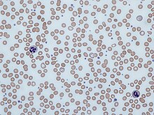
Polychromatic red blood cells appear bluish-gray on the blood smear.
62:
is a disorder where there is an abnormally high number of immature
188:
to expected hematocrit to get the 'corrected' reticulocyte count.
121:
115:, either from the bone marrow itself or as a consequence of
171:
The formation of red blood cells is commonly known as
438:
456:
442:
33:
28:
239:Thomas, Lyell J. (1937). "On the Life Cycle of
328:Camitta, Bruce M.; Slye, Rebecca Jean (2012).
8:
330:"Optimizing Use of the Complete Blood Count"
439:
375:. In Walker HK, Hall WD, Hurst JW (eds.).
44:
25:
345:
231:
175:. Up to the first 60 days of life, the
7:
308:The Lecturio Medical Concept Library
412:Boston Medical and Surgical Journal
14:
149:Association with reticulocytosis
119:from another part of the body.
381:(3rd ed.). Butterworths.
163:, or scarring, of the marrow.
1:
347:10.1016/S0031-3939(12)70597-8
424:10.1056/NEJM190910071611501
245:The Journal of Parasitology
509:
205:In 1890, research done by
15:
241:Contracaecum Spiculigerum
52:
43:
493:Red blood cell disorders
279:"What Is Polychromasia?"
70:during blood formation (
16:Not to be confused with
134:Association with anemia
130:
127:Iron-deficiency anemia
125:
18:Nuclear polychromasia
207:William Henry Howell
373:"156 Reticulocytes"
371:Bessman JD (1990).
457:External resources
304:"Anemia: Overview"
182:reticulocyte index
131:
38:Polychromatophilia
480:
479:
139:Normocytic anemia
57:
56:
23:Medical condition
500:
440:
428:
427:
407:
401:
400:
368:
362:
358:
352:
351:
349:
334:Pediatria Polska
325:
319:
318:
316:
314:
300:
294:
293:
291:
289:
275:
269:
268:
236:
48:
26:
508:
507:
503:
502:
501:
499:
498:
497:
483:
482:
481:
476:
475:
452:
451:
437:
432:
431:
409:
408:
404:
389:
370:
369:
365:
359:
355:
327:
326:
322:
312:
310:
302:
301:
297:
287:
285:
277:
276:
272:
257:10.2307/3272243
238:
237:
233:
228:
203:
194:
169:
151:
136:
100:
64:red blood cells
24:
21:
12:
11:
5:
506:
504:
496:
495:
485:
484:
478:
477:
474:
473:
461:
460:
458:
454:
453:
447:
446:
444:
443:Classification
436:
435:External links
433:
430:
429:
402:
387:
363:
353:
320:
295:
270:
230:
229:
227:
224:
220:lead poisoning
202:
199:
193:
190:
168:
165:
150:
147:
135:
132:
99:
96:
55:
54:
50:
49:
41:
40:
35:
31:
30:
22:
13:
10:
9:
6:
4:
3:
2:
505:
494:
491:
490:
488:
472:
468:
467:
463:
462:
459:
455:
450:
445:
441:
434:
425:
421:
418:(15): 493–9.
417:
413:
406:
403:
398:
394:
390:
388:0-409-90077-X
384:
380:
379:
374:
367:
364:
357:
354:
348:
343:
339:
335:
331:
324:
321:
309:
305:
299:
296:
284:
280:
274:
271:
266:
262:
258:
254:
251:(4): 429–31.
250:
246:
242:
235:
232:
225:
223:
221:
217:
213:
208:
200:
198:
191:
189:
187:
183:
178:
174:
173:hematopoiesis
166:
164:
162:
157:
148:
146:
144:
140:
133:
128:
124:
120:
118:
114:
110:
109:Erythropoetin
106:
97:
95:
93:
92:reticulocytes
89:
85:
81:
77:
73:
69:
65:
61:
60:Polychromasia
51:
47:
42:
39:
36:
32:
29:Polychromasia
27:
19:
464:
415:
411:
405:
377:
366:
356:
340:(1): 72–77.
337:
333:
323:
311:. Retrieved
307:
298:
286:. Retrieved
282:
273:
248:
244:
240:
234:
216:Max Askanazy
211:
204:
195:
170:
152:
137:
113:cancer cells
101:
88:Giemsa stain
83:
79:
75:
74:- refers to
71:
59:
58:
37:
466:MedlinePlus
156:Coombs test
68:bone marrow
34:Other names
226:References
186:hematocrit
167:Embryology
129:blood film
117:metastasis
80:-chromasia
399:. NBK264.
212:stippling
192:Diagnosis
143:hemolysis
487:Category
397:21250107
177:yolk sac
161:fibrosis
313:28 June
288:28 June
265:3272243
201:History
471:001318
395:
385:
361:Print.
263:
105:anemia
98:Causes
82:means
78:, and
283:WebMD
261:JSTOR
84:color
393:PMID
383:ISBN
315:2021
290:2021
76:many
72:poly
420:doi
416:161
342:doi
253:doi
243:".
489::
469::
414:.
391:.
338:87
336:.
332:.
306:.
281:.
259:.
249:23
247:.
107:.
449:D
426:.
422::
350:.
344::
317:.
292:.
267:.
255::
20:.
Text is available under the Creative Commons Attribution-ShareAlike License. Additional terms may apply.