816:. One systematic review found that among patients with COVID-19 and abnormal lung findings on CT, greater than 80% had GGOs, with greater than 50% having mixed GGOs and consolidation. GGOs with mixed consolidation has most often been found in elderly populations. Several studies have described a pattern among initial, intermediate, and hospital discharge imaging findings in the disease course of COVID-19. Most commonly, initial CT imaging reveals bilateral GGOs at the periphery of the lungs. During initial stages, this is most often found in the lower lobes, although involvement of the upper lobes and right middle lobe has also been reported early in the disease course. This is in contrast to the two similar coronaviruses,
612:
Differentiating between pre-malignancy and malignancy on the basis of CT alone can pose a challenge to radiologists; however, there are several features that are indicative of pre-malignant nodules. AAH is a pre-malignant cause of nodular GGO and is more commonly associated with lower attenuation on CT and smaller nodule size (<10 mm) compared to adenocarcinoma. In addition, AAH often lacks the solid features and spiculated appearance that are often associated with malignant growths. In contrast, as adenocarcinoma becomes invasive it will more often cause retraction of adjacent pleura and may show an increase in vascular markings. Nodules >15 mm almost always represent an invasive adenocarcinoma.
752:
824:, which more commonly involve only one lung on initial imaging. As the COVID-19 infection progresses, GGOs typically become more diffuse and often progress to consolidation. This is sometimes accompanied by the development of a crazy paving pattern and interlobular septal thickening. In many cases the most severe pulmonary CT abnormalities occurred within 2 weeks after symptoms began. At this point, many individuals begin showing resolution of consolidation and GGOs as symptoms improve. However, some patients have worsening symptoms and imaging findings, with further increase in septal thickening, GGOs, and consolidation. These patients may develop lung "white-out" with progression to
716:
765:
777:
161:
740:
789:
805:
356:
728:
274:
137:, and neoplasm. A correlation of imaging with a patient's clinical features is useful in narrowing the diagnosis. GGOs can be seen in normal lungs. Upon expiration there is less air in the lungs, leading to a relative increase in density of the tissue, and thus increased attenuation on CT. Furthermore, when a patient lays supine for a CT scan, the posterior lungs are in a dependent position, causing partial collapse of the posterior
33:
848:, a group of thoracic imaging radiologists. The original published definition read as: "Any extended, finely granular pattern of pulmonary opacity within which normal anatomic details are partly obscured; from a fancied resemblance to etched or abraded glass." It was again included in an updated glossary by the Fleischner Society in 2008 with a more detailed definition.
608:, organizing pneumonia, pulmonary contusion, pulmonary cryptococcus, and thoracic endometriosis. Focal interstitial fibrosis presents a unique challenge when differentiating from malignant nodular GGOs on CT imaging. It is typically persistent over long-term imaging follow-up and shares a similar appearance to malignant nodular GGOs.
751:
456:
650:
The crazy paving pattern may occur when there is both interlobular and intralobular widening. This sometimes resembles a road paved with irregular bricks or tiles. It is typically diffuse, involving larger areas of one or multiple lobes. There are a variety of potential causes, including
Pneumocystis
620:
Centrilobular GGOs refer to opacities occurring within one or multiple secondary lobules of the lung, which consist of a respiratory bronchiole, small pulmonary artery, and the surrounding tissue. A defining feature of these GGOs is the lack of involvement of the interlobular septum. Potential causes
554:
There are seven general patterns of ground-glass opacities. When combined with a patient's clinical signs and symptoms, the GGO pattern seen on imaging is useful in narrowing the differential diagnosis. It is important to note that while some disease processes present as only one pattern, many can
121:. This appears more grey, as opposed to the normally dark-appearing (air-filled) lung on CT imaging. In chest radiographs, the term refers to one or multiple areas in which the normally darker-appearing (air-filled) lung appears more opaque, hazy, or cloudy. Ground-glass opacity is in contrast to
641:
pattern of GGO refers to multiple irregular areas of both increased attenuation and decreased attenuation on CT. It is often the result of occlusion of small pulmonary arteries or obstruction of small airways leading to air trapping. Sarcoidosis is an additional cause of a mosaic GGOs due to the
831:
Preliminary reports have shown many patients have residual GGOs at time of discharge from the hospital. Due to the novelty of COVID-19, large studies investigating the long-term pulmonary CT changes have yet to be completed. However, long-term pulmonary changes have been seen in patients after
603:
There are numerous potential causes of nodular GGOs which can be broadly separated into benign and malignant conditions. Benign conditions potentially leading to the formation of nodular GGOs include aspergillosis, acute eosinophilic pneumonia, focal interstitial fibrosis, granulomatosis with
611:
Pre-malignant or malignant causes of nodular GGOs include adenocarcinoma, adenocarcinoma in situ, and atypical adenomatous hyperplasia (AAH). One large review study found that 80% of nodular GGOs which were present on repeated CT imaging represented either pre-malignant or malignant growths.
70:. When a substance other than air fills an area of the lung it increases that area's density. On both x-ray and CT, this appears more grey or hazy as opposed to the normally dark-appearing lungs. Although it can sometimes be seen in normal lungs, common pathologic causes include
659:
A halo sign refers to a GGO that fills the area around a consolidation or nodule. This is a most commonly seen in various types of pulmonary infections, including CMV pneumonia, tuberculosis, nocardia infection, some fungal pneumonias, and septic emboli. Schistosomiasis, a
651:
pneumonia, late-stage adenocarcinoma, pulmonary edema, some types of idiopathic interstitial pneumonias, diffuse alveolar hemorrhage, sarcoidosis, and pulmonary alveolar proteinosis. COVID-19 has also been shown to occasionally cause GGOs with a crazy paving pattern.
594:
individuals, is a classic cause of diffuse GGOs. Many viral pneumonias and idiopathic interstitial pneumonias can also lead to a diffuse GGO pattern. Radiation pneumonitis, a side effect of pulmonary radiation therapy, can lead to pulmonary fibrosis and diffuse GGOs.
125:, in which the pulmonary vascular markings are obscured. GGO can be used to describe both focal and diffuse areas of increased density. Subtypes of GGOs include diffuse, nodular, centrilobular, mosaic, crazy paving, halo sign, and reversed halo sign.
776:
739:
664:
infection, also commonly presents with the halo sign. Important non-infectious causes include granulomatosis with polyangiitis, metastatic disease with pulmonary hemorrhage, and some types of idiopathic interstitial pneumonias.
715:
36:
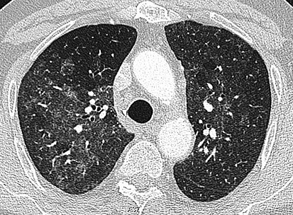
High-resolution CT image showing ground-glass opacities in the periphery of both lungs in a patient with COVID-19 (red arrows). The adjacent normal lung tissue with lower attenuation appears as darker areas.
757:
CT image showing mosaic attenuation pattern in patient with hypersensitivity pneumonitis. Note the alternating, patchy areas of increased and decreased attenuation, particularly in the left lung (screen
563:
The diffuse pattern typically refers to GGOs in multiple lobes of one or both lungs. Broadly, a diffuse pattern of GGO can be caused by displacement of air with fluid, inflammatory debris, or fibrosis.
764:
240:
642:
formation of granulomas in interstitial areas. This may coexist with granulomatosis with polyangiitis, leading to diffuse areas of increased attenuation with ground-glass appearance.
788:
1085:
El-Sherief AH, Gilman MD, Healey TT, Tambouret RH, Shepard JA, Abbott GF, Wu CC (2014). "Clear vision through the haze: a practical approach to ground-glass opacity".
1256:
Lee HY, Choi YL, Lee KS, Han J, Zo JI, Shim YM, Moon JW (March 2014). "Pure ground-glass opacity neoplastic lung nodules: histopathology, imaging, and management".
248:
677:. According to published criteria, the consolidation should form more than three-fourths of a circle and be at least 2 mm thick. It is often suggestive of
244:
94:
of air compared to the surrounding tissues. When air is replaced by another substance (e.g. fluid or fibrosis), the density of the area increases, causing the
745:
CT image showing centrilobular pattern of GGOs in patient with pulmonary tuberculosis. Note the small, nodular areas of increased attenuation in both lungs.
727:
832:
recovery from SARS and MERS, suggesting the possibility of similar long-term complications in patients who have recovered from acute COVID-19 infection.
157:
may cause GGOs. It is important to note that while many of the pulmonary infections listed below may lead to GGOs, this does not occur in every case.
1802:
Tuddenham WJ (September 1984). "Glossary of terms for thoracic radiology: recommendations of the
Nomenclature Committee of the Fleischner Society".
782:
CT image showing halo sign in patient with pulmonary aspergillosis. Note ground-glass opacification surrounding the area of consolidation (circled).
133:
The differential diagnosis for ground-glass opacities is broad. General etiologies include infections, interstitial lung diseases, pulmonary edema,
62:. It is typically defined as an area of hazy opacification (x-ray) or increased attenuation (CT) due to air displacement by fluid, airway collapse,
1172:
Rossi SE, Erasmus JJ, McAdams HP, Sporn TA, Goodman PC (1 September 2000). "Pulmonary drug toxicity: radiologic and pathologic manifestations".
153:, the presence of GGO (as opposed to consolidation) is a useful diagnostic clue. Most bacterial infections lead to lobar consolidation, while
1837:
Hansell DM, Bankier AA, MacMahon H, McLoud TC, Müller NL, Remy J (March 2008). "Fleischner
Society: glossary of terms for thoracic imaging".
1038:
1002:
969:
929:
893:
825:
626:
513:
437:
427:
102:
625:, some types of idiopathic interstitial pneumonias, hypersensitivity pneumonitis, aspiration pneumonitis, cholesterol granulomas, and
467:
432:
405:
1451:
1383:
817:
141:. This leads to an increase in density of the tissue, resulting increased attenuation and a possible ground-glass appearance on CT.
821:
698:
678:
495:
472:
442:
505:
422:
109:, although it is also used when describing chest radiographs. In CT, the term refers to one or multiple areas of increased
410:
484:
400:
1376:
Fundamentals of High-Resolution Lung CT: Common
Findings, Common Patterns, Common Diseases, and Differential Diagnosis
702:
500:
287:
462:
160:
1890:
572:
is a rarer cause of diffuse GGO seen in some types of vasculitis, autoimmune conditions, and bleeding disorders.
75:
840:
The first usage of "ground-glass opacity" by a major radiological society occurred in a 1984 publication of the
1216:"Nodular ground-glass opacity at thin-section CT: histologic correlation and evaluation of change at follow-up"
813:
459:
CT image showing diffuse GGOs throughout both lungs. An abscess is also noted in the right lung (screen left).
857:
674:
122:
164:
High-Resolution CT image in a patient with
Pneumocystis pneumonia infection showing ground-glass opacities.
808:
CT image in patient with COVID-19 showing bilateral ground-glass opacities at the periphery of both lungs.
804:
686:
579:
543:
332:
305:
292:
192:
455:
374:
264:
178:
118:
1701:
Carotti M, Salaffi F, Sarzi-Puttini P, Agostini A, Borgheresi A, Minorati D, et al. (July 2020).
862:
569:
528:
369:
259:
185:
134:
355:
518:
1750:
George PM, Barratt SL, Condliffe R, Desai SR, Devaraj A, Forrest I, et al. (November 2020).
1703:"Chest CT features of coronavirus disease 2019 (COVID-19) pneumonia: key points for radiologists"
845:
154:
138:
1854:
1819:
1781:
1732:
1683:
1642:
1597:
1576:"Coronavirus Disease 2019 (COVID-19): A Systematic Review of Imaging Findings in 919 Patients"
1549:
1528:"Coronavirus Disease 2019 (COVID-19): A Systematic Review of Imaging Findings in 919 Patients"
1503:
1447:
1441:
1422:
1379:
1325:
1273:
1235:
1189:
1154:
1102:
1044:
1034:
1008:
998:
975:
965:
935:
925:
899:
889:
721:
CT showing diffuse ground-glass opacities in periphery of both lungs in patient with COVID-19.
681:, but is only seen in about 20% of individuals with this condition. It can also be present in
661:
591:
273:
277:
CT image showing ground-glass opacification in the posterior of the right lung (screen left).
1846:
1811:
1771:
1763:
1722:
1714:
1673:
1632:
1624:
1587:
1539:
1493:
1485:
1412:
1347:
1315:
1307:
1265:
1227:
1181:
1144:
1136:
1094:
583:
378:
95:
51:
1185:
1125:"Review of the Chest CT Differential Diagnosis of Ground-Glass Opacities in the COVID Era"
812:
Ground-glass opacity is among the most common imaging findings in patients with confirmed
682:
565:
533:
523:
344:
254:
217:
79:
1876:
Ground-Glass
Opacity of the Lung Parenchyma: A Guide to Analysis with High-Resolution CT
1474:"Coronavirus Disease 2019 (COVID-19) CT Findings: A Systematic Review and Meta-analysis"
1296:"Chest CT manifestations of new coronavirus disease 2019 (COVID-19): a pictorial review"
32:
1776:
1751:
1727:
1702:
1637:
1616:
1498:
1473:
1320:
1295:
1149:
1124:
605:
1875:
1215:
1884:
1662:"Middle East Respiratory Syndrome Coronavirus: What Does a Radiologist Need to Know?"
1628:
694:
359:
CT image showing patchy areas of ground-glass opacities representing pulmonary edema.
312:
204:
90:
In both CT and chest radiographs, normal lungs appear dark due to the relative lower
1098:
690:
575:
390:
327:
322:
231:
41:
1767:
685:
where the halo consists of hemorrhage, as well as in infectious diseases such as
17:
1352:
706:
538:
317:
236:
110:
1718:
1489:
1311:
1850:
1140:
1048:
1012:
979:
939:
903:
888:(Fifth ed.). Philadelphia, PA: Elsevier. pp. Supplement 3, e36–e80.
770:
CT image showing crazy paving pattern of ground-glass opacities in both lungs.
622:
386:
382:
1426:
844:
It was published as part of a glossary of recommended nomenclature from the
673:
A reversed halo sign is a central ground-glass opacity surrounded by denser
394:
269:
150:
71:
1858:
1785:
1736:
1687:
1646:
1601:
1553:
1507:
1329:
1277:
1239:
1193:
1158:
1106:
883:
1823:
1815:
1417:
1401:"Medical image of the week: pulmonary infarction- the "reverse halo sign""
1400:
1678:
1661:
1592:
1575:
1544:
1527:
1269:
1231:
587:
211:
67:
63:
282:
91:
55:
638:
106:
794:
CT image of reversed halo sign in patient with organizing pneumonia.
1574:
Salehi S, Abedi A, Balakrishnan S, Gholamrezanezhad A (July 2020).
1526:
Salehi S, Abedi A, Balakrishnan S, Gholamrezanezhad A (July 2020).
1443:
High
Resolution Computed Tomography of the Lungs: A Practical Guide
31:
924:(Fourth ed.). Philadelphia, PA: Elsevier. pp. 299–331.
1214:
Park CM, Goo JM, Lee HJ, Lee CH, Chun EJ, Im JG (1 March 2007).
101:
Ground-glass opacity is most often used to describe findings in
59:
1033:(2nd ed.). Philadelphia, PA: Elsevier. pp. 109–137.
621:
of centrilobular GGOs include pulmonary calcifications from
27:
Radiologic sign on radiographs and computed tomography scans
1752:"Respiratory follow-up of patients with COVID-19 pneumonia"
1123:
Parekh M, Donuru A, Balasubramanya R, Kapur S (July 2020).
1294:
Ye Z, Zhang Y, Wang Y, Huang Z, Song B (August 2020).
997:(4th ed.). Philadelphia: Elsevier. pp. 2–4.
1660:Das KM, Lee EY, Langer RD, Larsson SG (June 2016).
568:and ARDS are common causes of a fluid-filled lung.
1472:Bao C, Liu X, Zhang H, Li Y, Liu J (June 2020).
1405:Southwest Journal of Pulmonary and Critical Care
995:Learning radiology : recognizing the basics
733:CT image showing ground-glass nodule (circled).
964:(Third ed.). Philadelphia, PA: Elsevier.
915:
913:
1399:Wu G, Schmit B, Arteaga V, Palacio D (2017).
1341:
1339:
8:
1478:Journal of the American College of Radiology
885:Felson's principles of chest roentgenology
826:acute respiratory distress syndrome (ARDS)
582:pneumonia, an infection typically seen in
578:and fibrosis can also cause diffuse GGOs.
1775:
1726:
1677:
1636:
1591:
1543:
1497:
1416:
1319:
1148:
1087:Current Problems in Diagnostic Radiology
803:
555:present with a mixture of GGO patterns.
454:
354:
272:
159:
874:
711:
1804:AJR. American Journal of Roentgenology
1797:
1795:
1666:AJR. American Journal of Roentgenology
1580:AJR. American Journal of Roentgenology
1532:AJR. American Journal of Roentgenology
1289:
1287:
1258:AJR. American Journal of Roentgenology
1186:10.1148/radiographics.20.5.g00se081245
1080:
1078:
1569:
1567:
1565:
1563:
1521:
1519:
1517:
1467:
1465:
1463:
1378:. Lippincott Williams & Wilkins.
1251:
1249:
1076:
1074:
1072:
1070:
1068:
1066:
1064:
1062:
1060:
1058:
7:
1209:
1207:
1205:
1203:
1118:
1116:
1024:
1022:
955:
953:
951:
949:
627:pulmonary capillary hemangiomastosis
514:Pulmonary capillary hemangiomatosis
438:Non-specific interstitial pneumonia
428:Desquamative interstitial pneumonia
1615:Ooi GC, Daqing M (November 2003).
842:American Journal of Roentgenology.
468:Adenocarcinoma in situ of the lung
433:Lymphocytic interstitial pneumonia
25:
417:Idiopathic interstitial pneumonia
288:Respiratory Syncytial Virus (RSV)
1629:10.1046/j.1440-1843.2003.00519.x
828:requiring treatment escalation.
787:
775:
763:
750:
738:
726:
714:
699:granulomatosis with polyangiitis
496:Granulomatosis with polyangiitis
473:Atypical adenomatous hyperplasia
443:Cryptogenic organizing pneumonia
98:to appear lighter or more grey.
1446:. JP Medical Ltd. p. 256.
1099:10.1067/j.cpradiol.2014.01.004
506:Pulmonary alveolar proteinosis
423:Acute interstitial pneumonitis
1:
1768:10.1136/thoraxjnl-2020-215314
1617:"SARS: radiological features"
1031:Müller's imaging of the chest
1374:Elicker BM, Webb WR (2012).
1348:"Reversed halo sign (lungs)"
1029:Walker CM, Chung JH (2019).
485:Acute eosinophilic pneumonia
401:Hypersensitivity pneumonitis
265:Human metapneumovirus (HMPV)
960:Sharma A, Abbott G (2019).
703:lymphomatoid granulomatosis
570:Diffuse alveolar hemorrhage
566:Cardiogenic pulmonary edema
492:Focal interstitial fibrosis
1907:
1719:10.1007/s11547-020-01237-4
1490:10.1016/j.jacr.2020.03.006
1312:10.1007/s00330-020-06801-0
260:Herpes Simplex Virus (HSV)
1851:10.1148/radiol.2462070712
1141:10.1148/radiol.2020202504
501:Lymphatoid granulomatosis
345:Pulmonary Schistosomiasis
76:interstitial lung disease
510:Pulmonary calcifications
186:Chlamydophila pneumoniae
56:computed tomography (CT)
1623:. 8 Suppl (s1): S15-9.
922:Essentials of radiology
858:Pulmonary consolidation
193:Legionella pneumophilia
50:) is a finding seen on
1440:Karthikeyan D (2013).
920:Mettler Jr FA (2019).
809:
687:paracoccidioidomycosis
544:Thoracic endometriosis
489:Cholesterol granulomas
460:
370:Aspiration pneumonitis
360:
333:Paracoccidioidomycosis
328:Pulmonary cryptococcus
313:Invasive aspergillosis
306:Pneumocystis jirovecii
278:
165:
37:
1816:10.2214/ajr.143.3.509
1418:10.13175/swjpcc124-17
1346:Foley R, et al.
807:
458:
411:Radiation pneumonitis
377:(most common include
358:
351:Non-infectious causes
276:
255:Cytomegalovirus (CMV)
179:Mycoplasma pneumoniae
163:
119:pulmonary vasculature
35:
1707:La Radiologia Medica
1679:10.2214/AJR.15.15363
1593:10.2214/AJR.20.23034
1545:10.2214/AJR.20.23034
1270:10.2214/AJR.13.11819
1232:10.1148/rg.272065061
863:Pulmonary infiltrate
679:organizing pneumonia
586:(e.g. patients with
534:Pulmonary infarction
529:Pulmonary hemorrhage
449:Neoplastic processes
135:pulmonary hemorrhage
882:Goodman LR (2015).
519:Pulmonary contusion
463:Lung adenocarcinoma
155:atypical pneumonias
117:concealment of the
1300:European Radiology
993:Herring W (2020).
846:Fleischner Society
810:
669:Reversed halo sign
623:metastatic disease
461:
361:
279:
166:
149:In the setting of
103:high-resolution CT
68:neoplastic process
38:
1762:(11): 1009–1016.
1040:978-0-323-53179-5
1004:978-0-323-56728-2
971:978-0-323-59699-2
931:978-0-323-56787-9
895:978-0-323-77795-7
584:immunocompromised
479:Additional causes
200:Focal or nodular
145:Infectious causes
18:Reverse halo sign
16:(Redirected from
1898:
1891:Radiologic signs
1863:
1862:
1834:
1828:
1827:
1799:
1790:
1789:
1779:
1747:
1741:
1740:
1730:
1698:
1692:
1691:
1681:
1657:
1651:
1650:
1640:
1612:
1606:
1605:
1595:
1571:
1558:
1557:
1547:
1523:
1512:
1511:
1501:
1469:
1458:
1457:
1437:
1431:
1430:
1420:
1396:
1390:
1389:
1371:
1365:
1364:
1362:
1360:
1343:
1334:
1333:
1323:
1306:(8): 4381–4389.
1291:
1282:
1281:
1253:
1244:
1243:
1211:
1198:
1197:
1169:
1163:
1162:
1152:
1135:(3): E289–E302.
1120:
1111:
1110:
1082:
1053:
1052:
1026:
1017:
1016:
990:
984:
983:
962:Thoracic imaging
957:
944:
943:
917:
908:
907:
879:
791:
779:
767:
754:
742:
730:
718:
697:, as well as in
592:immunosuppressed
379:cyclophosphamide
293:Varicella zoster
54:(radiograph) or
21:
1906:
1905:
1901:
1900:
1899:
1897:
1896:
1895:
1881:
1880:
1872:
1867:
1866:
1836:
1835:
1831:
1801:
1800:
1793:
1749:
1748:
1744:
1700:
1699:
1695:
1672:(6): 1193–201.
1659:
1658:
1654:
1614:
1613:
1609:
1573:
1572:
1561:
1525:
1524:
1515:
1471:
1470:
1461:
1454:
1439:
1438:
1434:
1398:
1397:
1393:
1386:
1373:
1372:
1368:
1358:
1356:
1345:
1344:
1337:
1293:
1292:
1285:
1255:
1254:
1247:
1213:
1212:
1201:
1171:
1170:
1166:
1122:
1121:
1114:
1084:
1083:
1056:
1041:
1028:
1027:
1020:
1005:
992:
991:
987:
972:
959:
958:
947:
932:
919:
918:
911:
896:
881:
880:
876:
871:
854:
838:
802:
795:
792:
783:
780:
771:
768:
759:
755:
746:
743:
734:
731:
722:
719:
683:lung infarction
671:
657:
648:
635:
618:
601:
561:
552:
524:Pulmonary edema
481:
451:
419:
366:
353:
341:
301:
228:
171:
147:
131:
105:imaging of the
88:
80:pulmonary edema
58:imaging of the
28:
23:
22:
15:
12:
11:
5:
1904:
1902:
1894:
1893:
1883:
1882:
1879:
1878:
1871:
1870:External links
1868:
1865:
1864:
1845:(3): 697–722.
1829:
1791:
1742:
1713:(7): 636–646.
1693:
1652:
1607:
1559:
1513:
1484:(6): 701–709.
1459:
1452:
1432:
1411:(4): 162–163.
1391:
1384:
1366:
1335:
1283:
1264:(3): W224-33.
1245:
1226:(2): 391–408.
1199:
1180:(5): 1245–59.
1164:
1112:
1054:
1039:
1018:
1003:
985:
970:
945:
930:
909:
894:
873:
872:
870:
867:
866:
865:
860:
853:
850:
837:
834:
801:
798:
797:
796:
793:
786:
784:
781:
774:
772:
769:
762:
760:
756:
749:
747:
744:
737:
735:
732:
725:
723:
720:
713:
670:
667:
656:
653:
647:
644:
634:
631:
617:
614:
606:IgA vasculitis
604:polyangiitis,
600:
597:
560:
557:
551:
548:
547:
546:
541:
536:
531:
526:
521:
516:
511:
508:
503:
498:
493:
490:
487:
480:
477:
476:
475:
470:
465:
450:
447:
446:
445:
440:
435:
430:
425:
418:
415:
414:
413:
408:
403:
398:
372:
365:
362:
352:
349:
348:
347:
340:
337:
336:
335:
330:
325:
320:
315:
310:
300:
297:
296:
295:
290:
285:
280:
267:
262:
257:
252:
234:
227:
224:
223:
222:
221:
220:
215:
208:
198:
197:
196:
189:
182:
170:
167:
146:
143:
130:
127:
87:
84:
26:
24:
14:
13:
10:
9:
6:
4:
3:
2:
1903:
1892:
1889:
1888:
1886:
1877:
1874:
1873:
1869:
1860:
1856:
1852:
1848:
1844:
1840:
1833:
1830:
1825:
1821:
1817:
1813:
1810:(3): 509–17.
1809:
1805:
1798:
1796:
1792:
1787:
1783:
1778:
1773:
1769:
1765:
1761:
1757:
1753:
1746:
1743:
1738:
1734:
1729:
1724:
1720:
1716:
1712:
1708:
1704:
1697:
1694:
1689:
1685:
1680:
1675:
1671:
1667:
1663:
1656:
1653:
1648:
1644:
1639:
1634:
1630:
1626:
1622:
1618:
1611:
1608:
1603:
1599:
1594:
1589:
1585:
1581:
1577:
1570:
1568:
1566:
1564:
1560:
1555:
1551:
1546:
1541:
1537:
1533:
1529:
1522:
1520:
1518:
1514:
1509:
1505:
1500:
1495:
1491:
1487:
1483:
1479:
1475:
1468:
1466:
1464:
1460:
1455:
1453:9789350904084
1449:
1445:
1444:
1436:
1433:
1428:
1424:
1419:
1414:
1410:
1406:
1402:
1395:
1392:
1387:
1385:9781469824796
1381:
1377:
1370:
1367:
1355:
1354:
1349:
1342:
1340:
1336:
1331:
1327:
1322:
1317:
1313:
1309:
1305:
1301:
1297:
1290:
1288:
1284:
1279:
1275:
1271:
1267:
1263:
1259:
1252:
1250:
1246:
1241:
1237:
1233:
1229:
1225:
1221:
1220:Radiographics
1217:
1210:
1208:
1206:
1204:
1200:
1195:
1191:
1187:
1183:
1179:
1175:
1174:Radiographics
1168:
1165:
1160:
1156:
1151:
1146:
1142:
1138:
1134:
1130:
1126:
1119:
1117:
1113:
1108:
1104:
1100:
1096:
1093:(3): 140–58.
1092:
1088:
1081:
1079:
1077:
1075:
1073:
1071:
1069:
1067:
1065:
1063:
1061:
1059:
1055:
1050:
1046:
1042:
1036:
1032:
1025:
1023:
1019:
1014:
1010:
1006:
1000:
996:
989:
986:
981:
977:
973:
967:
963:
956:
954:
952:
950:
946:
941:
937:
933:
927:
923:
916:
914:
910:
905:
901:
897:
891:
887:
886:
878:
875:
868:
864:
861:
859:
856:
855:
851:
849:
847:
843:
835:
833:
829:
827:
823:
819:
815:
806:
799:
790:
785:
778:
773:
766:
761:
753:
748:
741:
736:
729:
724:
717:
712:
710:
708:
704:
700:
696:
695:aspergillosis
692:
688:
684:
680:
676:
675:consolidation
668:
666:
663:
654:
652:
645:
643:
640:
632:
630:
628:
624:
616:Centrilobular
615:
613:
609:
607:
598:
596:
593:
589:
585:
581:
577:
573:
571:
567:
558:
556:
549:
545:
542:
540:
537:
535:
532:
530:
527:
525:
522:
520:
517:
515:
512:
509:
507:
504:
502:
499:
497:
494:
491:
488:
486:
483:
482:
478:
474:
471:
469:
466:
464:
457:
453:
452:
448:
444:
441:
439:
436:
434:
431:
429:
426:
424:
421:
420:
416:
412:
409:
407:
404:
402:
399:
396:
392:
388:
384:
380:
376:
375:Drug toxicity
373:
371:
368:
367:
363:
357:
350:
346:
343:
342:
338:
334:
331:
329:
326:
324:
321:
319:
316:
314:
311:
308:
307:
303:
302:
298:
294:
291:
289:
286:
284:
281:
275:
271:
268:
266:
263:
261:
258:
256:
253:
250:
246:
242:
238:
235:
233:
230:
229:
225:
219:
218:Septic emboli
216:
214:
213:
209:
207:
206:
205:Mycobacterium
202:
201:
199:
195:
194:
190:
188:
187:
183:
181:
180:
176:
175:
173:
172:
168:
162:
158:
156:
152:
144:
142:
140:
136:
128:
126:
124:
123:consolidation
120:
116:
112:
108:
104:
99:
97:
93:
85:
83:
81:
77:
73:
69:
65:
61:
57:
53:
49:
45:
43:
34:
30:
19:
1842:
1838:
1832:
1807:
1803:
1759:
1755:
1745:
1710:
1706:
1696:
1669:
1665:
1655:
1620:
1610:
1586:(1): 87–93.
1583:
1579:
1538:(1): 87–93.
1535:
1531:
1481:
1477:
1442:
1435:
1408:
1404:
1394:
1375:
1369:
1357:. Retrieved
1351:
1303:
1299:
1261:
1257:
1223:
1219:
1177:
1173:
1167:
1132:
1128:
1090:
1086:
1030:
994:
988:
961:
921:
884:
877:
841:
839:
830:
811:
691:tuberculosis
672:
658:
649:
646:Crazy paving
636:
619:
610:
602:
580:Pneumocystis
576:Inflammation
574:
562:
553:
391:methotrexate
323:Mucormycosis
304:
210:
203:
191:
184:
177:
148:
132:
114:
100:
89:
47:
42:Ground-glass
40:
39:
29:
1621:Respirology
1353:Radiopaedia
707:sarcoidosis
539:Sarcoidosis
318:Candidiasis
239:(including
237:Coronavirus
111:attenuation
52:chest x-ray
1049:1051135278
1013:1096282271
980:1022265855
940:1053711279
904:1134689400
869:References
387:carmustine
383:amiodarone
249:SARS-CoV-2
232:Adenovirus
113:(density)
86:Definition
72:infections
1839:Radiology
1427:2160-6773
1359:2 January
1129:Radiology
662:parasitic
655:Halo sign
395:bleomycin
364:Exposures
339:Parasitic
270:Influenza
169:Bacterial
151:pneumonia
1885:Category
1859:18195376
1786:32839287
1737:32500509
1688:26998804
1647:15018128
1602:32174129
1554:32174129
1508:32283052
1330:32193638
1278:24555618
1240:17374860
1194:10992015
1159:32633678
1107:24791617
852:See also
814:COVID-19
800:COVID-19
550:Patterns
245:SARS-CoV
241:MERS-CoV
212:Nocardia
174:Diffuse
64:fibrosis
1824:6380245
1777:7447111
1728:7270744
1638:7169195
1499:7151282
1321:7088323
1150:7350036
836:History
758:right).
599:Nodular
559:Diffuse
283:Measles
139:alveoli
115:without
92:density
66:, or a
44:opacity
1857:
1822:
1784:
1774:
1756:Thorax
1735:
1725:
1686:
1645:
1635:
1600:
1552:
1506:
1496:
1450:
1425:
1382:
1328:
1318:
1276:
1238:
1192:
1157:
1147:
1105:
1047:
1037:
1011:
1001:
978:
968:
938:
928:
902:
892:
705:, and
693:, and
639:mosaic
633:Mosaic
393:, and
299:Fungal
247:, and
129:Causes
107:thorax
96:tissue
78:, and
590:) or
406:EVALI
309:(PCP)
226:Viral
60:lungs
1855:PMID
1820:PMID
1782:PMID
1733:PMID
1684:PMID
1643:PMID
1598:PMID
1550:PMID
1504:PMID
1448:ISBN
1423:ISSN
1380:ISBN
1361:2018
1326:PMID
1274:PMID
1236:PMID
1190:PMID
1155:PMID
1103:PMID
1045:OCLC
1035:ISBN
1009:OCLC
999:ISBN
976:OCLC
966:ISBN
936:OCLC
926:ISBN
900:OCLC
890:ISBN
822:MERS
820:and
818:SARS
588:AIDS
1847:doi
1843:246
1812:doi
1808:143
1772:PMC
1764:doi
1723:PMC
1715:doi
1711:125
1674:doi
1670:206
1633:PMC
1625:doi
1588:doi
1584:215
1540:doi
1536:215
1494:PMC
1486:doi
1413:doi
1316:PMC
1308:doi
1266:doi
1262:202
1228:doi
1182:doi
1145:PMC
1137:doi
1133:297
1095:doi
48:GGO
1887::
1853:.
1841:.
1818:.
1806:.
1794:^
1780:.
1770:.
1760:75
1758:.
1754:.
1731:.
1721:.
1709:.
1705:.
1682:.
1668:.
1664:.
1641:.
1631:.
1619:.
1596:.
1582:.
1578:.
1562:^
1548:.
1534:.
1530:.
1516:^
1502:.
1492:.
1482:17
1480:.
1476:.
1462:^
1421:.
1409:15
1407:.
1403:.
1350:.
1338:^
1324:.
1314:.
1304:30
1302:.
1298:.
1286:^
1272:.
1260:.
1248:^
1234:.
1224:27
1222:.
1218:.
1202:^
1188:.
1178:20
1176:.
1153:.
1143:.
1131:.
1127:.
1115:^
1101:.
1091:43
1089:.
1057:^
1043:.
1021:^
1007:.
974:.
948:^
934:.
912:^
898:.
701:,
689:,
637:A
629:.
389:,
385:,
381:,
243:,
82:.
74:,
1861:.
1849::
1826:.
1814::
1788:.
1766::
1739:.
1717::
1690:.
1676::
1649:.
1627::
1604:.
1590::
1556:.
1542::
1510:.
1488::
1456:.
1429:.
1415::
1388:.
1363:.
1332:.
1310::
1280:.
1268::
1242:.
1230::
1196:.
1184::
1161:.
1139::
1109:.
1097::
1051:.
1015:.
982:.
942:.
906:.
709:.
397:)
251:)
46:(
20:)
Text is available under the Creative Commons Attribution-ShareAlike License. Additional terms may apply.