367:
742:
1997:
518:
499:
718:
483:
537:
40:
637:
730:
352:
337:
784:
688:
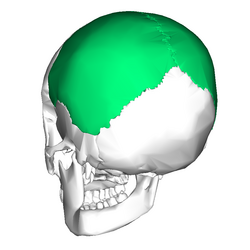
The parietal bone is usually present in the posterior end of the skull and is near the midline. This bone is part of the skull roof, which is a set of bones that cover the brain, eyes and nostrils. The parietal bones make contact with several other bones in the skull. The anterior part of the bone
604:
is truncated; it articulates with the occipital bone and with the mastoid portion of the temporal, and presents on its inner surface a broad, shallow groove which lodges part of the transverse sinus. The point of meeting of this angle with the occipital and the mastoid part of the temporal is named
705:
bone. The bone-supported neck frills of ceratopsians were formed by extensions of the parietal bone. These frills, which overhang the neck and extend past the rest of the skull is a diagnostic trait of ceratopsians. The recognizable skull domes present in pachycephalosaurs were formed by the
2017:
741:
582:, thin and acute, is received into the interval between the frontal bone and the great wing of the sphenoid. Its inner surface is marked by a deep groove, sometimes a canal, for the anterior divisions of the
366:
593:
is rounded and corresponds with the point of meeting of the sagittal and lambdoidal sutures—a point which is termed the lambda; in the fetus this part of the skull is membranous, and is called the
625:
Ossification gradually extends in a radial manner from the center toward the margins of the bone; the angles are consequently the parts last formed, and it is here that the fontanelles exist.
842:"The sixth sense in mammalians forerunners: variability of the parietal foramen and the evolution of the pineal eye in South African Permo-Triassic eutheriodont therapsids"
517:
498:
482:
298:
The internal surface is concave; it presents depressions corresponding to the cerebral convolutions, and numerous furrows (grooves) for the ramifications of the
536:
121:
717:
664:
that may be solely in the roof of the skull, or slope downwards to contribute to the back of the skull, depending on the species. In the living
960:
97:
2022:
1883:
1767:
1566:
1405:
390:, the longest and thickest, is dentated (has toothlike projections) and articulates with its fellow of the opposite side, forming the
567:
is practically a right angle, and corresponds with the point of meeting of the sagittal and coronal sutures; this point is named the
1486:
1468:
1415:
1299:
906:
824:
187:
923:
1907:
302:; the latter run upward and backward from the sphenoidal angle, and from the central and posterior part of the squamous border.
1083:
1061:
1007:
427:
is thin and pointed, bevelled at the expense of the outer surface, and overlapped by the tip of the great wing of the sphenoid;
216:, each bone is roughly quadrilateral in form, and has two surfaces, four borders, and four angles. It is named from the Latin
1902:
1135:
401:
is deeply serrated, and bevelled at the expense of the outer surface above and of the inner below; it articulates with the
1684:
1530:
766:
116:
1987:
351:
336:
2032:
1739:
1326:
1292:
901:
Martin, A.J. (2006). Introduction to the Study of
Dinosaurs. Second Edition. Oxford, Blackwell Publishing. pg. 299-300.
1963:
1707:
1626:
1071:
305:
Along the upper margin is a shallow groove, which, together with that on the opposite parietal, forms a channel, the
1968:
953:
1752:
1747:
1542:
1493:
1457:
1339:
1066:
1017:
128:
80:
676:), is present between the two parietal bones at the midline of the skull. This opening is the location of the
571:; in the fetal skull and for about a year and a half after birth this region is membranous, and is called the
1799:
1729:
747:
583:
310:
299:
283:
1996:
409:. The point where the coronal suture intersects with the sagittal suture forms a T-shape and is called the
1804:
1719:
1643:
628:
Occasionally the parietal bone is divided into two parts, upper and lower, by an antero-posterior suture.
321:
268:
104:
92:
2037:
1920:
1832:
1702:
1400:
946:
793:
252:
Crossing the middle of the bone in an arched direction are two curved lines, the superior and inferior
434:
is arched, bevelled at the expense of the outer surface, and overlapped by the squama of the temporal;
1945:
1915:
1547:
1503:
1390:
1282:
594:
2027:
1930:
1724:
1535:
1265:
1143:
572:
706:
fusion of the frontal and parietal bones and the addition of thick deposits of bone to that unit.
372:
Left parietal bone (shown in green). Animation stops for a few seconds at inner and outer surface.
1876:
1837:
1666:
1385:
1056:
990:
870:
729:
606:
1816:
1638:
1621:
1616:
1574:
1498:
1377:
1208:
1198:
1108:
1091:
902:
820:
798:
661:
619:
279:
238:
869:
Smith, Krister T.; Bhullar, Bhart-Anjan S.; Köhler, Gunther; Habersetzer, Jörg (April 2018).
1866:
1712:
1697:
1674:
1653:
1648:
1513:
1443:
1395:
1354:
1255:
1242:
1148:
1123:
1027:
882:
849:
694:
652:
In non-human vertebrates, the parietal bones typically form the rear or central part of the
523:
287:
152:
27:
Bone in the human skull which, when joined together, forms the sides and roof of the cranium
320:
Near the groove are several depressions, best marked in the skulls of old persons, for the
2018:
Knowledge (XXG) articles incorporating text from the 20th edition of Gray's
Anatomy (1918)
1925:
1811:
1782:
1633:
1428:
1423:
1344:
1334:
1309:
1213:
1118:
1096:
1039:
542:
489:
463:
391:
306:
261:
257:
39:
327:
In the groove is the internal opening of the parietal foramen when that aperture exists.
237:
The external surface is convex, smooth, and marked near the center by an eminence, the
2042:
2001:
1787:
1661:
1453:
1448:
1250:
1223:
1164:
1101:
981:
931:
841:
702:
698:
546:
504:
459:
406:
272:
205:
871:"The Only Known Jawed Vertebrate with Four Eyes and the Bauplan of the Pineal Complex"
636:
17:
2011:
1953:
1935:
1611:
1606:
1588:
1518:
1476:
1438:
1432:
1368:
1349:
1314:
1218:
1203:
1012:
789:
771:
680:(also called the pineal or third eye), which is much smaller than the two main eyes.
618:
The parietal bone is ossified in membrane from a single center, which appears at the
527:
446:
201:
466:. That point where the sagittal suture intersects the lambdoid suture is called the
1871:
1851:
1481:
1233:
1044:
1022:
969:
690:
677:
508:
402:
314:
246:
209:
85:
58:
Five bones: the opposite parietal, the occipital, frontal, temporal, and sphenoid
1757:
973:
267:
Above these lines the bone is covered by a tough layer of fibrous tissue – the
1287:
887:
653:
1174:
938:
668:
and some lizards, as well as in many fossil tetrapods, a small opening, the
109:
854:
750:. The bullet struck posterior part of his right parietal bone from behind.
1554:
1270:
657:
260:, and the latter indicates the upper limit of the muscular origin of the
840:
Benoit, Julien; Abdala, Fernando; Manger, Paul; Rubidge, Bruce (2016).
665:
641:
442:
1169:
568:
468:
134:
819:. Philadelphia, PA: Holt-Saunders International. pp. 217–244.
1003:
635:
290:; it is not constantly present, and its size varies considerably.
278:
At the back part and close to the upper or sagittal border is the
213:
197:
68:
761:
942:
179:
458:, deeply denticulated (finely toothed), articulates with the
748:
Trajectory of the missile through
President Kennedy's skull
167:
158:
648:), with the pineal foramen enclosed by the parietal bones
173:
656:, lying behind the frontal bones. In many non-mammalian
697:. The posterior part of the bone articulates with the
723:
Position of parietal bone (shown in green). Animation.
1985:
188:
176:
164:
155:
1944:
1892:
1859:
1850:
1825:
1766:
1738:
1683:
1596:
1587:
1565:
1467:
1414:
1376:
1367:
1325:
1241:
1232:
1189:
1157:
1134:
1082:
989:
980:
815:Romer, Alfred Sherwood; Parsons, Thomas S. (1977).
313:; the edges of the sulcus afford attachment to the
161:
115:
103:
91:
79:
67:
62:
54:
49:
32:
472:, because of its resemblance to the Greek letter.
275:, and affords attachment to the temporal muscle.
441:is thick and serrated for articulation with the
1557:in petrous part, but not part of temporal bone)
954:
660:, they are bordered to the rear by a pair of
8:
170:
1856:
1593:
1373:
1238:
986:
961:
947:
939:
545:. It separates the parietal bones and the
526:. It separates the parietal bones and the
507:. It separates the parietal bones and the
38:
886:
853:
1992:
807:
713:
492:separates left and right parietal bone.
478:
420:is divided into three parts: of these:
332:
788:This article incorporates text in the
286:, and sometimes a small branch of the
132:
29:
928:Roche Lexicon - illustrated navigator
622:about the eighth week of fetal life.
256:; the former gives attachment to the
7:
245:), which indicates the point where
360:Left parietal bone. Inner surface.
345:Left parietal bone. Outer surface.
271:; below them it forms part of the
25:
208:, form the sides and roof of the
1995:
782:
740:
728:
716:
535:
516:
497:
481:
365:
350:
335:
151:
1062:Internal occipital protuberance
1008:External occipital protuberance
924:"Anatomy diagram: 34256.000-1"
282:which transmits a vein to the
44:Position of the parietal bones
1:
846:Acta Palaeontologica Polonica
767:Terms for anatomical location
1627:Posterior clinoid processes
1072:Groove for transverse sinus
796:of the 20th edition of
2059:
2023:Bones of the head and neck
930:. Elsevier. Archived from
888:10.1016/j.cub.2018.02.021
127:
37:
1753:Anterior clinoid process
1748:Superior orbital fissure
1494:Internal auditory meatus
1340:Fossa for lacrimal gland
1067:Internal occipital crest
1018:External occipital crest
701:, and less commonly the
200:which, when joined at a
129:Anatomical terms of bone
1730:Sulcus of auditory tube
584:middle meningeal artery
311:superior sagittal sinus
300:middle meningeal artery
284:superior sagittal sinus
196:) are two bones in the
1644:Middle clinoid process
855:10.4202/app.00219.2015
649:
462:, forming half of the
405:, forming half of the
324:(Pacchionian bodies).
322:arachnoid granulations
269:epicranial aponeurosis
18:Superior temporal line
1921:Superior nasal concha
1401:Petrotympanic fissure
689:articulates with the
639:
488:Skull seen from top.
1916:Supreme nasal concha
1548:Petrosquamous suture
1504:Stylomastoid foramen
1391:Suprameatal triangle
1283:Supraorbital foramen
595:posterior fontanelle
2033:Human head and neck
1931:Middle nasal concha
1884:Perpendicular plate
1725:Infratemporal crest
1266:Superciliary arches
1144:Pharyngeal tubercle
881:(7): 1101–1107.e2.
817:The Vertebrate Body
646:Sphenodon punctatus
573:anterior fontanelle
1959:ethmoidal foramina
1877:Olfactory foramina
1838:Sphenoidal conchae
1675:Sphenoidal sinuses
1667:Sphenoidal lingula
1386:Articular tubercle
1057:Cruciform eminence
662:postparietal bones
650:
1983:
1982:
1979:
1978:
1846:
1845:
1672:Anterior surface:
1639:Chiasmatic groove
1622:Hypophysial fossa
1617:Tuberculum sellae
1604:Superior surface:
1583:
1582:
1575:Suprameatal spine
1531:Inferior tympanic
1499:Cochlear aqueduct
1406:Zygomatic process
1363:
1362:
1300:Zygomatic process
1199:Parietal eminence
1185:
1184:
1109:Hypoglossal canal
710:Additional images
672:(also called the
620:parietal eminence
239:parietal eminence
143:
142:
138:
16:(Redirected from
2050:
2000:
1999:
1991:
1908:Uncinate process
1867:Cribriform plate
1857:
1795:pterygoid plates
1659:Lateral surface:
1649:Petrosal process
1594:
1514:Subarcuate fossa
1444:Occipital groove
1396:Mandibular fossa
1374:
1355:Frontonasal duct
1256:Frontal eminence
1239:
1209:Parietal foramen
1124:Jugular tubercle
1028:Suprainiac fossa
987:
963:
956:
949:
940:
935:
910:
899:
893:
892:
890:
866:
860:
859:
857:
837:
831:
830:
812:
786:
785:
762:Bone terminology
744:
732:
720:
695:postorbital bone
670:parietal foramen
632:In other animals
580:sphenoidal angle
539:
524:Squamosal suture
520:
501:
485:
456:occipital border
369:
354:
339:
288:occipital artery
280:parietal foramen
192:
186:
185:
182:
181:
178:
175:
172:
169:
166:
163:
160:
157:
135:edit on Wikidata
42:
30:
21:
2058:
2057:
2053:
2052:
2051:
2049:
2048:
2047:
2008:
2007:
2006:
1994:
1986:
1984:
1975:
1940:
1926:Superior meatus
1900:Lateral surface
1888:
1842:
1821:
1812:Pterygoid canal
1769:
1762:
1734:
1679:
1634:Ethmoidal spine
1579:
1561:
1543:Styloid process
1463:
1429:Mastoid process
1424:Mastoid foramen
1410:
1359:
1345:Trochlear fovea
1335:Ethmoidal notch
1321:
1310:Sagittal sulcus
1228:
1214:Sagittal sulcus
1181:
1153:
1130:
1119:Jugular process
1097:Condyloid fossa
1078:
976:
967:
922:
919:
914:
913:
900:
896:
875:Current Biology
868:
867:
863:
839:
838:
834:
827:
814:
813:
809:
783:
780:
758:
751:
745:
736:
733:
724:
721:
712:
686:
634:
616:
591:occipital angle
560:
555:
554:
553:
550:
543:Lambdoid suture
540:
531:
521:
512:
502:
493:
490:Sagittal suture
486:
464:lambdoid suture
445:portion of the
418:squamous border
392:sagittal suture
388:sagittal border
383:
378:
377:
376:
373:
370:
361:
358:Figure 2 :
355:
346:
343:Figure 1 :
340:
307:sagittal sulcus
296:
262:temporal muscle
258:temporal fascia
243:tuber parietale
235:
230:
190:
154:
150:
139:
45:
28:
23:
22:
15:
12:
11:
5:
2056:
2054:
2046:
2045:
2040:
2035:
2030:
2025:
2020:
2010:
2009:
2005:
2004:
1981:
1980:
1977:
1976:
1974:
1973:
1972:
1971:
1966:
1956:
1950:
1948:
1942:
1941:
1939:
1938:
1933:
1928:
1923:
1918:
1913:Medial surface
1910:
1905:
1903:Orbital lamina
1896:
1894:
1890:
1889:
1887:
1886:
1881:
1880:
1879:
1874:
1863:
1861:
1854:
1848:
1847:
1844:
1843:
1841:
1840:
1835:
1829:
1827:
1823:
1822:
1820:
1819:
1814:
1809:
1808:
1807:
1802:
1792:
1791:
1790:
1785:
1774:
1772:
1764:
1763:
1761:
1760:
1755:
1750:
1744:
1742:
1736:
1735:
1733:
1732:
1727:
1722:
1717:
1716:
1715:
1710:
1705:
1700:
1689:
1687:
1681:
1680:
1678:
1677:
1669:
1664:
1662:Carotid groove
1656:
1651:
1646:
1641:
1636:
1631:
1630:
1629:
1624:
1619:
1614:
1600:
1598:
1591:
1585:
1584:
1581:
1580:
1578:
1577:
1571:
1569:
1563:
1562:
1560:
1559:
1550:
1545:
1540:
1539:
1538:
1533:
1523:
1522:
1521:
1516:
1506:
1501:
1496:
1491:
1490:
1489:
1479:
1473:
1471:
1465:
1464:
1462:
1461:
1454:Mastoid antrum
1451:
1449:Sigmoid sulcus
1446:
1441:
1436:
1426:
1420:
1418:
1412:
1411:
1409:
1408:
1403:
1398:
1393:
1388:
1382:
1380:
1371:
1365:
1364:
1361:
1360:
1358:
1357:
1352:
1347:
1342:
1337:
1331:
1329:
1323:
1322:
1320:
1319:
1318:
1317:
1312:
1302:
1297:
1296:
1295:
1290:
1285:
1275:
1274:
1273:
1268:
1258:
1253:
1251:Frontal suture
1247:
1245:
1236:
1230:
1229:
1227:
1226:
1224:Sagittal crest
1221:
1216:
1211:
1206:
1201:
1195:
1193:
1187:
1186:
1183:
1182:
1180:
1179:
1178:
1177:
1172:
1165:Foramen magnum
1161:
1159:
1155:
1154:
1152:
1151:
1146:
1140:
1138:
1132:
1131:
1129:
1128:
1127:
1126:
1121:
1111:
1106:
1105:
1104:
1102:Condylar canal
1099:
1088:
1086:
1080:
1079:
1077:
1076:
1075:
1074:
1069:
1064:
1059:
1049:
1048:
1047:
1042:
1032:
1031:
1030:
1025:
1020:
1015:
1010:
995:
993:
984:
978:
977:
968:
966:
965:
958:
951:
943:
937:
936:
934:on 2012-12-27.
918:
917:External links
915:
912:
911:
894:
861:
832:
825:
806:
805:
799:Gray's Anatomy
779:
776:
775:
774:
769:
764:
757:
754:
753:
752:
746:
739:
737:
734:
727:
725:
722:
715:
711:
708:
703:supraoccipital
699:squamosal bone
685:
682:
674:pineal foramen
633:
630:
615:
612:
611:
610:
598:
587:
576:
559:
556:
552:
551:
547:occipital bone
541:
534:
532:
522:
515:
513:
505:Coronal suture
503:
496:
494:
487:
480:
477:
476:
475:
474:
473:
460:occipital bone
452:
451:
450:
439:posterior part
435:
432:middle portion
428:
414:
407:coronal suture
399:frontal border
395:
382:
379:
375:
374:
371:
364:
362:
356:
349:
347:
341:
334:
331:
330:
329:
295:
292:
273:temporal fossa
254:temporal lines
234:
231:
229:
226:
206:cranial suture
147:parietal bones
141:
140:
131:
125:
124:
119:
113:
112:
107:
101:
100:
95:
89:
88:
83:
77:
76:
71:
65:
64:
60:
59:
56:
52:
51:
47:
46:
43:
35:
34:
26:
24:
14:
13:
10:
9:
6:
4:
3:
2:
2055:
2044:
2041:
2039:
2036:
2034:
2031:
2029:
2026:
2024:
2021:
2019:
2016:
2015:
2013:
2003:
1998:
1993:
1989:
1970:
1967:
1965:
1962:
1961:
1960:
1957:
1955:
1954:Ethmoid sinus
1952:
1951:
1949:
1947:
1943:
1937:
1936:Middle meatus
1934:
1932:
1929:
1927:
1924:
1922:
1919:
1917:
1914:
1911:
1909:
1906:
1904:
1901:
1898:
1897:
1895:
1891:
1885:
1882:
1878:
1875:
1873:
1870:
1869:
1868:
1865:
1864:
1862:
1858:
1855:
1853:
1849:
1839:
1836:
1834:
1831:
1830:
1828:
1824:
1818:
1815:
1813:
1810:
1806:
1803:
1801:
1798:
1797:
1796:
1793:
1789:
1786:
1784:
1781:
1780:
1779:
1776:
1775:
1773:
1771:
1765:
1759:
1756:
1754:
1751:
1749:
1746:
1745:
1743:
1741:
1737:
1731:
1728:
1726:
1723:
1721:
1718:
1714:
1711:
1709:
1706:
1704:
1701:
1699:
1696:
1695:
1694:
1691:
1690:
1688:
1686:
1682:
1676:
1673:
1670:
1668:
1665:
1663:
1660:
1657:
1655:
1652:
1650:
1647:
1645:
1642:
1640:
1637:
1635:
1632:
1628:
1625:
1623:
1620:
1618:
1615:
1613:
1612:Dorsum sellae
1610:
1609:
1608:
1607:Sella turcica
1605:
1602:
1601:
1599:
1595:
1592:
1590:
1586:
1576:
1573:
1572:
1570:
1568:
1567:Tympanic part
1564:
1558:
1556:
1551:
1549:
1546:
1544:
1541:
1537:
1534:
1532:
1529:
1528:
1527:
1524:
1520:
1519:Jugular fossa
1517:
1515:
1512:
1511:
1510:
1507:
1505:
1502:
1500:
1497:
1495:
1492:
1488:
1485:
1484:
1483:
1480:
1478:
1477:Carotid canal
1475:
1474:
1472:
1470:
1466:
1459:
1455:
1452:
1450:
1447:
1445:
1442:
1440:
1439:Mastoid notch
1437:
1434:
1433:Mastoid cells
1430:
1427:
1425:
1422:
1421:
1419:
1417:
1413:
1407:
1404:
1402:
1399:
1397:
1394:
1392:
1389:
1387:
1384:
1383:
1381:
1379:
1378:Squamous part
1375:
1372:
1370:
1366:
1356:
1353:
1351:
1350:Frontal sinus
1348:
1346:
1343:
1341:
1338:
1336:
1333:
1332:
1330:
1328:
1324:
1316:
1315:Frontal crest
1313:
1311:
1308:
1307:
1306:
1303:
1301:
1298:
1294:
1293:Foramen cecum
1291:
1289:
1286:
1284:
1281:
1280:
1279:
1276:
1272:
1269:
1267:
1264:
1263:
1262:
1259:
1257:
1254:
1252:
1249:
1248:
1246:
1244:
1243:Squamous part
1240:
1237:
1235:
1231:
1225:
1222:
1220:
1219:Sagittal keel
1217:
1215:
1212:
1210:
1207:
1205:
1204:Temporal line
1202:
1200:
1197:
1196:
1194:
1192:
1188:
1176:
1173:
1171:
1168:
1167:
1166:
1163:
1162:
1160:
1156:
1150:
1147:
1145:
1142:
1141:
1139:
1137:
1133:
1125:
1122:
1120:
1117:
1116:
1115:
1112:
1110:
1107:
1103:
1100:
1098:
1095:
1094:
1093:
1090:
1089:
1087:
1085:
1084:Lateral parts
1081:
1073:
1070:
1068:
1065:
1063:
1060:
1058:
1055:
1054:
1053:
1050:
1046:
1043:
1041:
1038:
1037:
1036:
1033:
1029:
1026:
1024:
1021:
1019:
1016:
1014:
1013:Occipital bun
1011:
1009:
1005:
1002:
1001:
1000:
997:
996:
994:
992:
991:Squamous part
988:
985:
983:
979:
975:
971:
964:
959:
957:
952:
950:
945:
944:
941:
933:
929:
925:
921:
920:
916:
908:
907:1-4051-3413-5
904:
898:
895:
889:
884:
880:
876:
872:
865:
862:
856:
851:
847:
843:
836:
833:
828:
826:0-03-910284-X
822:
818:
811:
808:
804:
803:
800:
797:
795:
791:
790:public domain
777:
773:
772:Parietal lobe
770:
768:
765:
763:
760:
759:
755:
749:
743:
738:
735:Parietal bone
731:
726:
719:
714:
709:
707:
704:
700:
696:
693:bone and the
692:
683:
681:
679:
675:
671:
667:
663:
659:
655:
647:
643:
640:Skull of the
638:
631:
629:
626:
623:
621:
613:
608:
603:
602:mastoid angle
599:
596:
592:
588:
585:
581:
577:
574:
570:
566:
565:frontal angle
562:
561:
557:
548:
544:
538:
533:
529:
528:temporal bone
525:
519:
514:
510:
506:
500:
495:
491:
484:
479:
471:
470:
465:
461:
457:
453:
448:
444:
440:
436:
433:
429:
426:
422:
421:
419:
415:
412:
408:
404:
400:
396:
393:
389:
385:
384:
380:
368:
363:
359:
353:
348:
344:
338:
333:
328:
325:
323:
318:
316:
312:
308:
303:
301:
293:
291:
289:
285:
281:
276:
274:
270:
265:
263:
259:
255:
250:
248:
244:
240:
232:
227:
225:
223:
219:
215:
211:
207:
203:
202:fibrous joint
199:
195:
194:
184:
148:
136:
130:
126:
123:
120:
118:
114:
111:
108:
106:
102:
99:
96:
94:
90:
87:
84:
82:
78:
75:
72:
70:
66:
61:
57:
55:Articulations
53:
48:
41:
36:
33:Parietal bone
31:
19:
2038:Neurosurgery
1958:
1912:
1899:
1872:Crista galli
1794:
1777:
1692:
1671:
1658:
1603:
1552:
1525:
1508:
1482:Facial canal
1469:Petrous part
1416:Mastoid part
1327:Orbital part
1304:
1277:
1260:
1190:
1136:Basilar part
1113:
1051:
1034:
1023:Nuchal lines
998:
970:Neurocranium
932:the original
927:
897:
878:
874:
864:
845:
835:
816:
810:
801:
787:
781:
687:
684:In dinosaurs
678:parietal eye
673:
669:
651:
645:
627:
624:
617:
614:Ossification
601:
590:
579:
564:
509:frontal bone
467:
455:
438:
431:
424:
417:
410:
403:frontal bone
398:
387:
357:
342:
326:
319:
315:falx cerebri
304:
297:
277:
266:
253:
251:
247:ossification
242:
236:
221:
217:
210:neurocranium
146:
144:
98:A02.1.02.001
74:os parietale
73:
1758:Optic canal
1740:Small wings
1685:Great wings
249:commenced.
204:known as a
63:Identifiers
2028:Flat bones
2012:Categories
1526:canaliculi
1288:Brow ridge
778:References
654:skull roof
309:, for the
1964:Posterior
1946:Labyrinth
1783:Pterygoid
1770:processes
1768:Pterygoid
1175:Opisthion
1040:Occipital
982:Occipital
658:tetrapods
224:), wall.
1969:Anterior
1893:Surfaces
1788:Scaphoid
1713:Spinosum
1698:Rotundum
1693:foramina
1597:Surfaces
1589:Sphenoid
1555:ossicles
1369:Temporal
1305:internal
1278:foramina
1271:Glabella
1261:external
1191:Parietal
1052:internal
999:external
794:page 133
756:See also
607:asterion
447:temporal
425:anterior
294:Internal
233:External
228:Surfaces
2002:Anatomy
1852:Ethmoid
1817:Hamulus
1800:Lateral
1708:Vesalii
1553:(note:
1536:Mastoid
1234:Frontal
1114:jugular
1092:Condyle
972:of the
691:frontal
666:tuatara
642:tuatara
443:mastoid
381:Borders
86:D010294
50:Details
1988:Portal
1860:Plates
1805:Medial
1778:fossae
1654:Clivus
1509:fossae
1487:Hiatus
1458:Aditus
1170:Basion
1149:Clivus
1045:Nuchal
1035:planes
905:
823:
802:(1918)
569:bregma
558:Angles
469:lambda
411:bregma
222:-ietis
218:paries
214:humans
193:-it-əl
2043:Skull
1826:Other
1720:Spine
1703:Ovale
1158:Other
1004:Inion
974:skull
792:from
212:. In
198:skull
133:[
69:Latin
1833:Body
903:ISBN
821:ISBN
605:the
600:The
589:The
578:The
563:The
454:The
437:the
430:the
423:the
416:The
397:The
386:The
145:The
122:9613
93:TA98
81:MeSH
883:doi
850:doi
189:pə-
117:FMA
110:504
105:TA2
2014::
926:.
879:28
877:.
873:.
848:.
844:.
317:.
264:.
191:RY
180:əl
168:aɪ
1990::
1460:)
1456:(
1435:)
1431:(
1006:/
962:e
955:t
948:v
909:.
891:.
885::
858:.
852::
829:.
644:(
609:.
597:.
586:.
575:.
549:.
530:.
511:.
449:.
413:.
394:.
241:(
220:(
183:/
177:t
174:ɪ
171:.
165:r
162:ˈ
159:ə
156:p
153:/
149:(
137:]
20:)
Text is available under the Creative Commons Attribution-ShareAlike License. Additional terms may apply.