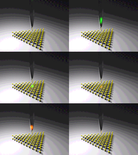
90:. The optical microscope is used to align the laser focal point with the tip coated with a SERS active metal. The three typical experimental configurations are bottom illumination, side illumination, and top illumination, depending on which direction the incident laser propagates towards the sample, with respect to the substrate. In the case of STM-TERS, only side and top illumination configurations can be applied, since the substrate is required to be conductive, therefore typically being non-transparent. In this case, the incident laser is usually linearly polarized and aligned parallel to the tip, in order to generate confined surface plasmon at the tip apex. The sample is moved rather than the tip so that the laser remains focused on the tip. The sample can be moved systematically to build up a series of tip enhanced Raman spectra from which a Raman map of the surface can be built allowing for surface heterogeneity to be assessed with up to 1.7 nm resolution. Subnanometer resolution has been demonstrated in certain cases allowing for submolecular features to be resolved.
1279:
94:
1291:
57:
Although the antennas' electric near-field distributions are commonly understood to determine the spatial resolution, recent experiments showing subnanometer-resolved optical images put this understanding into question. This is because such images enter a regime in which classical electrodynamical
105:
In 2019, Yan group and Liu group at
University of California, Riverside developed a lens-free nanofocusing technique, which concentrates the incident light from a tapered optical fiber to the tip apex of a metallic nanowire and collects the Raman signal through the same optical fiber.
1020:
He, Zhe; Han, Zehua; Kizer, Megan; Linhardt, Robert J.; Wang, Xing; Sinyukov, Alexander M.; Wang, Jizhou; Deckert, Volker; Sokolov, Alexei V. (2019-01-16). "Tip-Enhanced Raman
Imaging of Single-Stranded DNA with Single Base Resolution".
74:. Tip-enhanced Raman spectroscopy coupled with a scanning tunneling microscope (STM-TERS) has also become a reliable technique, since it utilizes the gap mode plasmon between the metallic probe and the metallic substrate.
46:, which is approximately half the wavelength of the incident light. Furthermore, with SERS spectroscopy the signal obtained is the sum of a relatively large number of molecules. TERS overcomes these limitations as the
1127:
731:
Hou, J. G.; Yang, J. L.; Luo, Y.; Aizpurua, J.; Y. Liao; Zhang, L.; Chen, L. G.; Zhang, C.; Jiang, S. (June 2013). "Chemical mapping of a single molecule by plasmon-enhanced Raman scattering".
961:
Lee, Joonhee; Crampton, Kevin T.; Tallarida, Nicholas; Apkarian, V. Ara (April 2019). "Visualizing vibrational normal modes of a single molecule with atomically confined light".
663:
Kim, Sanggon; Yu, Ning; Ma, Xuezhi; Zhu, Yangzhi; Liu, Qiushi; Liu, Ming; Yan, Ruoxue (2019). "High external-efficiency nanofocusing for lens-free near-field optical nanoscopy".
154:
Sonntag, Matthew D.; Pozzi, Eric A.; Jiang, Nan; Hersam, Mark C.; Van Duyne, Richard P. (18 September 2014). "Recent
Advances in Tip-Enhanced Raman Spectroscopy".
1089:
327:
Zhu, Wenqi; Esteban, Ruben; Borisov, Andrei G.; Baumberg, Jeremy J.; Nordlander, Peter; Lezec, Henri J.; Aizpurua, Javier; Crozier, Kenneth B. (2016-06-03).
119:
31:
TERS, with routine demonstrations of nanometer spatial resolution under ambient laboratory conditions, or better at ultralow temperatures and high pressure.
495:
Hayazawa, Norihiko; Inouye, Yasushi; Sekkat, Zouheir; Kawata, Satoshi (September 2000). "Metallized tip amplification of near-field Raman scattering".
460:
Stöckle, Raoul M.; Suh, Yung Doug; Deckert, Volker; Zenobi, Renato (February 2000). "Nanoscale chemical analysis by tip-enhanced Raman spectroscopy".
628:
Smolsky, Joseph; Krasnoslobodtsev, Alexey (8 August 2018). "Nanoscopic imaging of oxidized graphene monolayer using Tip-Enhanced Raman
Scattering".
1112:
98:
1142:
1132:
24:
1193:
1082:
123:
1152:
27:(SERS) that combines scanning probe microscopy with Raman spectroscopy. High spatial resolution chemical imaging is possible
1256:
1137:
1295:
1122:
1329:
1283:
1075:
707:
798:
Lee, Joonhee; Tallarida, Nicholas; Chen, Xing; Liu, Pengchong; Jensen, Lasse; Apkarian, Vartkess Ara (2017-10-12).
376:
Barbry, M.; Koval, P.; Marchesin, F.; Esteban, R.; Borisov, A. G.; Aizpurua, J.; Sánchez-Portal, D. (2015-05-04).
1324:
800:"Tip-Enhanced Raman Spectromicroscopy of Co(II)-Tetraphenylporphyrin on Au(111): Toward the Chemists' Microscope"
87:
114:
Several research have used TERS to image single atoms and the internal structure of the molecules. In 2019, the
1334:
1263:
1234:
58:
descriptions might no longer be applicable and quantum plasmonic and atomistic effects could become relevant.
425:
Anderson, Mark S. (2000). "Locally enhanced Raman spectroscopy with an atomic force microscope (AFM-TERS)".
71:
1117:
532:"A 1.7 nm resolution chemical analysis of carbon nanotubes by tip-enhanced Raman imaging in the ambient"
233:"A 1.7 nm resolution chemical analysis of carbon nanotubes by tip-enhanced Raman imaging in the ambient"
115:
1209:
970:
909:
740:
543:
504:
469:
434:
1339:
1229:
1168:
579:
Jiang, S.; Zhang, X.; Zhang, Y.; Hu, Ch.; Zhang, R.; Liao, Y.; Smith, Z.; Dong, Zh. (6 June 2017).
83:
1098:
1054:
1002:
780:
688:
645:
309:
35:
1046:
1038:
994:
986:
943:
925:
878:
870:
829:
821:
772:
764:
680:
610:
561:
407:
348:
301:
262:
254:
213:
171:
581:"Subnanometer-resolved chemical imaging via multivariate analysis of tip-enhanced Raman maps"
1224:
1219:
1214:
1183:
1030:
978:
933:
917:
896:
Lee, Joonhee; Tallarida, Nicholas; Chen, Xing; Jensen, Lasse; Apkarian, V. Ara (June 2018).
860:
849:"Tip-Enhanced Raman Spectromicroscopy on the Angstrom Scale: Bare and CO-Terminated Ag Tips"
811:
756:
748:
672:
637:
600:
592:
551:
512:
477:
442:
397:
389:
356:
340:
293:
244:
205:
163:
67:
39:
282:"Visualizing vibrational normal modes of a single molecule with atomically confined light"
974:
913:
744:
547:
508:
473:
438:
938:
897:
605:
580:
361:
193:
47:
516:
481:
280:
Lee, Joonhee; Crampton, Kevin T.; Tallarida, Nicholas; Apkarian, V. Ara (April 2019).
1318:
1173:
784:
692:
649:
378:"Atomistic Near-Field Nanoplasmonics: Reaching Atomic-Scale Resolution in Nanooptics"
1058:
1006:
313:
281:
1302:
1239:
377:
393:
1178:
209:
127:
982:
676:
641:
297:
93:
43:
1042:
990:
929:
874:
825:
768:
684:
411:
352:
305:
258:
232:
217:
192:
Shi, Xian; Coca-López, Nicolás; Janik, Julia; Hartschuh, Achim (2017-04-12).
865:
848:
816:
799:
131:
51:
1050:
998:
947:
921:
882:
833:
776:
614:
565:
329:"Quantum mechanical effects in plasmonic structures with subnanometre gaps"
266:
175:
1034:
328:
194:"Advances in Tip-Enhanced Near-Field Raman Microscopy Using Nanoantennas"
66:
The earliest reports of tip enhanced Raman spectroscopy typically used a
847:
Tallarida, Nicholas; Lee, Joonhee; Apkarian, Vartkess Ara (2017-10-09).
760:
752:
596:
402:
344:
1188:
556:
531:
249:
167:
1067:
446:
530:
Chen, Chi; Hayazawa, Norihiko; Kawata, Satoshi (12 February 2014).
92:
1071:
135:
1128:
Rotating-polarization coherent anti-Stokes Raman spectroscopy
231:
Chen, Chi; Hayazawa, Norihiko; Kawata, Satoshi (2014-02-12).
898:"Microscopy with a single-molecule scanning electrometer"
1248:
1202:
1161:
1105:
106:Fiber-in-fiber-out NSOM-TERS has been developed.
1083:
101:probe design for lens-free TERS measurement.
99:near-field scanning optical microscopy (NSOM)
8:
120:Center for Chemistry at the Space-Time Limit
54:within a few tens of nanometers of the tip.
708:"Fiber-optic probe can see molecular bonds"
82:Tip-enhanced Raman spectroscopy requires a
34:The maximum resolution achievable using an
1090:
1076:
1068:
937:
864:
815:
604:
555:
401:
360:
248:
156:The Journal of Physical Chemistry Letters
1023:Journal of the American Chemical Society
1113:Coherent anti-Stokes Raman spectroscopy
146:
138:sequencing has also been demonstrated.
50:obtained originates primarily from the
7:
1290:
187:
185:
1143:Surface-enhanced Raman spectroscopy
1133:Spatially offset Raman spectroscopy
25:surface-enhanced Raman spectroscopy
1194:Stimulated Raman adiabatic passage
14:
134:molecules using TERS. TERS-based
1289:
1278:
1277:
124:University of California, Irvine
1153:Transmission Raman spectroscopy
1148:Tip-enhanced Raman spectroscopy
17:Tip-enhanced Raman spectroscopy
1:
1257:Journal of Raman Spectroscopy
1138:Stimulated Raman spectroscopy
517:10.1016/S0030-4018(00)00894-4
482:10.1016/S0009-2614(99)01451-7
1123:Resonance Raman spectroscopy
394:10.1021/acs.nanolett.5b00759
210:10.1021/acs.chemrev.6b00640
1356:
1273:
983:10.1038/s41586-019-1059-9
677:10.1038/s41566-019-0456-9
642:10.1007/s12274-018-2158-x
298:10.1038/s41586-019-1059-9
88:scanning probe microscope
1264:Vibrational Spectroscopy
1235:Rule of mutual exclusion
462:Chemical Physics Letters
128:vibrational normal modes
866:10.1021/acsnano.7b06022
817:10.1021/acsnano.7b06183
427:Applied Physics Letters
72:atomic force microscope
1118:Raman optical activity
922:10.1126/sciadv.aat5472
102:
536:Nature Communications
497:Optics Communications
333:Nature Communications
237:Nature Communications
97:A fiber-in-fiber-out
96:
1210:Depolarization ratio
1035:10.1021/jacs.8b11506
42:, is limited by the
1230:Rayleigh scattering
1169:Raman amplification
975:2019Natur.568...78L
914:2018SciA....4.5472L
859:(11): 11393–11401.
810:(11): 11466–11474.
753:10.1038/nature12151
745:2013Natur.498...82Z
597:10.1038/lsa.2017.98
548:2014NatCo...5.3312C
509:2000OptCo.183..333H
474:2000CPL...318..131S
439:2000ApPhL..76.3130A
345:10.1038/ncomms11495
84:confocal microscope
1330:Raman spectroscopy
1099:Raman spectroscopy
557:10.1038/ncomms4312
250:10.1038/ncomms4312
103:
36:optical microscope
23:) is a variant of
1312:
1311:
712:UC Riverside News
636:(12): 6346–6359.
168:10.1021/jz5015746
162:(18): 3125–3130.
40:Raman microscopes
1347:
1325:Raman scattering
1293:
1292:
1281:
1280:
1225:Raman scattering
1220:Nonlinear optics
1215:Four-wave mixing
1184:Raman microscope
1092:
1085:
1078:
1069:
1063:
1062:
1017:
1011:
1010:
958:
952:
951:
941:
902:Science Advances
893:
887:
886:
868:
844:
838:
837:
819:
795:
789:
788:
728:
722:
721:
719:
718:
703:
697:
696:
665:Nature Photonics
660:
654:
653:
625:
619:
618:
608:
576:
570:
569:
559:
527:
521:
520:
503:(1–4): 333–336.
492:
486:
485:
468:(1–3): 131–136.
457:
451:
450:
447:10.1063/1.126546
422:
416:
415:
405:
388:(5): 3410–3419.
373:
367:
366:
364:
324:
318:
317:
277:
271:
270:
252:
228:
222:
221:
204:(7): 4945–4960.
198:Chemical Reviews
189:
180:
179:
151:
70:coupled with an
68:Raman microscope
1355:
1354:
1350:
1349:
1348:
1346:
1345:
1344:
1335:Surface science
1315:
1314:
1313:
1308:
1269:
1244:
1198:
1157:
1101:
1096:
1066:
1019:
1018:
1014:
969:(7750): 78–82.
960:
959:
955:
908:(6): eaat5472.
895:
894:
890:
846:
845:
841:
797:
796:
792:
739:(7452): 82–86.
730:
729:
725:
716:
714:
705:
704:
700:
662:
661:
657:
627:
626:
622:
578:
577:
573:
529:
528:
524:
494:
493:
489:
459:
458:
454:
424:
423:
419:
375:
374:
370:
326:
325:
321:
292:(7750): 78–82.
279:
278:
274:
230:
229:
225:
191:
190:
183:
153:
152:
148:
144:
112:
80:
64:
12:
11:
5:
1353:
1351:
1343:
1342:
1337:
1332:
1327:
1317:
1316:
1310:
1309:
1307:
1306:
1299:
1287:
1274:
1271:
1270:
1268:
1267:
1260:
1252:
1250:
1246:
1245:
1243:
1242:
1237:
1232:
1227:
1222:
1217:
1212:
1206:
1204:
1200:
1199:
1197:
1196:
1191:
1186:
1181:
1176:
1171:
1165:
1163:
1159:
1158:
1156:
1155:
1150:
1145:
1140:
1135:
1130:
1125:
1120:
1115:
1109:
1107:
1103:
1102:
1097:
1095:
1094:
1087:
1080:
1072:
1065:
1064:
1029:(2): 753–757.
1012:
953:
888:
839:
790:
723:
698:
671:(9): 636–643.
655:
620:
591:(11): e17098.
585:Light Sci Appl
571:
522:
487:
452:
417:
368:
319:
272:
223:
181:
145:
143:
140:
111:
108:
79:
76:
63:
60:
48:Raman spectrum
13:
10:
9:
6:
4:
3:
2:
1352:
1341:
1338:
1336:
1333:
1331:
1328:
1326:
1323:
1322:
1320:
1305:
1304:
1300:
1298:
1297:
1288:
1286:
1285:
1276:
1275:
1272:
1266:
1265:
1261:
1259:
1258:
1254:
1253:
1251:
1247:
1241:
1238:
1236:
1233:
1231:
1228:
1226:
1223:
1221:
1218:
1216:
1213:
1211:
1208:
1207:
1205:
1201:
1195:
1192:
1190:
1187:
1185:
1182:
1180:
1177:
1175:
1174:Raman cooling
1172:
1170:
1167:
1166:
1164:
1160:
1154:
1151:
1149:
1146:
1144:
1141:
1139:
1136:
1134:
1131:
1129:
1126:
1124:
1121:
1119:
1116:
1114:
1111:
1110:
1108:
1104:
1100:
1093:
1088:
1086:
1081:
1079:
1074:
1073:
1070:
1060:
1056:
1052:
1048:
1044:
1040:
1036:
1032:
1028:
1024:
1016:
1013:
1008:
1004:
1000:
996:
992:
988:
984:
980:
976:
972:
968:
964:
957:
954:
949:
945:
940:
935:
931:
927:
923:
919:
915:
911:
907:
903:
899:
892:
889:
884:
880:
876:
872:
867:
862:
858:
854:
850:
843:
840:
835:
831:
827:
823:
818:
813:
809:
805:
801:
794:
791:
786:
782:
778:
774:
770:
766:
762:
758:
754:
750:
746:
742:
738:
734:
727:
724:
713:
709:
706:Ober, Holly.
702:
699:
694:
690:
686:
682:
678:
674:
670:
666:
659:
656:
651:
647:
643:
639:
635:
631:
630:Nano Research
624:
621:
616:
612:
607:
602:
598:
594:
590:
586:
582:
575:
572:
567:
563:
558:
553:
549:
545:
541:
537:
533:
526:
523:
518:
514:
510:
506:
502:
498:
491:
488:
483:
479:
475:
471:
467:
463:
456:
453:
448:
444:
440:
436:
432:
428:
421:
418:
413:
409:
404:
399:
395:
391:
387:
383:
379:
372:
369:
363:
358:
354:
350:
346:
342:
338:
334:
330:
323:
320:
315:
311:
307:
303:
299:
295:
291:
287:
283:
276:
273:
268:
264:
260:
256:
251:
246:
242:
238:
234:
227:
224:
219:
215:
211:
207:
203:
199:
195:
188:
186:
182:
177:
173:
169:
165:
161:
157:
150:
147:
141:
139:
137:
133:
129:
125:
121:
118:group at the
117:
109:
107:
100:
95:
91:
89:
85:
77:
75:
73:
69:
61:
59:
55:
53:
49:
45:
41:
37:
32:
30:
26:
22:
18:
1303:Spectroscopy
1301:
1294:
1282:
1262:
1255:
1240:Stokes shift
1162:Applications
1147:
1026:
1022:
1015:
966:
962:
956:
905:
901:
891:
856:
852:
842:
807:
803:
793:
761:10261/102366
736:
732:
726:
715:. Retrieved
711:
701:
668:
664:
658:
633:
629:
623:
588:
584:
574:
539:
535:
525:
500:
496:
490:
465:
461:
455:
433:(21): 3130.
430:
426:
420:
403:10261/136309
385:
382:Nano Letters
381:
371:
336:
332:
322:
289:
285:
275:
240:
236:
226:
201:
197:
159:
155:
149:
116:Ara Apkarian
113:
110:Applications
104:
81:
65:
56:
38:, including
33:
28:
20:
16:
15:
1179:Raman laser
243:(1): 3312.
1340:Plasmonics
1319:Categories
1106:Techniques
717:2020-01-10
142:References
130:of single
44:Abbe limit
1043:0002-7863
991:0028-0836
930:2375-2548
875:1936-0851
826:1936-0851
785:205233946
769:1476-4687
693:195093429
685:1749-4893
650:139119548
412:1530-6984
353:2041-1723
306:1476-4687
259:2041-1723
218:0009-2665
132:porphyrin
78:Equipment
52:molecules
1284:Category
1249:Journals
1059:58552541
1051:30586988
1007:92998248
999:30944493
948:29963637
883:28980800
853:ACS Nano
834:28976729
804:ACS Nano
777:23739426
615:30167216
566:24518208
542:: 3312.
314:92998248
267:24518208
176:26276323
86:, and a
1296:Commons
1189:SHERLOC
971:Bibcode
939:6025905
910:Bibcode
741:Bibcode
606:6062048
544:Bibcode
505:Bibcode
470:Bibcode
435:Bibcode
362:4895716
126:imaged
62:History
1203:Theory
1057:
1049:
1041:
1005:
997:
989:
963:Nature
946:
936:
928:
881:
873:
832:
824:
783:
775:
767:
733:Nature
691:
683:
648:
613:
603:
564:
410:
359:
351:
312:
304:
286:Nature
265:
257:
216:
174:
1055:S2CID
1003:S2CID
781:S2CID
689:S2CID
646:S2CID
339:(1).
310:S2CID
1047:PMID
1039:ISSN
995:PMID
987:ISSN
944:PMID
926:ISSN
879:PMID
871:ISSN
830:PMID
822:ISSN
773:PMID
765:ISSN
681:ISSN
611:PMID
562:PMID
408:ISSN
349:ISSN
302:ISSN
263:PMID
255:ISSN
214:ISSN
172:PMID
21:TERS
1031:doi
1027:141
979:doi
967:568
934:PMC
918:doi
861:doi
812:doi
757:hdl
749:doi
737:498
673:doi
638:doi
601:PMC
593:doi
552:doi
513:doi
501:183
478:doi
466:318
443:doi
398:hdl
390:doi
357:PMC
341:doi
294:doi
290:568
245:doi
206:doi
202:117
164:doi
136:DNA
29:via
1321::
1053:.
1045:.
1037:.
1025:.
1001:.
993:.
985:.
977:.
965:.
942:.
932:.
924:.
916:.
904:.
900:.
877:.
869:.
857:11
855:.
851:.
828:.
820:.
808:11
806:.
802:.
779:.
771:.
763:.
755:.
747:.
735:.
710:.
687:.
679:.
669:13
667:.
644:.
634:11
632:.
609:.
599:.
587:.
583:.
560:.
550:.
538:.
534:.
511:.
499:.
476:.
464:.
441:.
431:76
429:.
406:.
396:.
386:15
384:.
380:.
355:.
347:.
335:.
331:.
308:.
300:.
288:.
284:.
261:.
253:.
239:.
235:.
212:.
200:.
196:.
184:^
170:.
158:.
122:,
1091:e
1084:t
1077:v
1061:.
1033::
1009:.
981::
973::
950:.
920::
912::
906:4
885:.
863::
836:.
814::
787:.
759::
751::
743::
720:.
695:.
675::
652:.
640::
617:.
595::
589:6
568:.
554::
546::
540:5
519:.
515::
507::
484:.
480::
472::
449:.
445::
437::
414:.
400::
392::
365:.
343::
337:7
316:.
296::
269:.
247::
241:5
220:.
208::
178:.
166::
160:5
19:(
Text is available under the Creative Commons Attribution-ShareAlike License. Additional terms may apply.
