176:
and/or low crystallographic symmetry, such as nano-crystalline materials or materials with defects. Off-axis TKD is often preferred for materials science research because it provides more information about the crystallographic orientation and microstructure of the sample, especially in samples with a high density of defects or a high degree of lattice strain. However, on-axis TKD can still be useful for studying samples with high crystallographic symmetry or for verifying the crystallographic orientation of a sample before performing off-axis TKD. The on-axis technique can speed up acquisition by more than 20 times, and a low scattering angle setup also gives rise to higher quality patterns.
30:
44:
1522:
1534:
168:. However, such machines are expensive and their operation requires particular skills and training. Additionally, the diffraction patterns obtained from TKD can be more complex to interpret than those obtained from conventional EBSD techniques due to the complex geometry of the diffracted electrons.
179:
EBSD resolution is influenced by multiple factors including the beam size, electron accelerating voltage, the material's atomic mass and the specimen's thickness. Out of these variables, sample thickness has the greatest effect on the pattern quality and resolution of the image. An increase in the
98:
is that in TEM, discrete diffraction spots arise from coherent scattering of the incident beam, while the formation of
Kikuchi bands is described as a two-step process consisting of incoherent scattering of the primary beam followed by coherent scattering of these forward biased electrons. TKD has
175:
In off-axis TKD, the sample is tilted with respect to the incident electron beam, typically at an angle of several degrees. This results in a diffraction pattern that is shifted away from the transmitted beam direction. Off-axis TKD is typically used for analysing samples with high lattice strain
171:
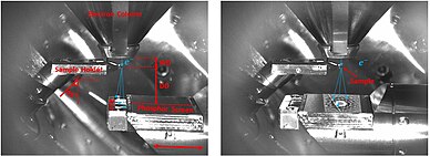
On-axis and off-axis TKD methods differ in the sample's orientation with respect to the electron beam. In on-axis TKD, the sample is oriented so that the incident electron beam is nearly perpendicular to the sample surface. This results in a diffraction pattern that is nearly centred around the
157:(STEM). Another advantage of TKD is its high sensitivity to local variations in crystallographic orientation. This is because the transmitted electrons in TKD are diffracted at very small angles, which makes the diffraction pattern highly sensitive to local variations in the crystal lattice.
694:
Meisnar, Martina; Vilalta-Clemente, Arantxa; Gholinia, Ali; Moody, Michael; Wilkinson, Angus J.; Huin, Nicolas; Lozano-Perez, Sergio (2015). "Using transmission
Kikuchi diffraction to study intergranular stress corrosion cracking in type 316 stainless steels 5000700".
781:
Yuan, H.; Brodu, E.; Chen, C.; Bouzy, E.; Fundenberger, J-J.; Toth, L.S. (2017). "On-axis versus off-axis
Transmission Kikuchi Diffraction technique: application to the characterisation of severe plastic deformation-induced ultrafine-grained microstructures".
82:
TKD offers improved spatial resolution, enabling effective characterization of nanocrystalline materials and heavily deformed samples where high dislocation densities can prevent successful characterization using conventional
606:
Brosusch, N.; Demers, H.; Gauvin, R. (2013). "Nanometres-resolution
Kikuchi patterns from materials science specimens with transmission electron forward scatter diffraction in the scanning electron microscope".
650:
Liang, X. Z.; Dodge, M. F.; Jiang, J.; Dong, H. B. (2019). "Using transmission
Kikuchi diffraction in a scanning electron microscope to quantify geometrically necessary dislocation density at the nanoscale".
148:
that can reach a few nanometres. This is achieved by using a small electron beam spot size, typically less than 10 nanometres in diameter, and by collecting the transmitted electrons with a small-angle
371:
172:
transmitted beam direction. On-axis TKD is typically used for analysing samples with low lattice strain and high crystallographic symmetry, such as single crystals or large grains.
1166:
550:
Liu, Junliang; Lozano-Perez, Sergio; Wilkinson, Angus J.; Grovenor, Chris R. M. (2019). "On the depth resolution of transmission
Kikuchi diffraction (TKD) analysis 9300920".
130:, which has been introduced around the 1970s, and has since become increasingly popular in materials science research, especially for studying materials at the nanoscale.
1149:
1144:
1293:
996:
1283:
886:
79:(SEM). This technique has been widely utilised in the characterization of nano-crystalline materials, including oxides, superconductors, and metallic alloys.
1186:
1159:
106:
samples in a scanning electron microscope. The preparation of TKD samples can be done with standard methods used for transmission electron microscopy (TEM).
1094:
512:
Trimby, Patrick W. (2012). "Orientation mapping of nanostructured materials using transmission
Kikuchi diffraction in the scanning electron microscope".
141:. The diffraction pattern is then collected by a detector and analysed to determine the crystallographic orientation and microstructure of the sample.
1154:
825:
Rice, K.P.; Keller, R.R.; Stoykovich, M.P. (2014). "Specimen-thickness effects on transmission
Kikuchi patterns in the scanning electron microscope".
154:
1298:
431:
Sneddon, Glenn C.; Trimby, Patrick W.; Cairney, Julie M. (2016). "Transmission
Kikuchi diffraction in a scanning electron microscope: A review".
1114:
1238:
1104:
1051:
160:
TKD can also be used to study nano-sized materials, such as nanoparticles and thin films. Thin foil samples can be prepared for TKD using a
1493:
1221:
1206:
1132:
127:
393:
Keller, R.R.; Geiss, R.H. (2012). "Transmission EBSD from 10 nm domains in a scanning electron microscope: Transmission EBSD in the SEM".
732:"Mapping the full lattice strain tensor of a single dislocation by high angular resolution transmission Kikuchi diffraction (HR-TKD)"
1243:
1076:
929:
879:
1226:
1124:
1099:
1066:
138:
115:
84:
72:
1303:
1288:
1268:
1006:
1565:
1071:
914:
1538:
1248:
1211:
1056:
1526:
1086:
872:
468:
134:
76:
137:. The electron beam is then focused on a small spot on the sample, and the crystal lattice of the sample diffracts the
1273:
1191:
1498:
1452:
1278:
343:
1488:
71:), is a method for orientation mapping at the nanoscale. It’s used for analysing the microstructures of thin
1201:
1137:
1011:
970:
95:
1391:
119:
1570:
1396:
1216:
150:
29:
257:
224:
191:
1560:
1401:
960:
91:
43:
1366:
1351:
1258:
1253:
1196:
1061:
1043:
975:
965:
909:
895:
165:
1437:
1016:
980:
850:
807:
763:
743:
676:
632:
585:
559:
410:
145:
1381:
1376:
842:
799:
712:
668:
624:
577:
529:
491:
363:
324:
277:
244:
211:
1331:
1263:
834:
791:
753:
704:
660:
616:
569:
521:
483:
440:
402:
355:
316:
305:"Transmission Kikuchi diffraction study of submicrotexture within ultramylonitic peridotite"
269:
236:
203:
161:
730:
Yu, Hongbing; Liu, Junliang; Karamched, Phani; Wilkinson, Angus J.; Hofmann, Felix (2019).
223:
Fundenberger, J. J.; Bouzy, E.; Goran, D.; Guyon, J.; Yuan, H.; Morawiec, A. (2016-02-01).
1386:
133:
In TKD, a thin foil sample is prepared and placed perpendicular to the electron beam of a
342:
van Bremen, R.; Ribas Gomes, D.; de Jeer, L.T.H.; Ocelík, V.; De Hosson, J.Th.M. (2016).
1341:
955:
344:"On the optimum resolution of transmission-electron backscattered diffraction (t-EBSD)"
123:
1554:
1417:
1361:
1021:
767:
758:
731:
680:
589:
406:
100:
1468:
854:
636:
414:
1478:
1442:
1336:
1326:
1001:
950:
811:
664:
573:
525:
487:
359:
273:
240:
469:"A systematic comparison of on-axis and off-axis transmission Kikuchi diffraction"
258:"A systematic comparison of on-axis and off-axis transmission Kikuchi diffraction"
180:
sample thickness broadens the beam, thus reducing the lateral spatial resolution.
708:
1422:
1356:
1346:
444:
320:
207:
1503:
924:
919:
192:"Transmission Kikuchi diffraction in a scanning electron microscope: A review"
103:
328:
304:
281:
248:
215:
1371:
1026:
846:
803:
716:
672:
628:
581:
533:
495:
367:
35:
Off-axis TKD with an example EBSP. Right: On-axis TKD with an example EBSP
1473:
939:
1483:
1427:
1109:
190:
Sneddon, Glenn C.; Trimby, Patrick W.; Cairney, Julie M. (2016-12-01).
864:
838:
795:
620:
1447:
748:
564:
49:
Imaging using diodes in on-axis TKD setup. Right: on-axis TKD setup
256:
Niessen, F.; Burrows, A.; Fanta, A. Bastos da Silva (2018-03-01).
225:"Orientation mapping by transmission-SEM with an on-axis detector"
1432:
126:
of materials at a high spatial resolution. It is a variation of
868:
87:. Many studies have reported sub-10 nm resolution using TKD.
467:
Niessen, F.; Burrows, A.; Fanta, A. Bastos da Silva (2018).
114:
Transmission
Kikuchi diffraction (TKD or t-EBSD) is an
303:
Igami, Yohei; Michibayashi, Katsuyoshi (2021-09-29).
1461:
1410:
1319:
1312:
1179:
1123:
1085:
1042:
1035:
989:
938:
902:
1294:Serial block-face scanning electron microscopy
997:Detectors for transmission electron microscopy
880:
433:Materials Science and Engineering: R: Reports
196:Materials Science and Engineering: R: Reports
144:One of the key advantages of TKD is its high
118:(EBSD) technique that is used to analyse the
65:transmission-electron backscatter diffraction
8:
1316:
1039:
887:
873:
865:
99:also been applied to analyse fine-grained
757:
747:
563:
155:scanning transmission electron microscope
295:
22:Transmission Kikuchi diffraction setup
601:
599:
545:
543:
507:
505:
462:
460:
458:
456:
454:
426:
424:
7:
1533:
128:convergent-beam electron diffraction
16:Nanoscale orientation mapping method
374:from the original on 25 March 2023
14:
930:Timeline of microscope technology
309:Physics and Chemistry of Minerals
1532:
1521:
1520:
759:10.1016/j.scriptamat.2018.12.039
407:10.1111/j.1365-2818.2011.03566.x
116:Electron backscatter diffraction
85:Electron backscatter diffraction
73:transmission electron microscopy
57:Transmission Kikuchi Diffraction
42:
28:
1289:Precession electron diffraction
665:10.1016/j.ultramic.2018.11.011
574:10.1016/j.ultramic.2019.06.003
526:10.1016/j.ultramic.2012.06.004
488:10.1016/j.ultramic.2017.12.017
360:10.1016/j.ultramic.2015.10.025
274:10.1016/j.ultramic.2017.12.017
241:10.1016/j.ultramic.2015.11.002
1:
709:10.1016/j.micron.2015.04.011
135:scanning electron microscope
120:crystallographic orientation
90:The main difference between
77:scanning electron microscope
151:annular dark-field detector
1587:
1274:Immune electron microscopy
1192:Annular dark-field imaging
1007:Everhart–Thornley detector
445:10.1016/j.mser.2016.10.001
321:10.1007/s00269-021-01161-7
208:10.1016/j.mser.2016.10.001
1516:
1428:Hitachi High-Technologies
63:), also sometimes called
1453:Thermo Fisher Scientific
1279:Geometric phase analysis
1167:Aberration-Corrected TEM
1202:Charge contrast imaging
1012:Field electron emission
75:(TEM) specimens in the
1392:Thomas Eugene Everhart
1566:Scientific techniques
1397:Vernon Ellis Cosslett
1217:Dark-field microscopy
827:Journal of Microscopy
784:Journal of Microscopy
609:Journal of Microscopy
395:Journal of Microscopy
139:transmitted electrons
1402:Vladimir K. Zworykin
1052:Correlative light EM
961:Electron diffraction
1367:Manfred von Ardenne
1352:Gerasimos Danilatos
1259:Electron tomography
1254:Electron holography
1197:Cathodoluminescence
976:Secondary electrons
966:Electron scattering
910:Electron microscopy
896:Electron microscopy
166:ion milling machine
1489:Digital Micrograph
1095:Environmental SEM
1017:Field emission gun
981:X-ray fluorescence
736:Scripta Materialia
146:spatial resolution
1548:
1547:
1512:
1511:
1382:Nestor J. Zaluzec
1377:Maximilian Haider
1175:
1174:
839:10.1111/jmi.12124
796:10.1111/jmi.12548
621:10.1111/jmi.12007
1578:
1536:
1535:
1524:
1523:
1332:Bodo von Borries
1317:
1077:Photoemission EM
1040:
889:
882:
875:
866:
859:
858:
822:
816:
815:
778:
772:
771:
761:
751:
727:
721:
720:
691:
685:
684:
647:
641:
640:
603:
594:
593:
567:
547:
538:
537:
509:
500:
499:
473:
464:
449:
448:
428:
419:
418:
390:
384:
383:
381:
379:
339:
333:
332:
300:
285:
252:
219:
162:Focused ion beam
153:(STEM-ADF) in a
46:
32:
1586:
1585:
1581:
1580:
1579:
1577:
1576:
1575:
1551:
1550:
1549:
1544:
1508:
1457:
1406:
1387:Ondrej Krivanek
1308:
1171:
1119:
1081:
1067:Liquid-Phase EM
1031:
990:Instrumentation
985:
943:
934:
898:
893:
863:
862:
824:
823:
819:
780:
779:
775:
729:
728:
724:
693:
692:
688:
653:Ultramicroscopy
649:
648:
644:
605:
604:
597:
552:Ultramicroscopy
549:
548:
541:
514:Ultramicroscopy
511:
510:
503:
476:Ultramicroscopy
471:
466:
465:
452:
430:
429:
422:
392:
391:
387:
377:
375:
348:Ultramicroscopy
341:
340:
336:
302:
301:
297:
292:
262:Ultramicroscopy
255:
229:Ultramicroscopy
222:
189:
186:
184:Further reading
112:
54:
53:
52:
51:
50:
47:
38:
37:
36:
33:
24:
23:
17:
12:
11:
5:
1584:
1582:
1574:
1573:
1568:
1563:
1553:
1552:
1546:
1545:
1543:
1542:
1530:
1517:
1514:
1513:
1510:
1509:
1507:
1506:
1501:
1496:
1494:Direct methods
1491:
1486:
1481:
1476:
1471:
1465:
1463:
1459:
1458:
1456:
1455:
1450:
1445:
1440:
1435:
1430:
1425:
1420:
1414:
1412:
1408:
1407:
1405:
1404:
1399:
1394:
1389:
1384:
1379:
1374:
1369:
1364:
1359:
1354:
1349:
1344:
1342:Ernst G. Bauer
1339:
1334:
1329:
1323:
1321:
1314:
1310:
1309:
1307:
1306:
1301:
1296:
1291:
1286:
1281:
1276:
1271:
1266:
1261:
1256:
1251:
1246:
1241:
1236:
1235:
1234:
1224:
1219:
1214:
1209:
1204:
1199:
1194:
1189:
1183:
1181:
1177:
1176:
1173:
1172:
1170:
1169:
1164:
1163:
1162:
1152:
1147:
1142:
1141:
1140:
1129:
1127:
1121:
1120:
1118:
1117:
1112:
1107:
1102:
1097:
1091:
1089:
1083:
1082:
1080:
1079:
1074:
1069:
1064:
1059:
1054:
1048:
1046:
1037:
1033:
1032:
1030:
1029:
1024:
1019:
1014:
1009:
1004:
999:
993:
991:
987:
986:
984:
983:
978:
973:
968:
963:
958:
956:Bremsstrahlung
953:
947:
945:
936:
935:
933:
932:
927:
922:
917:
912:
906:
904:
900:
899:
894:
892:
891:
884:
877:
869:
861:
860:
833:(3): 129–136.
817:
773:
722:
686:
642:
595:
539:
501:
450:
420:
401:(3): 245–251.
385:
334:
294:
293:
291:
288:
287:
286:
253:
220:
185:
182:
124:microstructure
111:
108:
48:
41:
40:
39:
34:
27:
26:
25:
21:
20:
19:
18:
15:
13:
10:
9:
6:
4:
3:
2:
1583:
1572:
1569:
1567:
1564:
1562:
1559:
1558:
1556:
1541:
1540:
1531:
1529:
1528:
1519:
1518:
1515:
1505:
1502:
1500:
1497:
1495:
1492:
1490:
1487:
1485:
1482:
1480:
1477:
1475:
1472:
1470:
1467:
1466:
1464:
1460:
1454:
1451:
1449:
1446:
1444:
1441:
1439:
1436:
1434:
1431:
1429:
1426:
1424:
1421:
1419:
1418:Carl Zeiss AG
1416:
1415:
1413:
1411:Manufacturers
1409:
1403:
1400:
1398:
1395:
1393:
1390:
1388:
1385:
1383:
1380:
1378:
1375:
1373:
1370:
1368:
1365:
1363:
1362:James Hillier
1360:
1358:
1355:
1353:
1350:
1348:
1345:
1343:
1340:
1338:
1335:
1333:
1330:
1328:
1325:
1324:
1322:
1318:
1315:
1311:
1305:
1302:
1300:
1297:
1295:
1292:
1290:
1287:
1285:
1282:
1280:
1277:
1275:
1272:
1270:
1267:
1265:
1262:
1260:
1257:
1255:
1252:
1250:
1247:
1245:
1242:
1240:
1237:
1233:
1230:
1229:
1228:
1225:
1223:
1220:
1218:
1215:
1213:
1210:
1208:
1205:
1203:
1200:
1198:
1195:
1193:
1190:
1188:
1185:
1184:
1182:
1178:
1168:
1165:
1161:
1158:
1157:
1156:
1153:
1151:
1148:
1146:
1143:
1139:
1136:
1135:
1134:
1131:
1130:
1128:
1126:
1122:
1116:
1115:Ultrafast SEM
1113:
1111:
1108:
1106:
1103:
1101:
1098:
1096:
1093:
1092:
1090:
1088:
1084:
1078:
1075:
1073:
1072:Low-energy EM
1070:
1068:
1065:
1063:
1060:
1058:
1055:
1053:
1050:
1049:
1047:
1045:
1041:
1038:
1034:
1028:
1025:
1023:
1022:Magnetic lens
1020:
1018:
1015:
1013:
1010:
1008:
1005:
1003:
1000:
998:
995:
994:
992:
988:
982:
979:
977:
974:
972:
971:Kikuchi lines
969:
967:
964:
962:
959:
957:
954:
952:
949:
948:
946:
941:
937:
931:
928:
926:
923:
921:
918:
916:
913:
911:
908:
907:
905:
901:
897:
890:
885:
883:
878:
876:
871:
870:
867:
856:
852:
848:
844:
840:
836:
832:
828:
821:
818:
813:
809:
805:
801:
797:
793:
789:
785:
777:
774:
769:
765:
760:
755:
750:
745:
741:
737:
733:
726:
723:
718:
714:
710:
706:
702:
698:
690:
687:
682:
678:
674:
670:
666:
662:
658:
654:
646:
643:
638:
634:
630:
626:
622:
618:
614:
610:
602:
600:
596:
591:
587:
583:
579:
575:
571:
566:
561:
557:
553:
546:
544:
540:
535:
531:
527:
523:
519:
515:
508:
506:
502:
497:
493:
489:
485:
481:
477:
470:
463:
461:
459:
457:
455:
451:
446:
442:
438:
434:
427:
425:
421:
416:
412:
408:
404:
400:
396:
389:
386:
373:
369:
365:
361:
357:
353:
349:
345:
338:
335:
330:
326:
322:
318:
314:
310:
306:
299:
296:
289:
283:
279:
275:
271:
267:
263:
259:
254:
250:
246:
242:
238:
234:
230:
226:
221:
217:
213:
209:
205:
201:
197:
193:
188:
187:
183:
181:
177:
173:
169:
167:
163:
158:
156:
152:
147:
142:
140:
136:
131:
129:
125:
121:
117:
109:
107:
105:
102:
101:ultramylonite
97:
96:Kikuchi bands
93:
88:
86:
80:
78:
74:
70:
66:
62:
58:
45:
31:
1571:Spectroscopy
1537:
1525:
1479:EM Data Bank
1443:Nion Company
1337:Dennis Gabor
1327:Albert Crewe
1231:
1105:Confocal SEM
1002:Electron gun
951:Auger effect
830:
826:
820:
790:(1): 70–80.
787:
783:
776:
739:
735:
725:
700:
696:
689:
656:
652:
645:
612:
608:
555:
551:
517:
513:
479:
475:
436:
432:
398:
394:
388:
376:. Retrieved
351:
347:
337:
312:
308:
298:
265:
261:
232:
228:
199:
195:
178:
174:
170:
159:
143:
132:
113:
89:
81:
68:
64:
60:
56:
55:
1561:Diffraction
1423:FEI Company
1357:Harald Rose
1347:Ernst Ruska
1036:Microscopes
944:with matter
942:interaction
615:(1): 1–14.
482:: 158–170.
354:: 256–264.
268:: 158–170.
110:Description
92:diffraction
1555:Categories
1504:Multislice
1320:Developers
1180:Techniques
925:Microscope
920:Micrograph
749:1808.10055
565:1904.04140
315:(10): 38.
290:References
104:peridotite
94:spots and
1372:Max Knoll
1027:Stigmator
768:119075799
742:: 36–41.
681:205526130
659:: 39–45.
590:102350683
520:: 16–24.
329:1432-2021
282:0304-3991
249:0304-3991
235:: 17–22.
216:0927-796X
164:(FIB) or
1527:Category
1474:CrysTBox
1462:Software
1133:Cryo-TEM
940:Electron
855:25006562
847:24660836
804:28328010
717:25974882
703:: 1–10.
673:30496887
637:20435127
629:23346885
582:31234103
558:: 5–12.
534:22796555
496:29335225
439:: 1–12.
415:39521418
378:20 March
372:Archived
368:26579885
202:: 1–12.
1539:Commons
1187:4D STEM
1160:4D STEM
1138:Cryo-ET
1110:SEM-XRF
1100:CryoSEM
1057:Cryo-EM
915:History
812:7988685
1484:EMsoft
1469:CASINO
1448:TESCAN
1313:Others
1212:cryoEM
903:Basics
853:
845:
810:
802:
766:
715:
697:Micron
679:
671:
635:
627:
588:
580:
532:
494:
413:
366:
327:
280:
247:
214:
69:t-EBSD
1438:Leica
1284:PINEM
1150:HRTEM
1145:EFTEM
851:S2CID
808:S2CID
764:S2CID
744:arXiv
677:S2CID
633:S2CID
586:S2CID
560:arXiv
472:(PDF)
411:S2CID
1499:IUCr
1433:JEOL
1304:WBDF
1299:WDXS
1249:EBIC
1244:EELS
1239:ECCI
1227:EBSD
1207:CBED
1155:STEM
843:PMID
800:PMID
713:PMID
669:PMID
625:PMID
578:PMID
530:PMID
492:PMID
380:2023
364:PMID
325:ISSN
278:ISSN
245:ISSN
212:ISSN
122:and
1269:FEM
1264:FIB
1232:TKD
1222:EDS
1125:TEM
1087:SEM
1062:EMP
835:doi
831:254
792:doi
788:267
754:doi
740:164
705:doi
661:doi
657:197
617:doi
613:250
570:doi
556:205
522:doi
518:120
484:doi
480:186
441:doi
437:110
403:doi
399:245
356:doi
352:160
317:doi
270:doi
266:186
237:doi
233:161
204:doi
200:110
61:TKD
1557::
1044:EM
849:.
841:.
829:.
806:.
798:.
786:.
762:.
752:.
738:.
734:.
711:.
701:75
699:.
675:.
667:.
655:.
631:.
623:.
611:.
598:^
584:.
576:.
568:.
554:.
542:^
528:.
516:.
504:^
490:.
478:.
474:.
453:^
435:.
423:^
409:.
397:.
370:.
362:.
350:.
346:.
323:.
313:48
311:.
307:.
276:.
264:.
260:.
243:.
231:.
227:.
210:.
198:.
194:.
888:e
881:t
874:v
857:.
837::
814:.
794::
770:.
756::
746::
719:.
707::
683:.
663::
639:.
619::
592:.
572::
562::
536:.
524::
498:.
486::
447:.
443::
417:.
405::
382:.
358::
331:.
319::
284:.
272::
251:.
239::
218:.
206::
67:(
59:(
Text is available under the Creative Commons Attribution-ShareAlike License. Additional terms may apply.

