174:
212:
158:
193:
617:
718:
31:
730:
68:
of the transmitted light in dense areas of the sample. Bright-field microscopy is the simplest of a range of techniques used for illumination of samples in light microscopes, and its simplicity makes it a popular technique. The typical appearance of a bright-field microscopy image is a dark sample on
285:
Samples that are naturally colorless and transparent cannot be seen well, e.g. many types of mammalian cells. These samples often have to be stained before viewing. Samples that do have their own color can be seen without preparation, e.g. the observation of
173:
211:
143:
is often required to increase contrast, which prevents use on live cells in many situations. Bright-field illumination is useful for samples that have an intrinsic color, for example mitochondria found in cells.
274:
The practical limit to magnification with a light microscope is around 1300X. Although higher magnifications are possible, it becomes increasingly difficult to maintain image clarity as the magnification
157:
83:
The light path of a bright-field microscope is extremely simple, no additional components are required beyond the normal light-microscope setup. The light path therefore consists of:
192:
660:
571:
499:
756:
703:
698:
566:
359:
665:
400:
601:
581:
429:
645:
641:
492:
445:
221:
670:
734:
722:
485:
345:
on the light source to highlight features not visible under white light. The use of filters is especially useful with
688:
378:
179:
561:
649:
631:
312:
65:
57:
551:
124:
556:
315:
lens and a special immersion oil placed on a glass cover over the specimen. Immersion oil has the same
287:
198:
120:
388:
Advanced Light
Microscopy vol. 1 Principles and Basic Properties by Maksymilian Pluta, Elsevier (1988)
60:
techniques. Sample illumination is transmitted (i.e., illuminated from below and observed from above)
183:
636:
576:
397:
Microbiology: Principles and
Explorations by Jacquelyn G. Black, John Wiley & Sons, Inc. (2005)
202:
394:
Introduction to Light
Microscopy by S. Bradbury, B. Bracegirdle, BIOS Scientific Publishers (1998)
527:
279:
234:
54:
425:
373:
268:
136:
616:
606:
78:
61:
693:
391:
Advanced Light
Microscopy vol. 2 Specialised Methods by Maksymilian Pluta, Elsevier (1989)
342:
39:
586:
335:
327:
305:
217:
102:
95:
17:
750:
655:
242:
230:
456:
148:
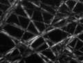
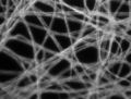
Comparison of transillumination techniques used to generate contrast in a sample of
323:
291:
149:
88:
338:) and differential stains (negative stains, flagellar stains, endospore stains).
229:
Bright-field microscopy is a standard light-microscopy technique, and therefore
591:
520:
508:
316:
238:
164:
35:
30:
331:
140:
108:
346:
596:
477:
139:
with most biological samples, as few absorb light to a great extent.
112:
29:
481:
105:, which collects light from the sample and magnifies the image;
319:
as glass and improves the resolution of the observed specimen.
304:
Reducing or increasing the amount of the light source by the
98:, which focuses light from the light source onto the sample;
182:
illumination, sample contrast comes from the rotation of
661:
Total internal reflection fluorescence microscopy (TIRF)
257:
Living cells can be seen with bright-field microscopes.
254:
Simplicity of setup with only basic equipment required.
163:
Bright-field illumination, sample contrast comes from
224:
of different path lengths of light through the sample
699:
Photo-activated localization microscopy (PALM/STORM)
679:
624:
537:
201:illumination, sample contrast comes from light
341:Use of a colored (usually blue) or polarizing
602:Interference reflection microscopy (IRM/RICM)
493:
87:a transillumination light source, commonly a
8:
424:(4th ed.). New York: Garland Science.
500:
486:
478:
322:Use of sample-staining methods for use in
135:Bright-field microscopy typically has low
64:, and contrast in the sample is caused by
38:. This image shows a cross-section of the
282:due to the blur of out-of-focus material;
220:illumination, sample contrast comes from
27:Optical microscopy illumination technique
572:Differential interference contrast (DIC)
412:
146:
567:Quantitative phase-contrast microscopy
69:a bright background, hence the name.
7:
729:
694:Stimulated emission depletion (STED)
420:Alberts, Bruce; et al. (2002).
25:
666:Lightsheet microscopy (LSFM/SPIM)
446:"Microscopy: Types of Microscopy"
401:Microscopy and Imaging Literature
728:
717:
716:
615:
210:
191:
172:
156:
119:Bright-field microscopy may use
671:Lattice light-sheet microscopy
582:Second harmonic imaging (SHIM)
453:Hillsborough Community College
1:
757:Optical microscopy techniques
422:Molecular biology of the cell
53:) is the simplest of all the
379:Resources in other libraries
271:of most biological samples;
773:
127:to illuminate the sample.
76:
712:
613:
515:
374:Resources in your library
326:, such as simple stains (
115:to view the sample image.
186:light through the sample
91:in the microscope stand;
34:An example bright-field
632:Fluorescence microscopy
592:Structured illumination
547:Bright-field microscopy
365:Bright-field microscopy
313:oil-immersion objective
47:Bright-field microscopy
704:Near-field (NSOM/SNOM)
642:Multiphoton microscopy
167:of light in the sample
43:
18:Brightfield microscopy
557:Dark-field microscopy
288:cytoplasmic streaming
180:Cross-polarized light
33:
625:Fluorescence methods
656:Image deconvolution
637:Confocal microscopy
577:Dispersion staining
552:Köhler illumination
125:Köhler illumination
528:Optical microscopy
509:Optical microscopy
280:optical resolution
237:possible with the
233:is limited by the
55:optical microscopy
44:
744:
743:
689:Diffraction limit
360:Library resources
16:(Redirected from
764:
732:
731:
720:
719:
682:limit techniques
619:
540:contrast methods
538:Illumination and
502:
495:
488:
479:
472:
471:
469:
467:
462:on 20 April 2017
461:
455:. Archived from
450:
442:
436:
435:
417:
214:
195:
176:
160:
152:(1.559 μm/pixel)
79:light microscope
42:in a plant stem.
21:
772:
771:
767:
766:
765:
763:
762:
761:
747:
746:
745:
740:
708:
681:
680:Sub-diffraction
675:
620:
611:
539:
533:
511:
506:
476:
475:
465:
463:
459:
448:
444:
443:
439:
432:
419:
418:
414:
409:
385:
384:
383:
368:
367:
363:
356:
301:
264:
251:
235:resolving power
225:
215:
206:
196:
187:
177:
168:
161:
133:
81:
75:
40:vascular tissue
28:
23:
22:
15:
12:
11:
5:
770:
768:
760:
759:
749:
748:
742:
741:
739:
738:
726:
713:
710:
709:
707:
706:
701:
696:
691:
685:
683:
677:
676:
674:
673:
668:
663:
658:
653:
639:
634:
628:
626:
622:
621:
614:
612:
610:
609:
604:
599:
594:
589:
587:4Pi microscope
584:
579:
574:
569:
564:
562:Phase contrast
559:
554:
549:
543:
541:
535:
534:
532:
531:
524:
516:
513:
512:
507:
505:
504:
497:
490:
482:
474:
473:
437:
430:
411:
410:
408:
405:
404:
403:
398:
395:
392:
389:
382:
381:
376:
370:
369:
358:
357:
355:
352:
351:
350:
339:
336:crystal violet
328:methylene blue
320:
309:
306:iris diaphragm
300:
297:
296:
295:
283:
276:
272:
263:
260:
259:
258:
255:
250:
247:
227:
226:
218:Phase-contrast
216:
209:
207:
197:
190:
188:
178:
171:
169:
162:
155:
153:
132:
129:
117:
116:
106:
103:objective lens
99:
96:condenser lens
92:
77:Main article:
74:
71:
26:
24:
14:
13:
10:
9:
6:
4:
3:
2:
769:
758:
755:
754:
752:
737:
736:
727:
725:
724:
715:
714:
711:
705:
702:
700:
697:
695:
692:
690:
687:
686:
684:
678:
672:
669:
667:
664:
662:
659:
657:
654:
651:
647:
643:
640:
638:
635:
633:
630:
629:
627:
623:
618:
608:
605:
603:
600:
598:
595:
593:
590:
588:
585:
583:
580:
578:
575:
573:
570:
568:
565:
563:
560:
558:
555:
553:
550:
548:
545:
544:
542:
536:
530:
529:
525:
523:
522:
518:
517:
514:
510:
503:
498:
496:
491:
489:
484:
483:
480:
458:
454:
447:
441:
438:
433:
431:0-8153-3218-1
427:
423:
416:
413:
406:
402:
399:
396:
393:
390:
387:
386:
380:
377:
375:
372:
371:
366:
361:
353:
348:
344:
340:
337:
333:
329:
325:
321:
318:
314:
310:
307:
303:
302:
298:
293:
289:
284:
281:
278:Low apparent
277:
273:
270:
266:
265:
261:
256:
253:
252:
248:
246:
244:
243:visible light
240:
236:
232:
231:magnification
223:
219:
213:
208:
205:by the sample
204:
200:
194:
189:
185:
181:
175:
170:
166:
159:
154:
151:
147:
145:
142:
138:
130:
128:
126:
122:
114:
110:
107:
104:
100:
97:
93:
90:
86:
85:
84:
80:
72:
70:
67:
63:
59:
56:
52:
48:
41:
37:
32:
19:
733:
721:
650:Three-photon
546:
526:
519:
464:. Retrieved
457:the original
452:
440:
421:
415:
364:
324:microbiology
299:Enhancements
228:
222:interference
150:tissue paper
134:
118:
89:halogen lamp
82:
58:illumination
50:
46:
45:
262:Limitations
131:Performance
66:attenuation
62:white light
646:Two-photon
521:Microscope
354:References
317:refraction
311:Use of an
275:increases;
249:Advantages
239:wavelength
199:Dark-field
165:absorbance
73:Light path
36:micrograph
267:Very low
203:scattered
184:polarized
111:and/or a
751:Category
723:Category
466:19 April
349:samples.
332:safranin
269:contrast
141:Staining
137:contrast
121:critical
735:Commons
347:mineral
109:oculars
597:Sarfus
428:
362:about
343:filter
294:cells.
113:camera
607:Raman
460:(PDF)
449:(PDF)
407:Notes
292:Chara
468:2017
426:ISBN
290:in
241:of
123:or
101:an
753::
648:,
451:.
334:,
330:,
245:.
94:a
51:BF
652:)
644:(
501:e
494:t
487:v
470:.
434:.
308:.
49:(
20:)
Text is available under the Creative Commons Attribution-ShareAlike License. Additional terms may apply.